





Tested Applications
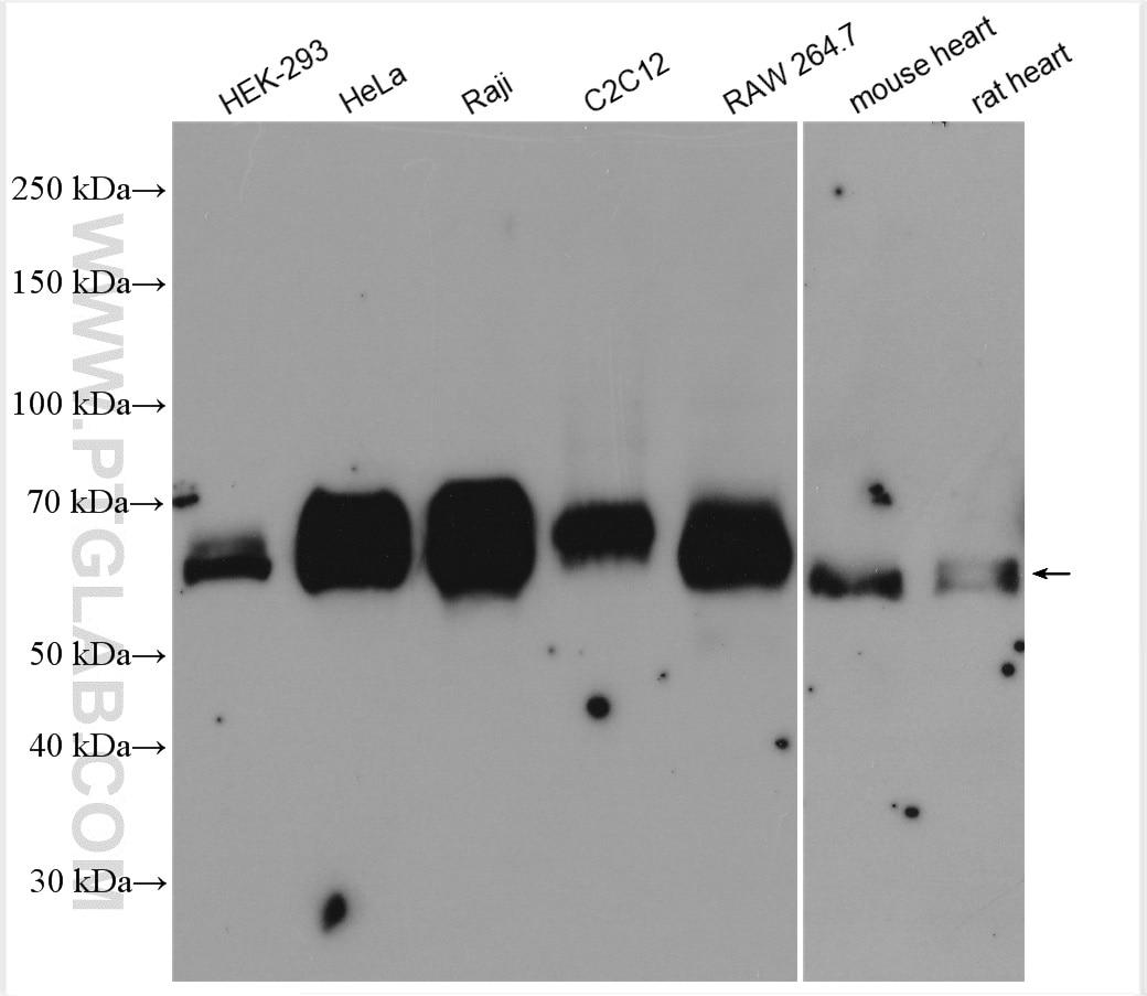
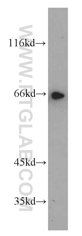
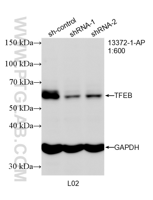
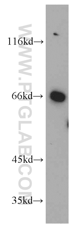
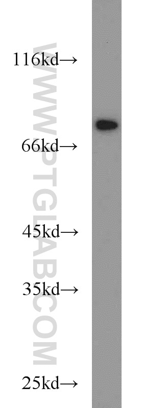
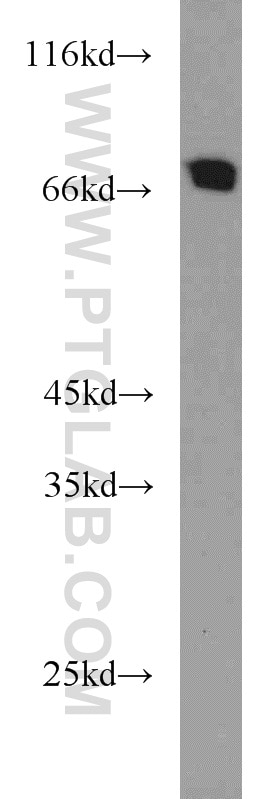
| Positive WB detected in | A549 cells, Jurkat cells, human placenta tissue, NIH/3T3 cells, mouse brain tissue, L02 cells, HEK-293 cells, HeLa cells, Raji cells, C2C12 cells, RAW 264.7 cells, mouse heart tissue, rat heart tissue, rat brain tissue |
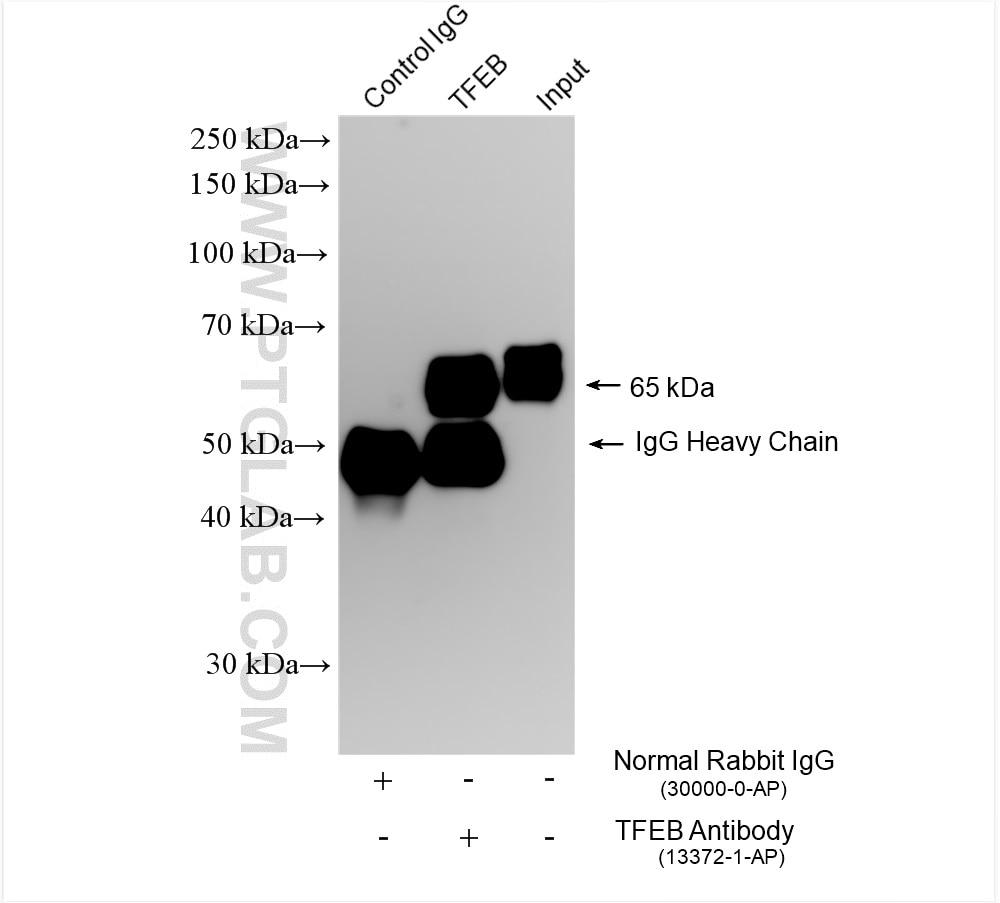
| Positive IP detected in | Raji cells |
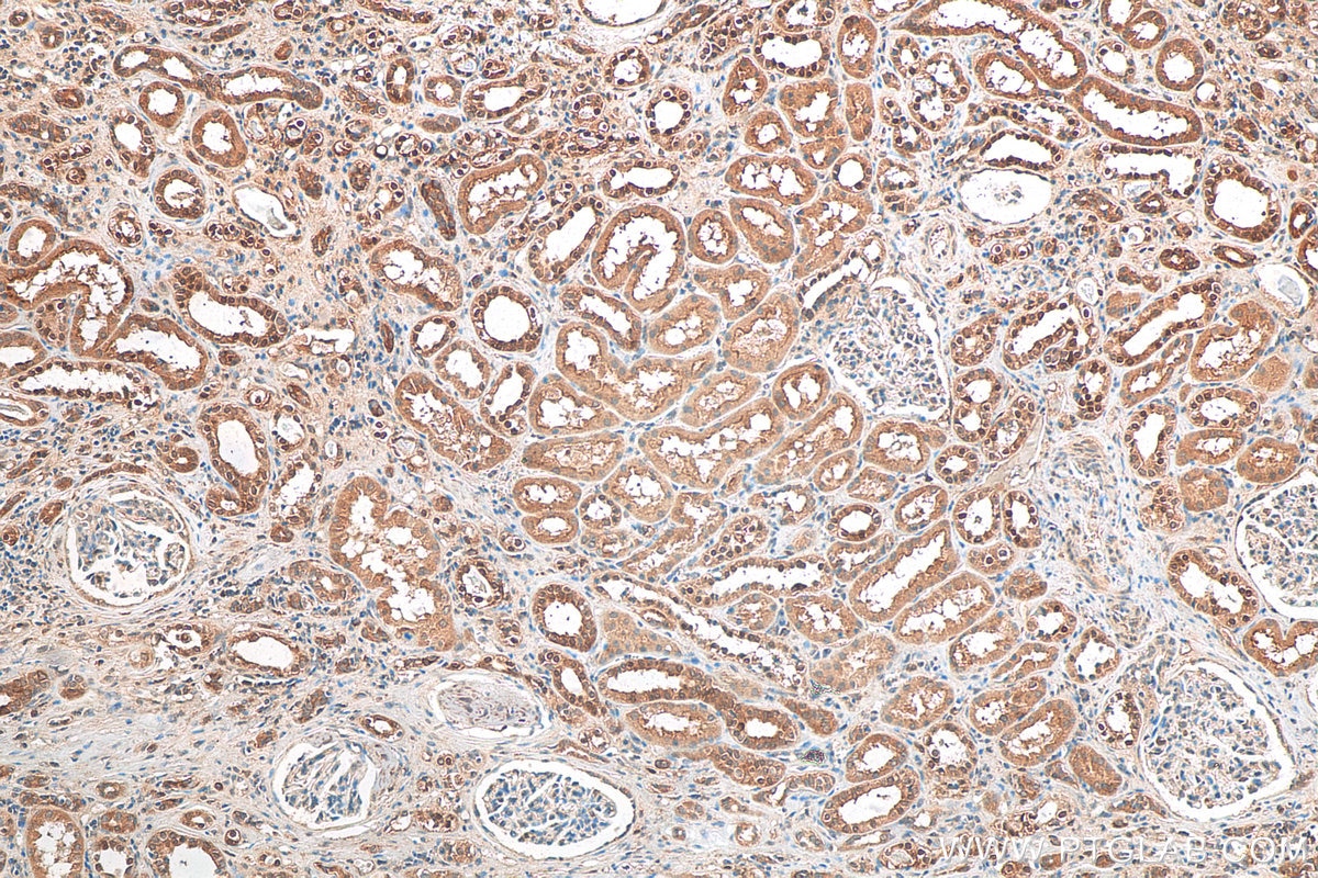
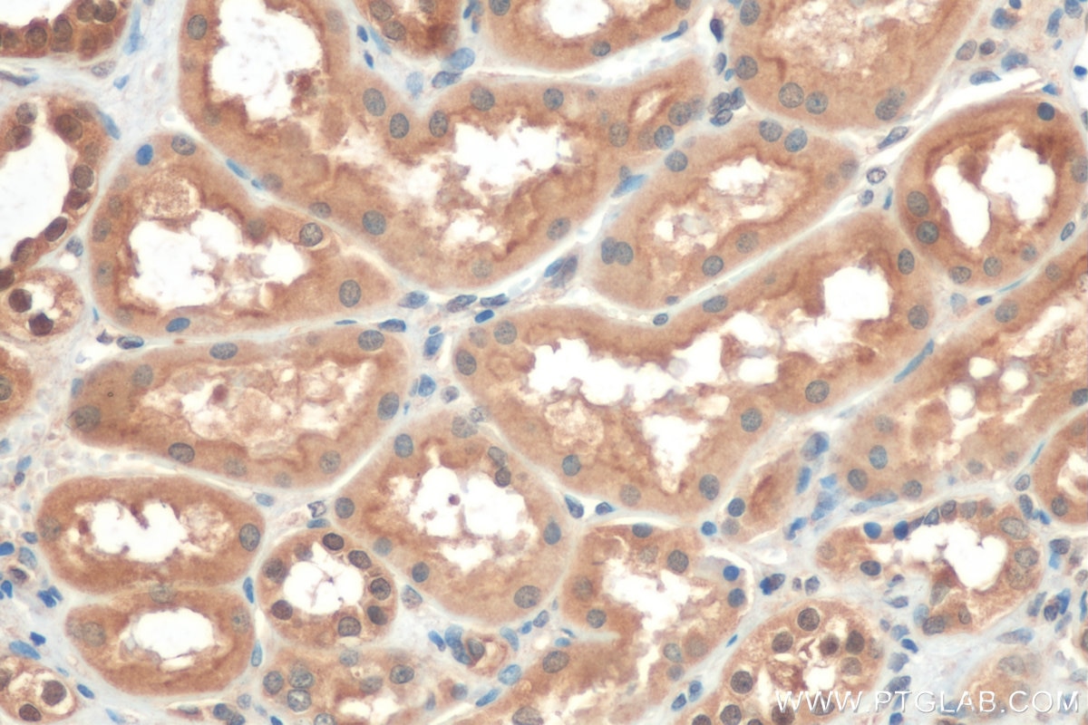
| Positive IHC detected in | human renal cell carcinoma tissue, rat spleen tissue Note: suggested antigen retrieval with TE buffer pH 9.0; (*) Alternatively, antigen retrieval may be performed with citrate buffer pH 6.0 |
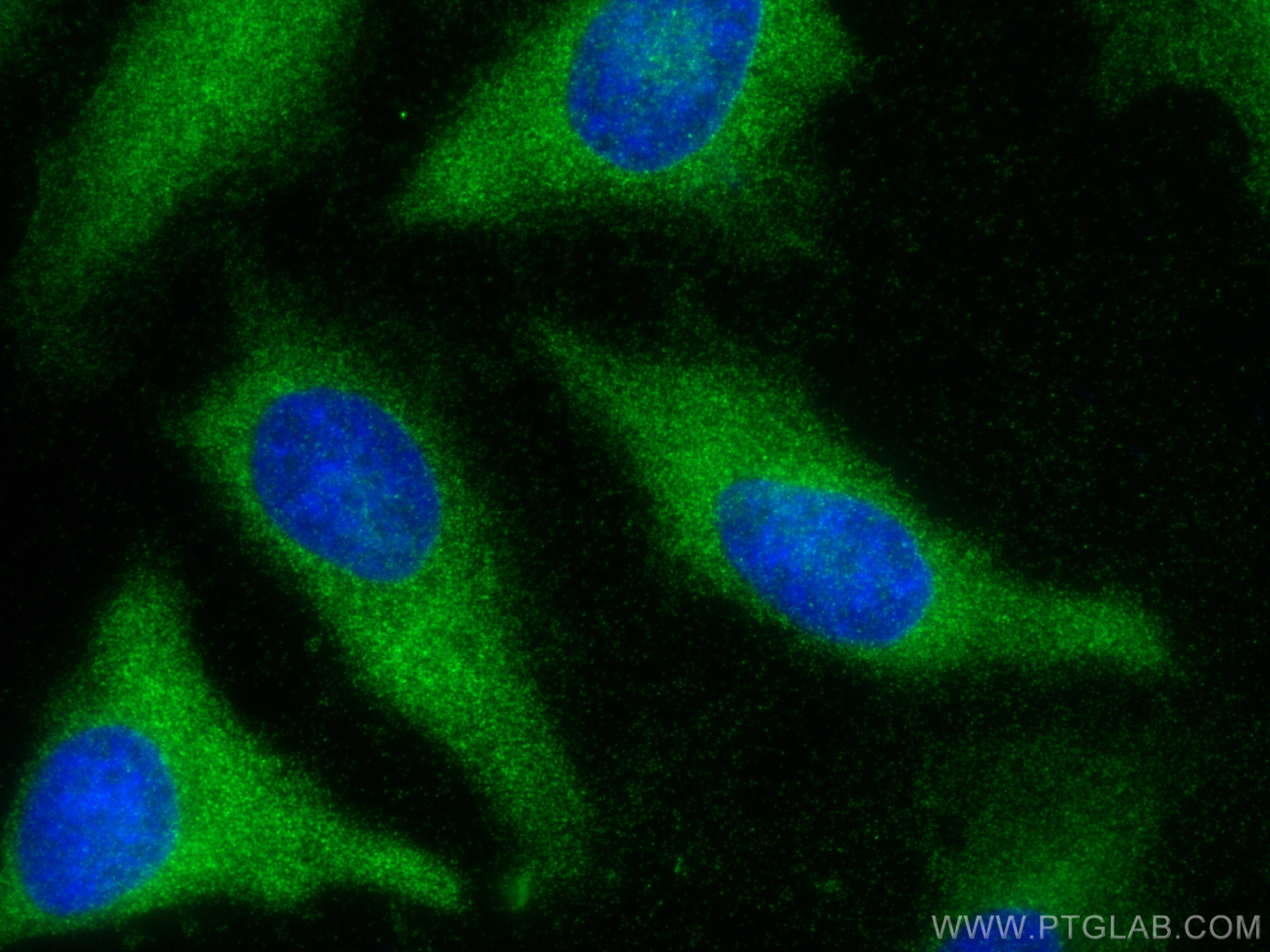
| Positive IF/ICC detected in | HeLa cells, HepG2 cells |
Recommended dilution
| Application | Dilution |
|---|---|
| Western Blot (WB) | WB : 1:1000-1:6000 |
| Immunoprecipitation (IP) | IP : 0.5-4.0 ug for 1.0-3.0 mg of total protein lysate |
| Immunohistochemistry (IHC) | IHC : 1:50-1:500 |
| Immunofluorescence (IF)/ICC | IF/ICC : 1:50-1:500 |
| It is recommended that this reagent should be titrated in each testing system to obtain optimal results. | |
| Sample-dependent, Check data in validation data gallery. | |
Product Information
13372-1-AP targets TFEB in WB, IHC, IF/ICC, IP, CoIP, ELISA applications and shows reactivity with human, mouse, rat samples.
| Tested Reactivity | human, mouse, rat |
| Cited Reactivity | human, mouse, rat, pig, monkey, zebrafish, bovine, goat |
| Host / Isotype | Rabbit / IgG |
| Class | Polyclonal |
| Type | Antibody |
| Immunogen |
CatNo: Ag4194 Product name: Recombinant human TFEB protein Source: e coli.-derived, PGEX-4T Tag: GST Domain: 1-476 aa of BC032448 Sequence: MASRIGLRMQLMREQAQQEEQRERMQQQAVMHYMQQQQQQQQQQLGGPPTPAINTPVHFQSPPPVPGEVLKVQSYLENPTSYHLQQSQHQKVREYLSETYGNKFAAHISPAQGSPKPPPAASPGVRAGHVLSSSAGNSAPNSPMAMLHIGSNPERELDDVIDNIMRLDDVLGYINPEMQMPNTLPLSSSHLNVYSSDPQVTASLVGVTSSSCPADLTQKRELTDAESRALAKERQKKDNHNLIERRRRFNINDRIKELGMLIPKANDLDVRWNKGTILKASVDYIRRMQKDLQKSRELENHSRRLEMTNKQLWLRIQELEMQARVHGLPTTSPSGMNMAELAQQVVKQELPSEEGPGEALMLGAEVPDPEPLPALPPQAPLPLPTQPPSPFHHLDFSHSLSFGGREDEGPPGYPEPLAPGHGSPFPSLSKKDLDLMLLDDSLLPLASDPLLSTMSPEASKASSRRSSFSMEEGDVL Predict reactive species |
| Full Name | transcription factor EB |
| Calculated Molecular Weight | 476 aa, 53 kDa |
| Observed Molecular Weight | 65-70 kDa |
| GenBank Accession Number | BC032448 |
| Gene Symbol | TFEB |
| Gene ID (NCBI) | 7942 |
| RRID | AB_2199611 |
| Conjugate | Unconjugated |
| Form | Liquid |
| Purification Method | Antigen affinity purification |
| UNIPROT ID | P19484 |
| Storage Buffer | PBS with 0.02% sodium azide and 50% glycerol, pH 7.3. |
| Storage Conditions | Store at -20°C. Stable for one year after shipment. Aliquoting is unnecessary for -20oC storage. 20ul sizes contain 0.1% BSA. |
Background Information
Transcription factor EB(TFEB) is one of the transcription factors that contain a basic HLH-ZIP domain(for DNA binding ) and HLH and ZIP domains( for loigomerization). TFEB specifically recognizes and binds to E-box sequences, which required dimerization with itself or another MiT/TFE family member. It plays a role in T-cell-dependent antibody responses to activated CD4+ T-cells and thymus-dependent humoral immunity once associated with TFE3. It also activates lysosomal gene by binding the CLEAR-box sequence in the regulatory region. It involves in the signal transduction processes required for normal vascularization of the placenta. This is a rabbit polyclonal antibody raised against the full length of human TFEB. The calculated molecular weight of TFEB is 53 kDa, but modified TFEB is about 65-70 kDa (PMID: 12837690). TFEB mainly present in the cytoplasm (PubMed:23434374). Under aberrant lysosomal storage conditions, it translocates from the cytoplasm to the nucleus (PubMed:23434374). Fisetin increases the levels of TFEB localized in nuclei (PMID: 27112200).TFEB protein has double bands in the range of 65-70 kDa, which is mainly related to its post-translation modification (especially phosphorylation). The 70 kDa band represents phosphorylated TFEB, while the 65 kDa band represents dephosphorylated TFEB, and this phosphorylation state is regulated by mTORC1(PMID: 32950675).
Protocols
| Product Specific Protocols | |
|---|---|
| IF protocol for TFEB antibody 13372-1-AP | Download protocol |
| IHC protocol for TFEB antibody 13372-1-AP | Download protocol |
| IP protocol for TFEB antibody 13372-1-AP | Download protocol |
| WB protocol for TFEB antibody 13372-1-AP | Download protocol |
| Standard Protocols | |
|---|---|
| Click here to view our Standard Protocols |
Publications
What published studies show
The TFEB Polyclonal antibody (Cat# 13372-1-AP) has 340 total citations. These appear across high-impact journals including Autophagy, Cell Metabolism, Nature Communications, Cell Death & Differentiation, Cell Death & Disease, Molecular Cell, Redox Biology, Free Radical Biology and Medicine, Acta Neuropathologica, and Theranostics. Research fields span autophagy and lysosomal biogenesis, neurodegenerative diseases (Parkinson's, Alzheimer's, Huntington's), cancer, diabetic nephropathy, cardiovascular disease, ferroptosis, and innate immunity. Applications include WB, IF, IHC, IP, and CoIP across human, mouse, rat, and other species.
| Species | Application | Title |
|---|---|---|
Mol Cancer tRNA m(2)G methyltransferase complex THUMPD3-TRMT112 promotes pancreatic cancer progression and autophagy via modulating TFEB translation. | ||
Cell Metab ABCG2 is an itaconate exporter that limits antibacterial innate immunity by alleviating TFEB-dependent lysosomal biogenesis | ||
Cell Metab Hepatic lipid remodeling in cold exposure uncovers direct regulation of bis(monoacylglycero)phosphate lipids by phospholipase A2 group XV | ||
Immunity CRISPR screens unveil nutrient-dependent lysosomal and mitochondrial nodes impacting intestinal tissue-resident memory CD8+ T cell formation | ||
Cancer Commun (Lond) HMMR alleviates endoplasmic reticulum stress by promoting autophagolysosomal activity during endoplasmic reticulum stress-driven hepatocellular carcinoma progression | ||
Bioact Mater Oxygen-vacancy-engineered t-ZnO-CeO(2) Schottky junction enhanced infectious bone regeneration via photoelectric-photocatalytic effects-induced mitochondrial quality control. |
Reviews
What customers say
Customers rate this TFEB antibody favorably, with most reviews giving 5 stars. It performs well in immunofluorescence and immunohistochemistry, producing strong, clean signals with good subcellular localization, and works in organoids, mouse kidney sections, and mouse BMDMs. Western blot results are generally positive, with specific bands in HeLa and HEK293 lysates, often working on the first try. A few users noted drawbacks: some WB bands appeared blurry or multiple, and one reviewer saw two weak bands of unclear identity in primary mouse microglia. Overall, it is considered reliable for mouse applications and IF/IHC, with mixed results in certain WB contexts.
The reviews below have been submitted by verified Proteintech customers who received an incentive for providing their feedback.
Sheeny (Verified Customer) (09-02-2025) | Used in organoids and it works well. The specificity of TFEB is very good with the subcellular localization.
|
Flaviane (Verified Customer) (07-30-2025) | Very strong and clean signal.
 |
Stanislas (Verified Customer) (08-05-2024) | WB on HeLa lysates using 15µg of protein and nitrocellulose membranes, blockage in TBST-5%Milk 1h, Incubation in 2%BSA with TFEB antibody (1:3000) overnight at 4°C. Worked on the first trial. IF on HeLa fixed in 4%PFA for 15min, blocked 1h in 1XPBS, 5%NGS, 0.3% Triton X-100, incubates with TFEB antibody (1:100) in 1XPBS, 1%BSA, 0.3% Triton X-100 overnight at 4°C. Worked on first trial.
|
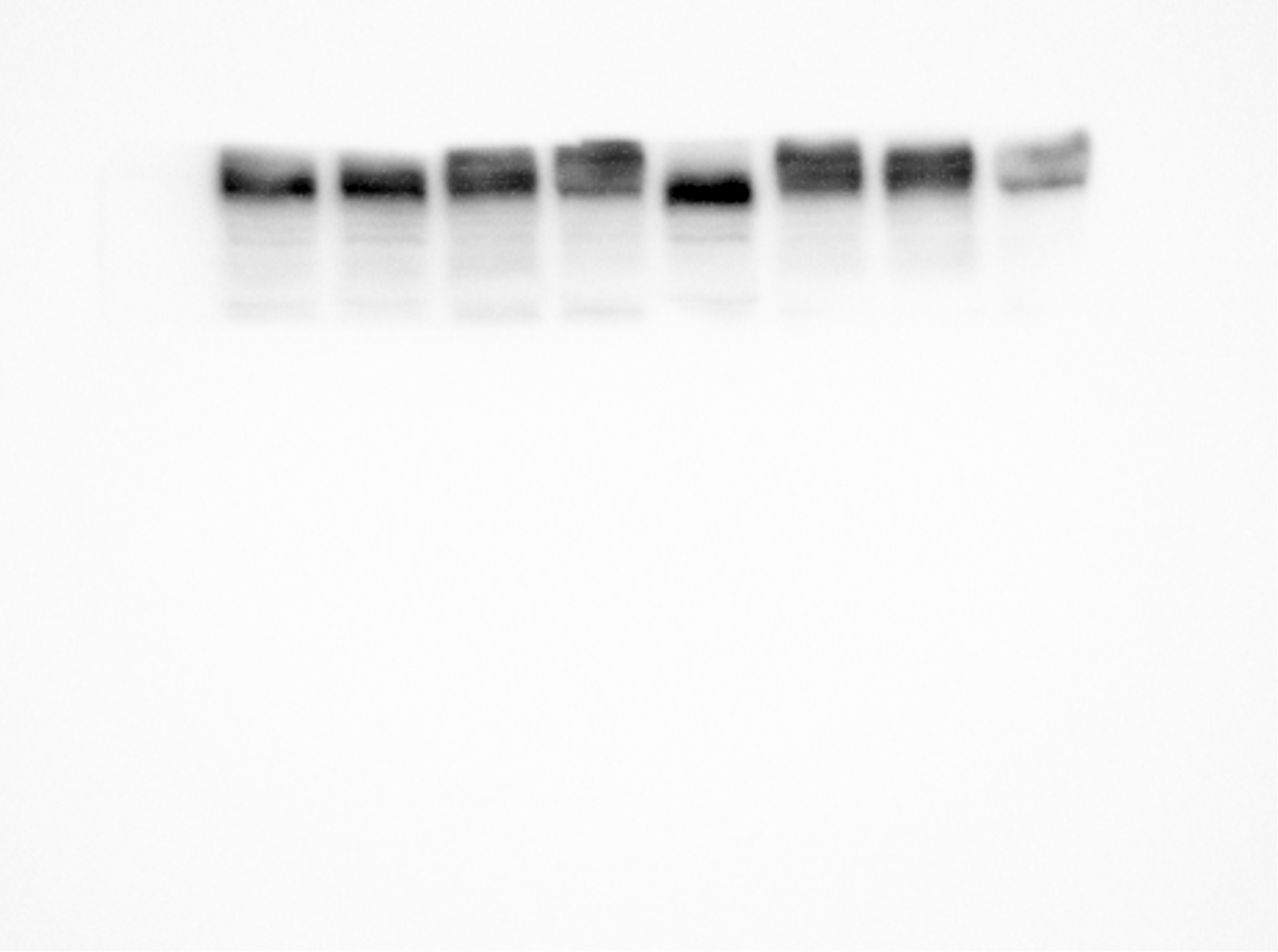
Shunbin (Verified Customer) (07-17-2023) | bands look specific, but blurry and multiple.
 |
Weihao (Verified Customer) (04-11-2022) | Good antibody for mouse BMDM! not for human MDM 1:500 for IF 1:2000 for WB
|
Marina (Verified Customer) (11-22-2021) | 2 bands visible, unclear which is the right one, although size is ok-ish for both. Both bands are quite weak.
|
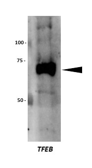
Tom (Verified Customer) (02-19-2021) | HEK293 lysate (10ug) subjected to SDS-PAGE and transferred to nitrocellullose. Membrane blocked in 5% BSA for 1 hour prior to incubation with antibody (1:1000 in block) overnight at 4 degrees. Band detected using anti-rabbit HRP secondary.
 |















