



Tested Applications
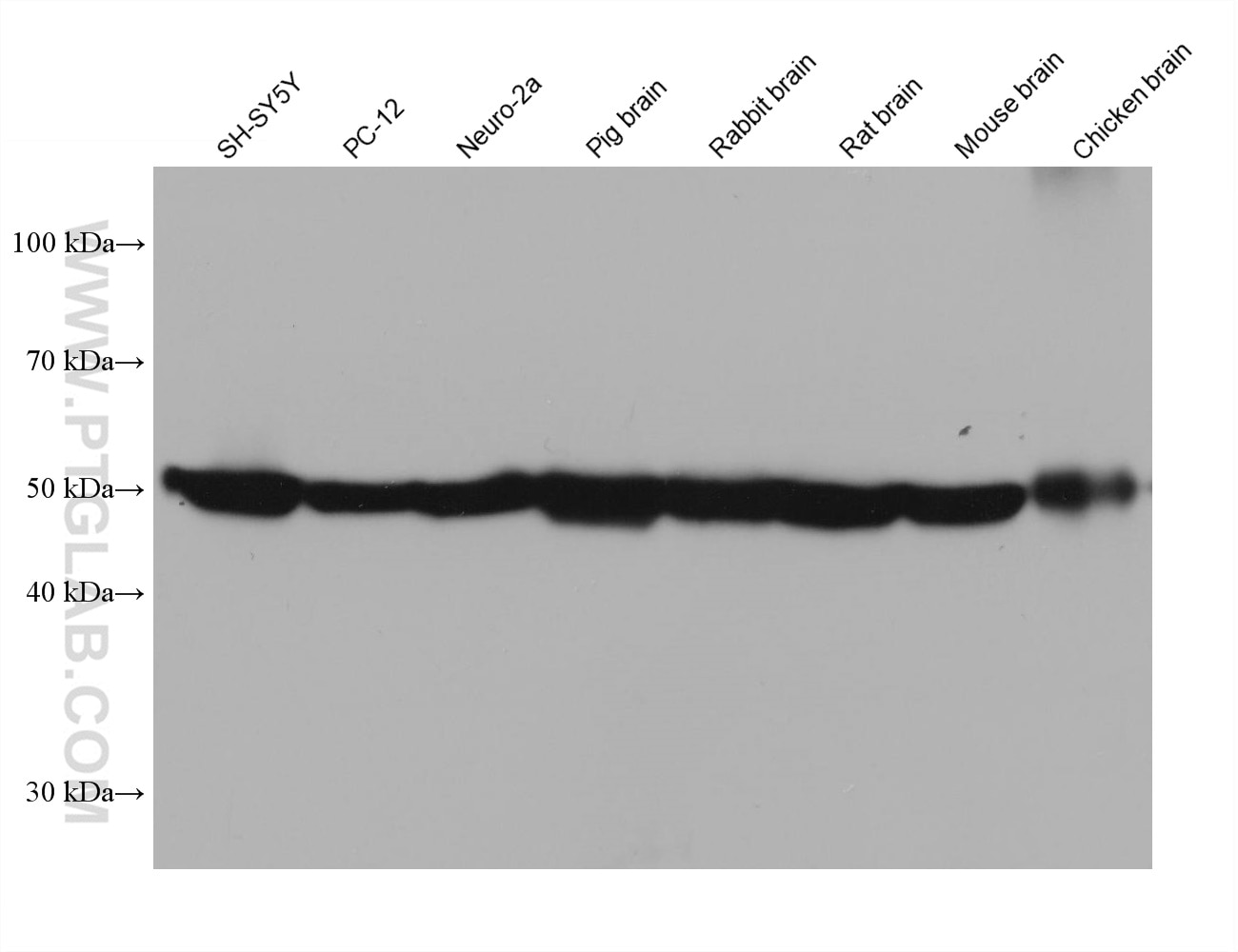
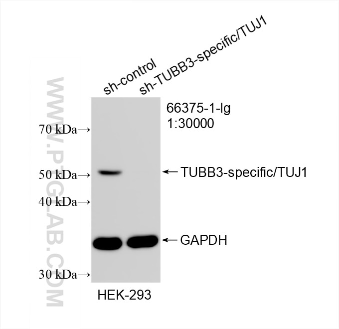
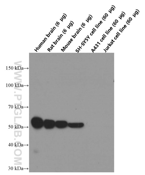
| Positive WB detected in | SH-SY5Y cells, fetal human brain tissue, HEK-293 cells, PC-12 cells, Neuro-2a cells, Pig brain, Rabbit brain, Rat brain Mouse brain, Chicken brain |
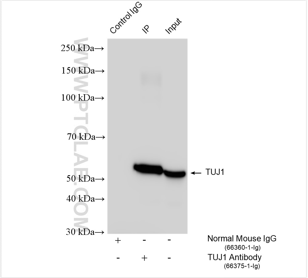
| Positive IP detected in | SH-SY5Y cells |
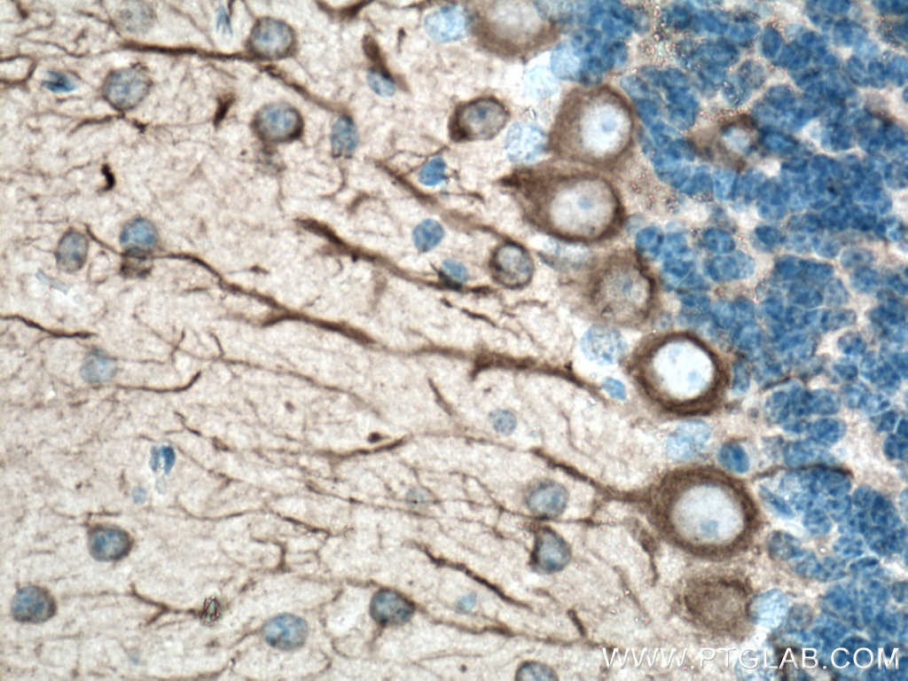
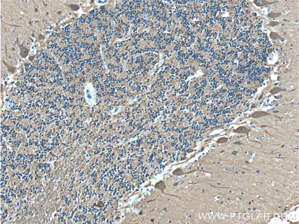
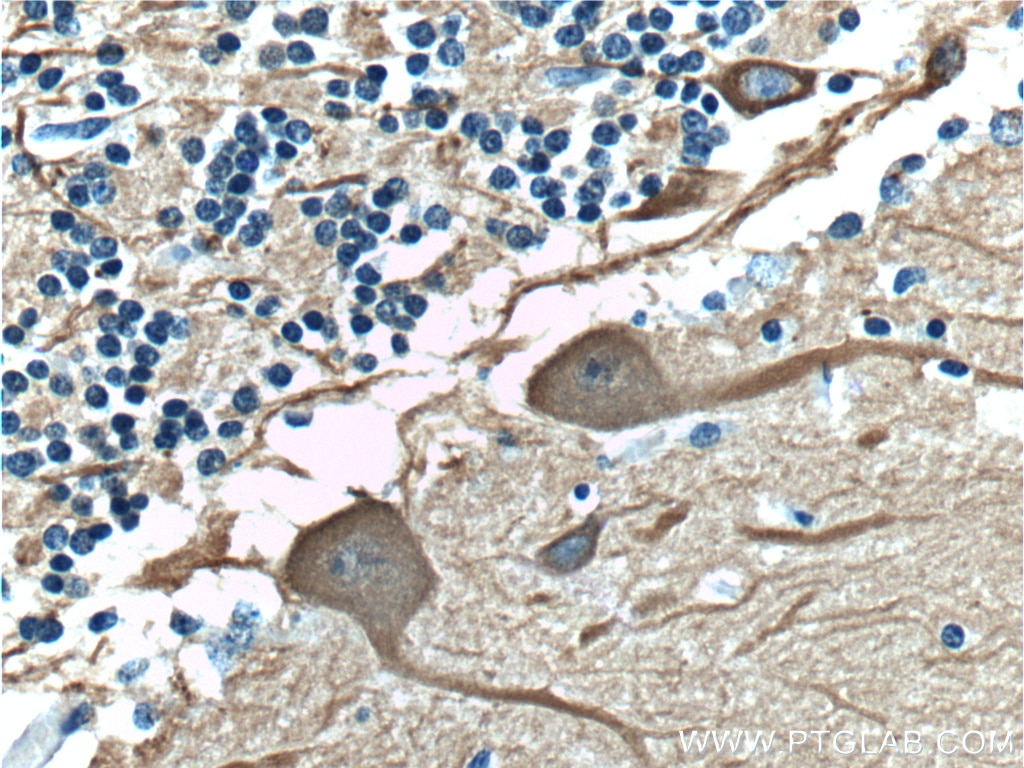
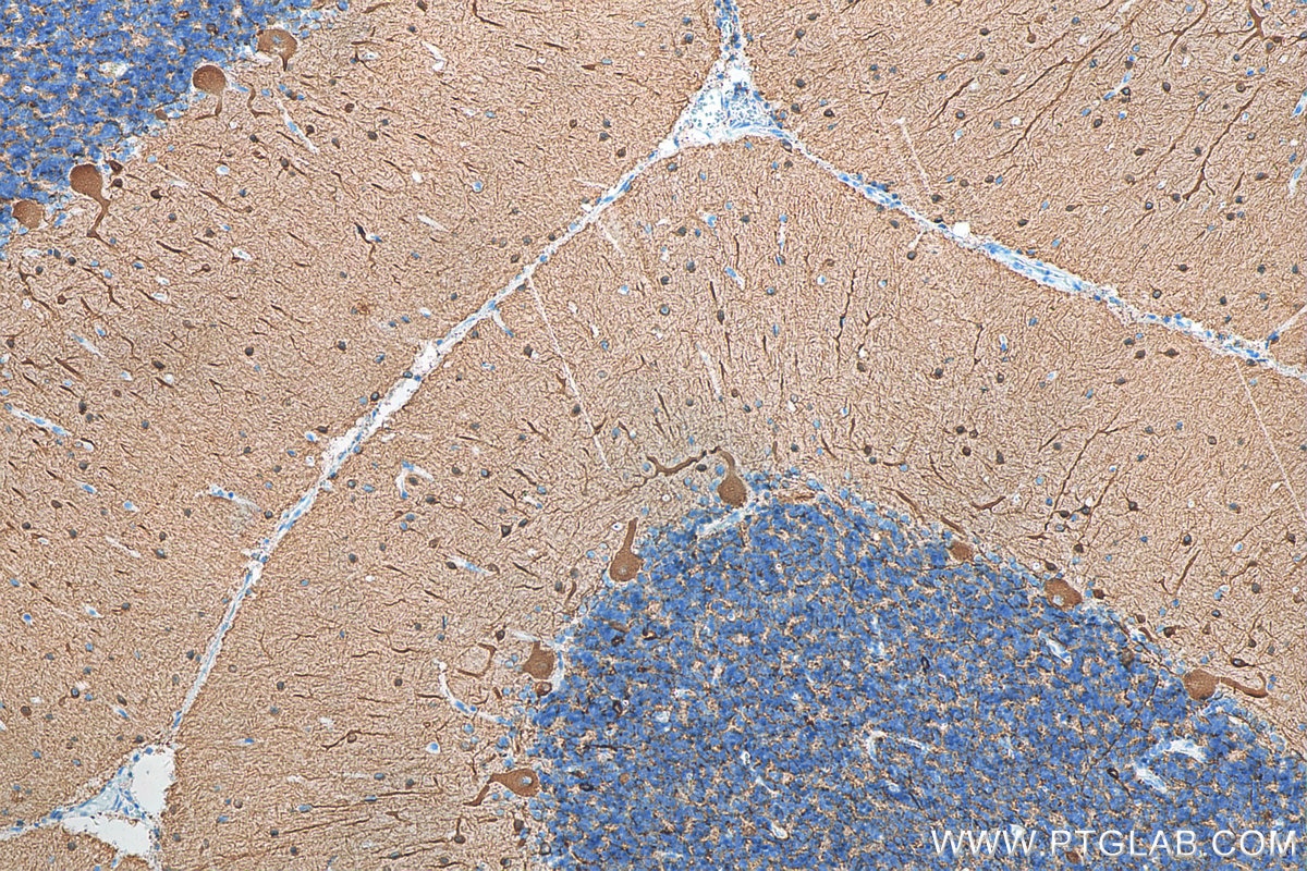
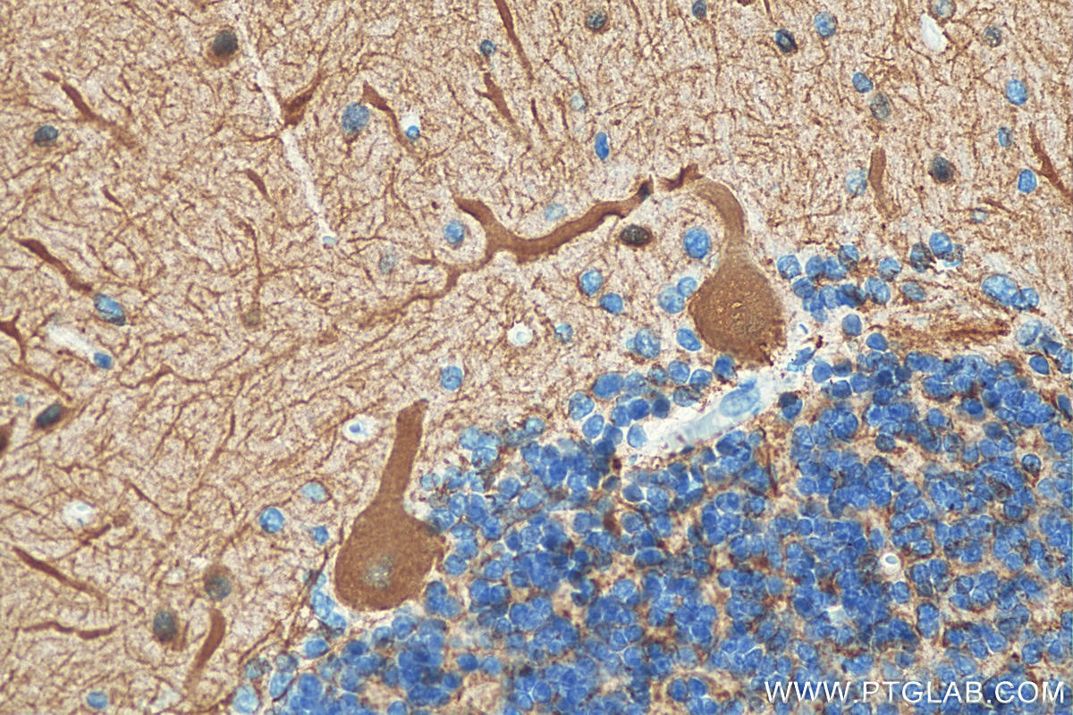
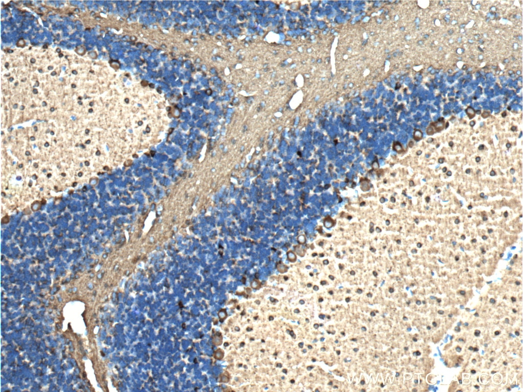
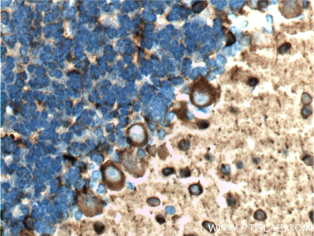
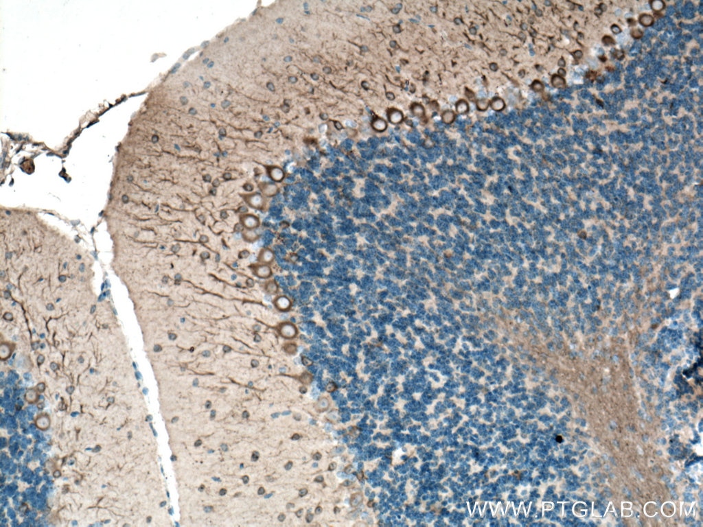
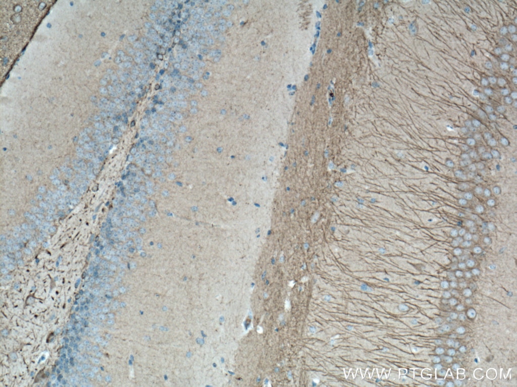
| Positive IHC detected in | human cerebellum tissue, mouse brain tissue, rat cerebellum tissue, mouse cerebellum tissue, rat brain tissue, human cerebellum tissue Note: suggested antigen retrieval with TE buffer pH 9.0; (*) Alternatively, antigen retrieval may be performed with citrate buffer pH 6.0 |
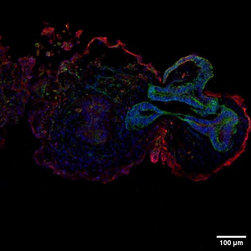
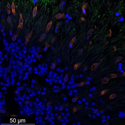
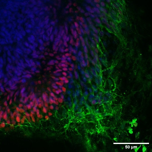
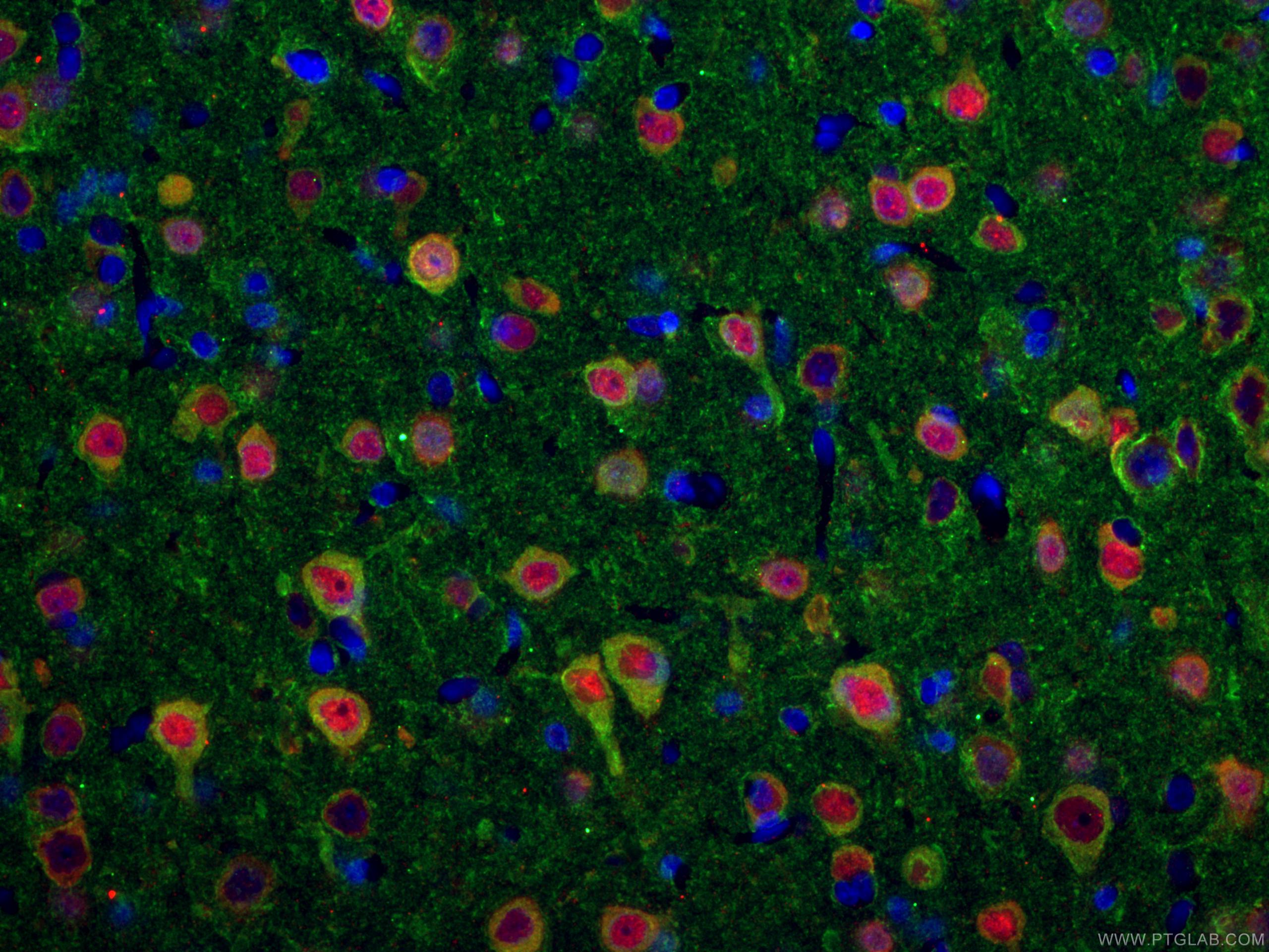
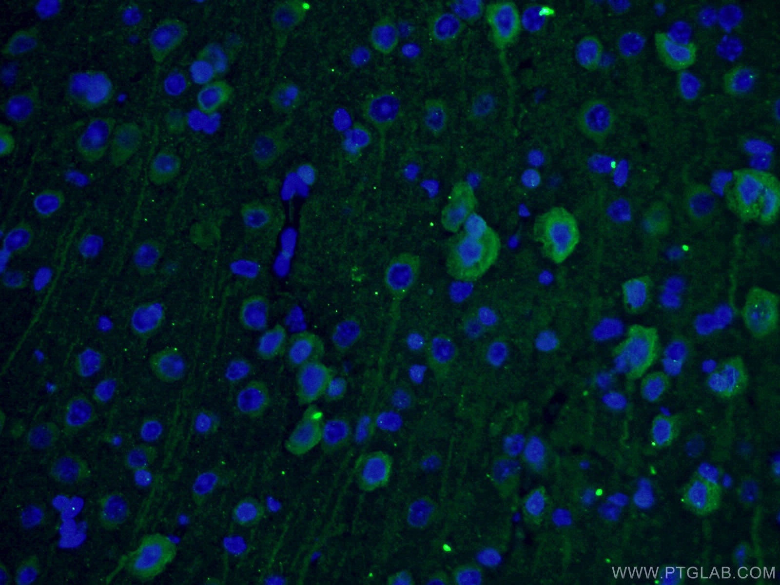
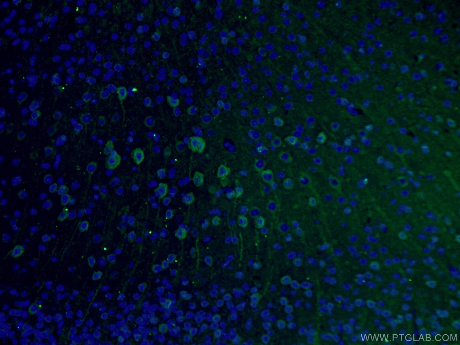
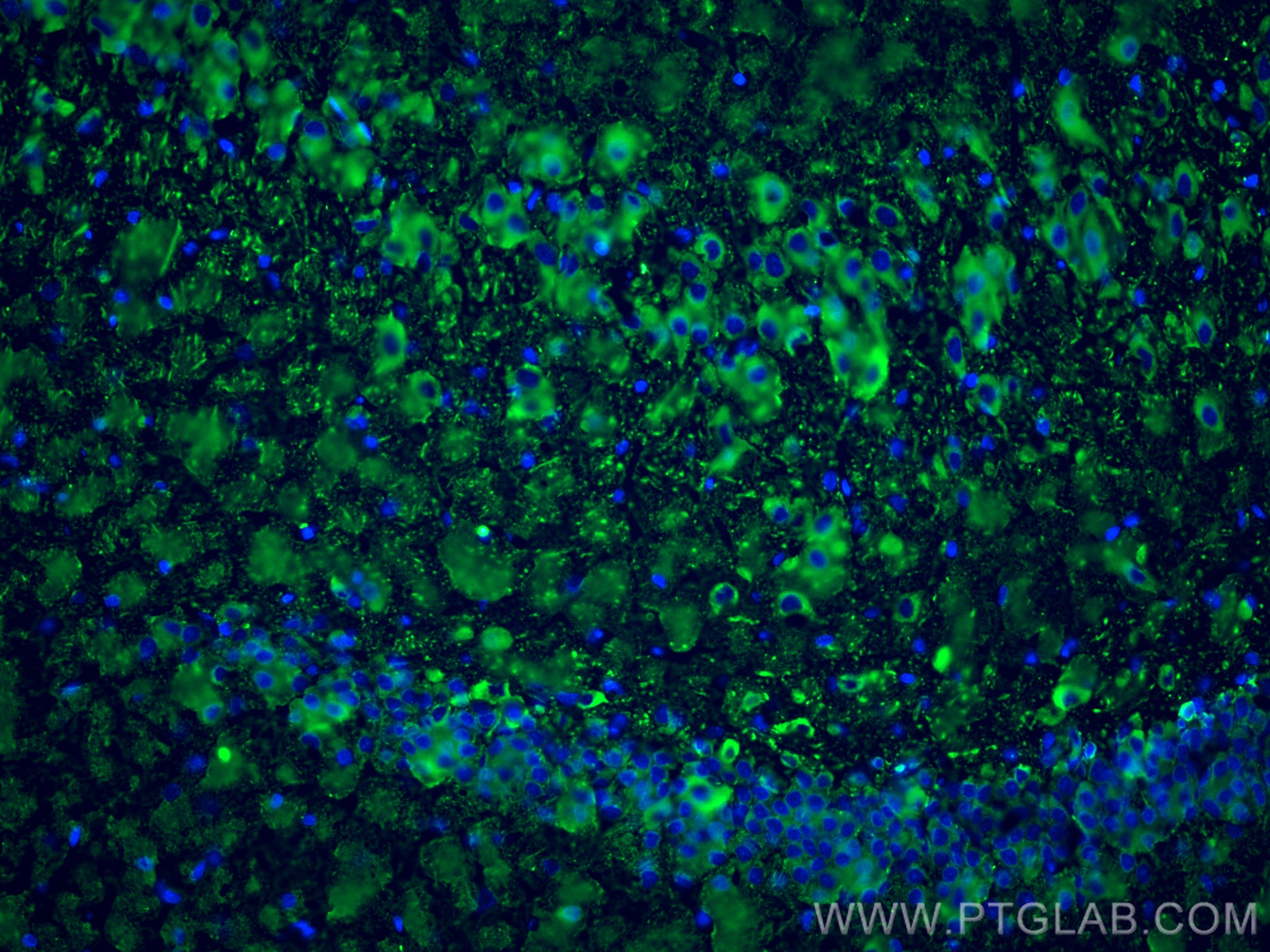
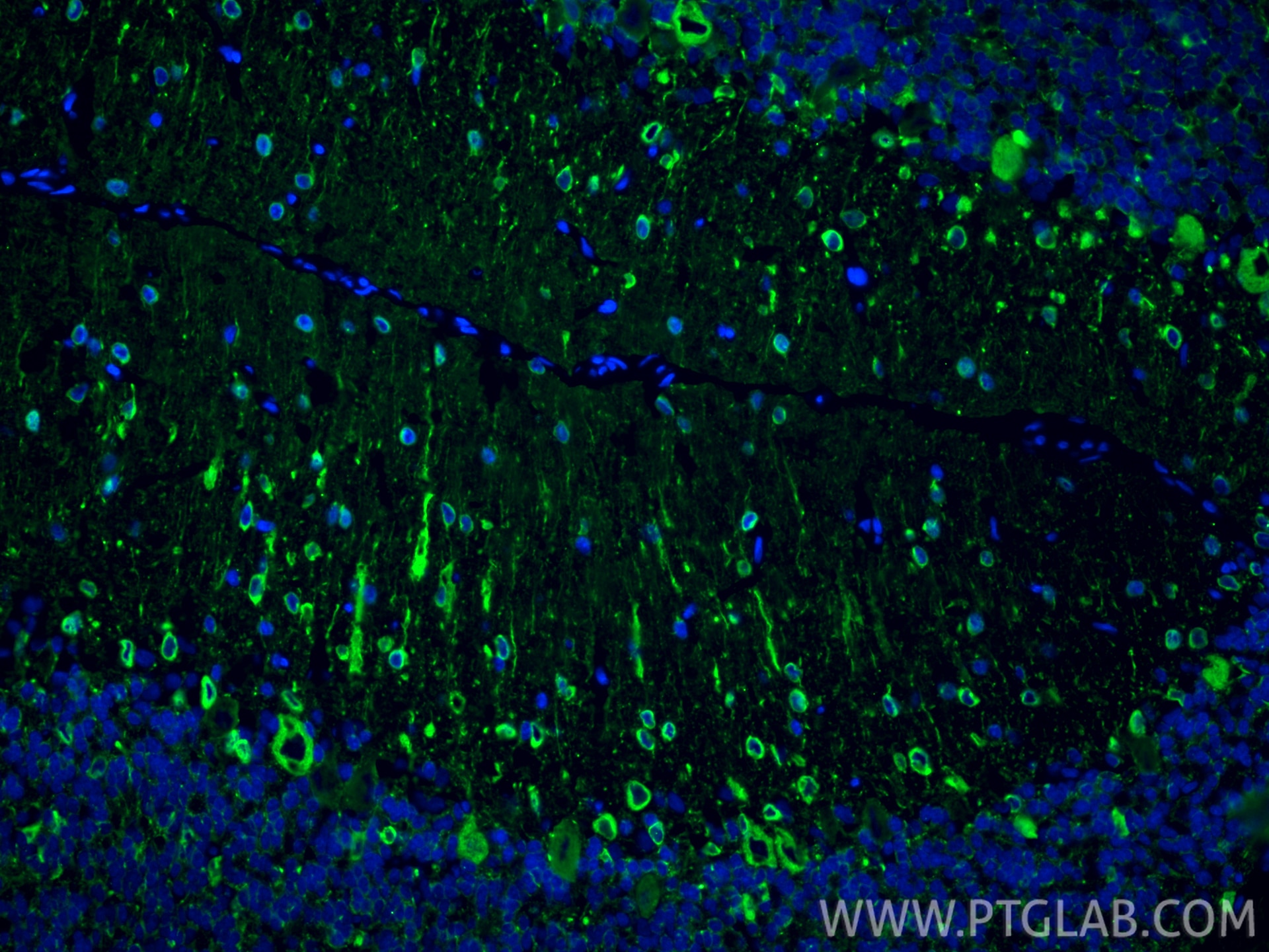
| Positive IF-P detected in | rat brain tissue, Retinal organoids, mouse brain tissue, human neuron |
| Positive IF-Fro detected in | rat brain tissue, rat cerebellum tissue |
| Positive IF/ICC detected in | iPS cells |
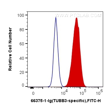
| Positive FC (Intra) detected in | SH-SY5Y cells |
Recommended dilution
| Application | Dilution |
|---|---|
| Western Blot (WB) | WB : 1:5000-1:50000 |
| Immunoprecipitation (IP) | IP : 0.5-4.0 ug for 1.0-3.0 mg of total protein lysate |
| Immunohistochemistry (IHC) | IHC : 1:400-1:20000 |
| Immunofluorescence (IF)-P | IF-P : 1:50-1:500 |
| Immunofluorescence (IF)-FRO | IF-FRO : 1:400-1:1600 |
| Immunofluorescence (IF)/ICC | IF/ICC : 1:125-1:500 |
| Flow Cytometry (FC) (INTRA) | FC (INTRA) : 0.20 ug per 10^6 cells in a 100 µl suspension |
| It is recommended that this reagent should be titrated in each testing system to obtain optimal results. | |
| Sample-dependent, Check data in validation data gallery. | |
Product Information
66375-1-Ig targets TUBB3-specific/TUJ1 in WB, IHC, IF/ICC, IF-P, IF-Fro, FC (Intra), IP, ELISA applications and shows reactivity with human, mouse, rat, pig, rabbit, chicken samples.
| Tested Reactivity | human, mouse, rat, pig, rabbit, chicken |
| Cited Reactivity | human, mouse, rat, rabbit, chicken |
| Host / Isotype | Mouse / IgG1 |
| Class | Monoclonal |
| Type | Antibody |
| Immunogen |
Peptide Predict reactive species |
| Full Name | tubulin, beta 3 |
| Calculated Molecular Weight | 55 kDa |
| Observed Molecular Weight | 50-55 kDa |
| GenBank Accession Number | NM_001197181 |
| Gene Symbol | TUBB3 |
| Gene ID (NCBI) | 10381 |
| RRID | AB_2814998 |
| Conjugate | Unconjugated |
| Form | Liquid |
| Purification Method | Protein G purification |
| UNIPROT ID | Q13509 |
| Storage Buffer | PBS with 0.02% sodium azide and 50% glycerol, pH 7.3. |
| Storage Conditions | Store at -20°C. Stable for one year after shipment. Aliquoting is unnecessary for -20oC storage. 20ul sizes contain 0.1% BSA. |
Background Information
TUBB3, the class III β tubulin or Tuj1, is selectively expressed in testis and neurons of the central and peripheral nervous system. It has been widely used as a marker for neurons. Aberrant expression of TUBB3 has also been found in various tumors of non-neural origin and can be used as a biomarker for cancer aggressiveness and a marker for the tendency to respond poorly to chemotherapy. This antibody is specific to TUBB3 but not cross-react with other tubulin isoforms.
Protocols
| Product Specific Protocols | |
|---|---|
| FC protocol for TUBB3-specific/TUJ1 antibody 66375-1-Ig | Download protocol |
| IF protocol for TUBB3-specific/TUJ1 antibody 66375-1-Ig | Download protocol |
| IHC protocol for TUBB3-specific/TUJ1 antibody 66375-1-Ig | Download protocol |
| IP protocol for TUBB3-specific/TUJ1 antibody 66375-1-Ig | Download protocol |
| WB protocol for TUBB3-specific/TUJ1 antibody 66375-1-Ig | Download protocol |
| Standard Protocols | |
|---|---|
| Click here to view our Standard Protocols |
Publications
What published studies show
The TUBB3-specific/TUJ1 Monoclonal antibody (Cat# 66375-1-Ig) has 165 citations. These appear across journals including Nature Cell Biology, Nature Communications, Neuron, Cell Reports, EMBO Reports, eLife, Bioactive Materials, Theranostics, Advanced Science, Biomaterials, Small, Cell Death & Disease, and Stem Cell Research. Research fields span neuroscience (neural differentiation, spinal cord injury, neurodegeneration, neuroinflammation), stem cell/iPSC line generation, tissue engineering and biomaterials, cancer, and pain research.
| Species | Application | Title |
|---|---|---|
Bioact Mater Biodegradable Mg(2+)-releasing piezoelectric scaffold for segmental bone defect repair. | ||
Bioact Mater An intelligent nanoliposome alleviates disc degeneration and discogenic pain by inhibiting neurovascular ingrowth via a "Soil-conditioning, seed-modulating, and weeds-suppressing" strategy. | ||
Bioact Mater Bioprinting of live platelet-loaded nerve conduit using energy-dissipative hydrogel. | ||
Nat Cell Biol RNA-binding proteins mediate the maturation of chromatin topology during differentiation | ||
Nat Commun Unified bilayer membranes with mechanically reinforced interface for stage-adaptive bone regeneration. | ||
Nat Commun An implantable mechano-electro cascade platform synchronizes neuro-muscle repair. |
Reviews
What customers say
Users consistently rate this TUBB3 antibody highly across Western Blot, Immunofluorescence, and Immunohistochemistry. Common dilutions range from 1:400 to 1:1000. Reviewers praise strong signal-to-noise ratio, minimal background, and reliable performance in neuronal cell lines, brain organoids, and colon cancer tissue. One user noted slight colocalization with astrocytic processes at 1:500, suggesting it works best as part of a multi-marker panel. Overall, the antibody is described as a dependable neuronal differentiation marker with clean staining and good reproducibility across multiple lots.
The reviews below have been submitted by verified Proteintech customers who received an incentive for providing their feedback.
Iram (Verified Customer) (03-23-2026) | Very good TUBB3 antibody
|
Emile (Verified Customer) (02-27-2026) | This antibody works perfectly in IF at low concentrations.
 |
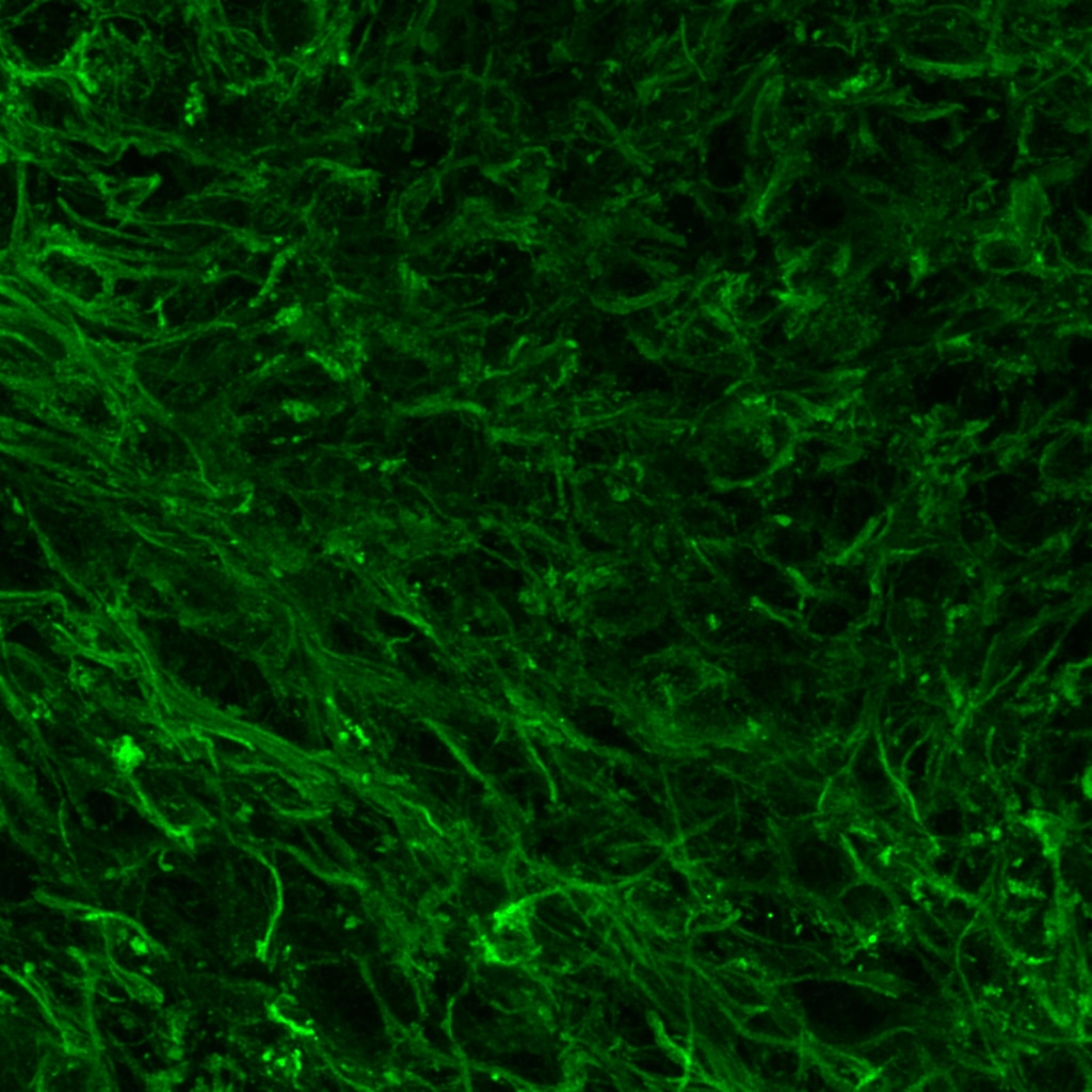
Ankush (Verified Customer) (05-02-2025) | worked fine
 |
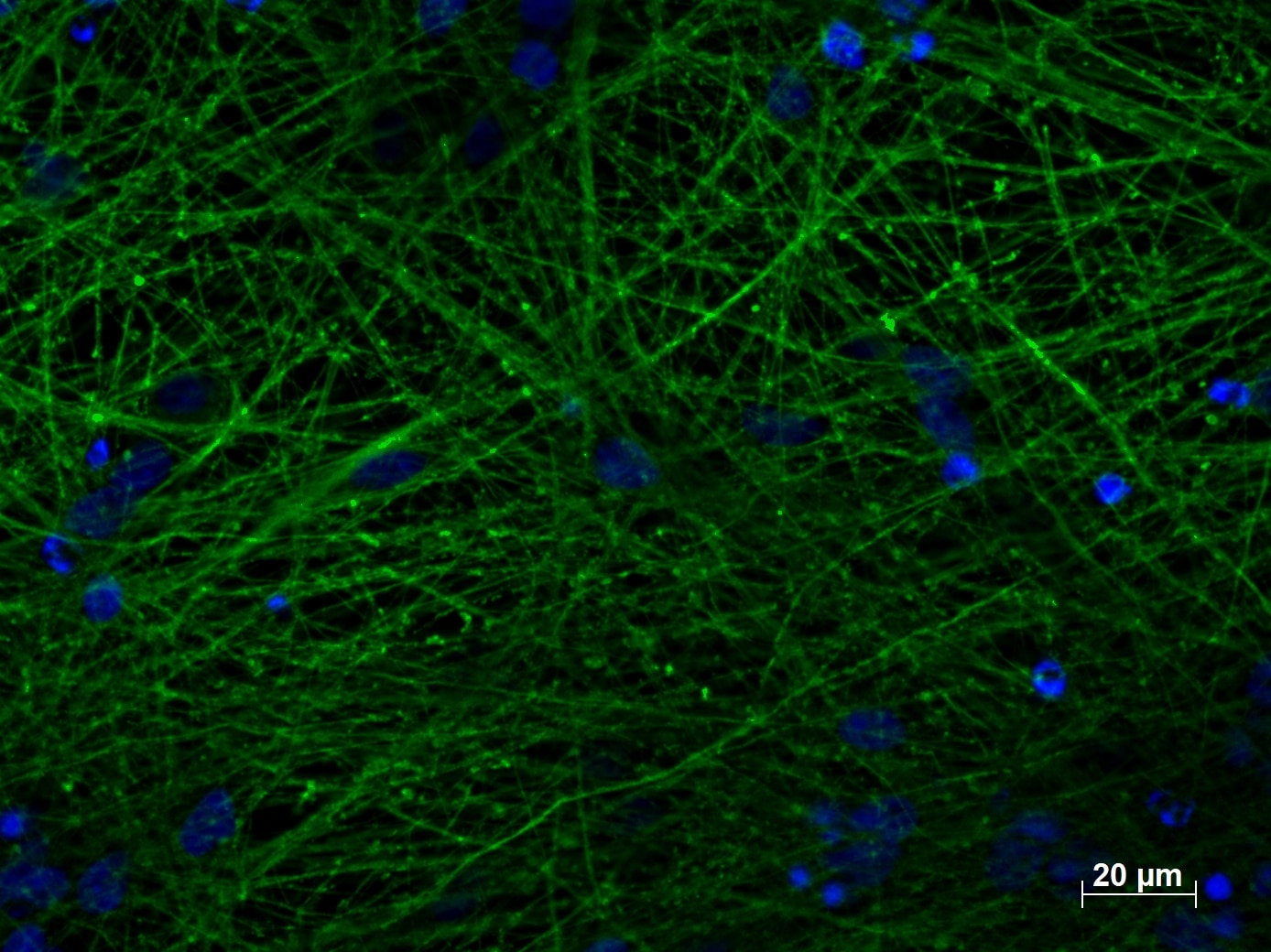
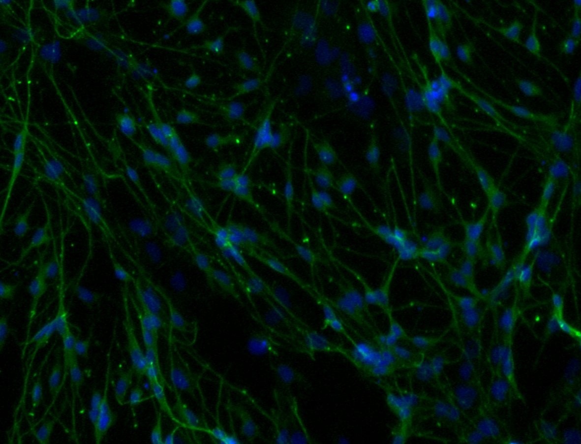
Gabriele (Verified Customer) (01-12-2023) | The antibody works very well with our IF protocol. Maybe the dilution could be pushed to 1:500-1:600. TUBB3 1:400 (green), Hoechst (Blue)
 |
Alessandro (Verified Customer) (07-27-2022) | No aspecific staining, great outcome
|
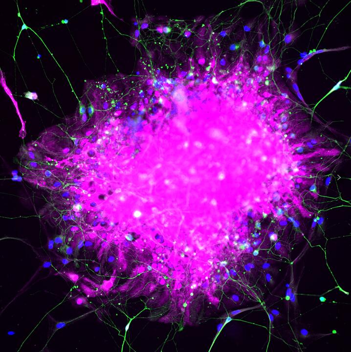
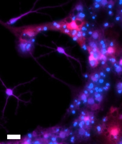
Josh (Verified Customer) (12-18-2018) | BIII tubulin antibody signal nicely fills neuronal cell bodies and processes with good signal to noise at a reasonable dilution (1:500 in 1% NDS, 0.3% Triton-X, PBS with donkey anti-mouse 647 secondary, 1:500). However, BIII tubulin antibody signal also colocalized with S100 immunoreactive astrocytic processes, though with a lesser intensity. Overall, the antibody is a useful marker of neuronal differentiation when used in combination with other cell-selective antibodies and morphology. Image caption: Differentiated adult hippocampal neural stem cell culture. BIII tubulin 1:500 (purple), S100 1:500 (red), Hoechst (blue). Scale bar = 20 um.
 |



























