Tested Applications
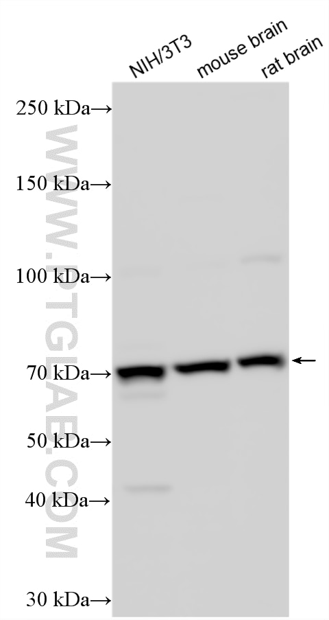
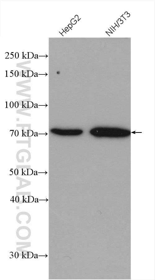
| Positive WB detected in | NIH/3T3 cells, HepG2 cells, mouse brain tissue, rat brain tissue |
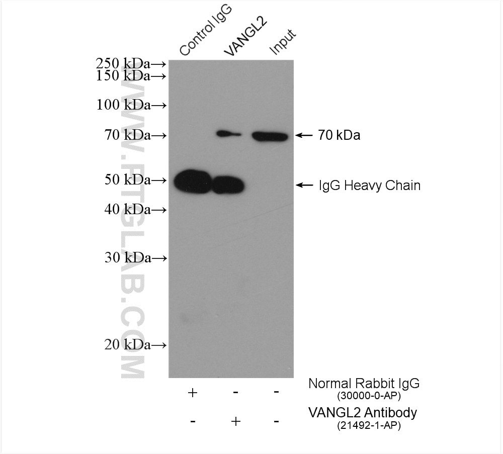
| Positive IP detected in | HepG2 cells |
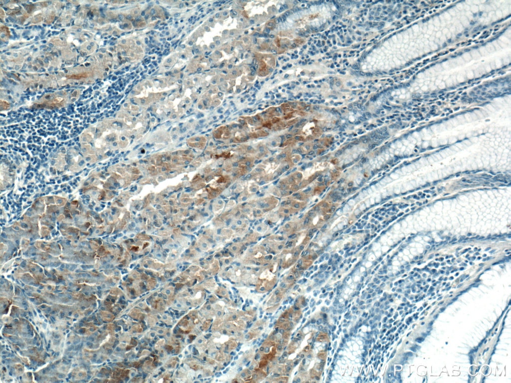
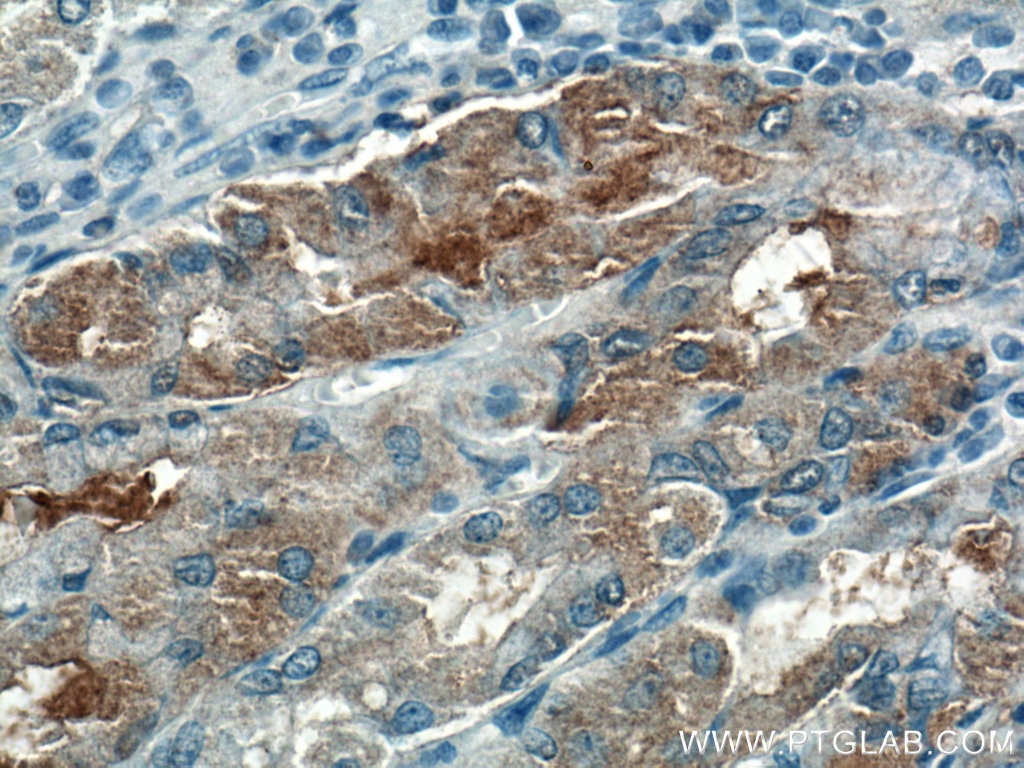
| Positive IHC detected in | human stomach tissue Note: suggested antigen retrieval with TE buffer pH 9.0; (*) Alternatively, antigen retrieval may be performed with citrate buffer pH 6.0 |
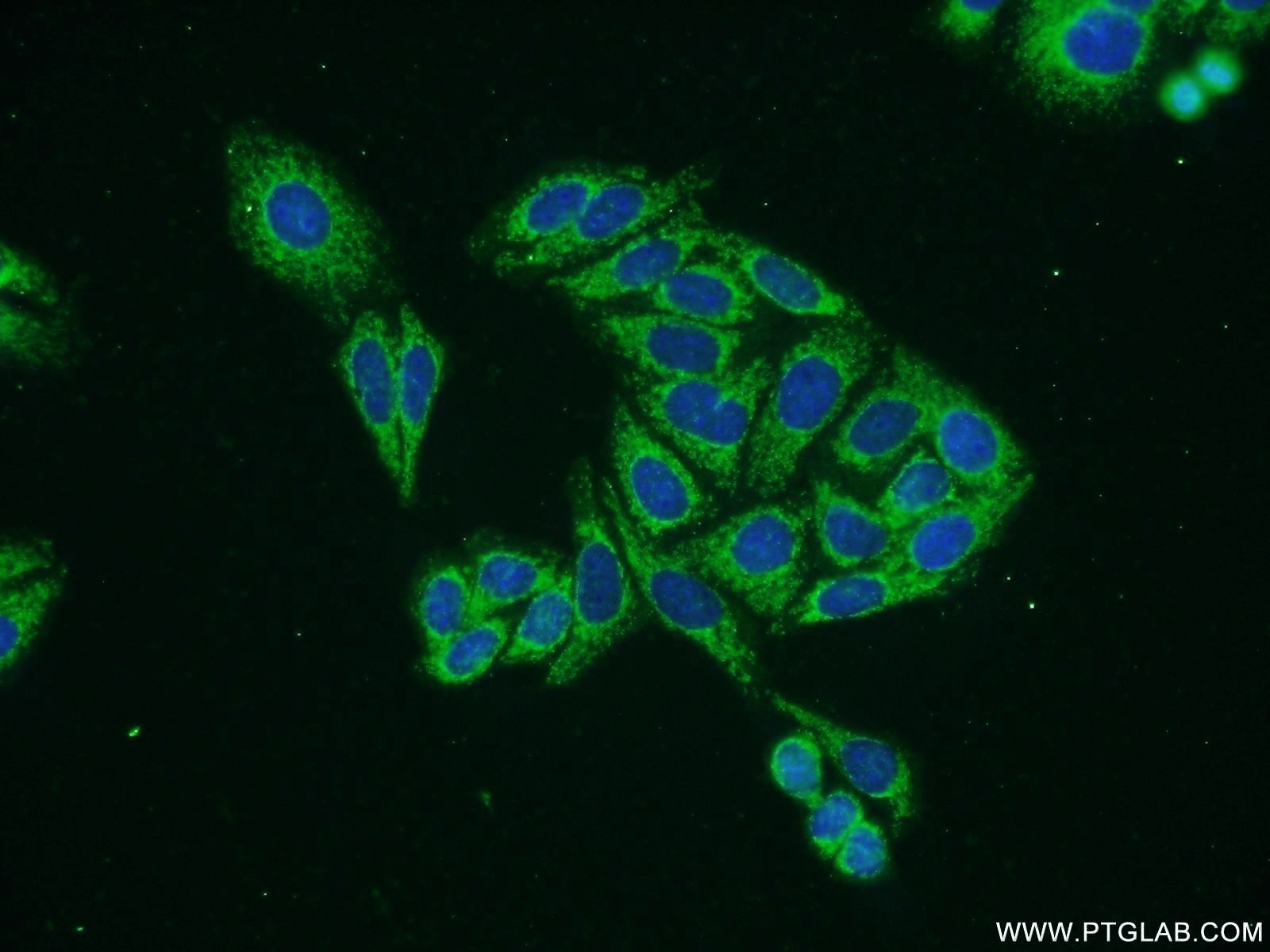
| Positive IF/ICC detected in | HepG2 cells |
Recommended dilution
| Application | Dilution |
|---|---|
| Western Blot (WB) | WB : 1:1000-1:6000 |
| Immunoprecipitation (IP) | IP : 0.5-4.0 ug for 1.0-3.0 mg of total protein lysate |
| Immunohistochemistry (IHC) | IHC : 1:50-1:500 |
| Immunofluorescence (IF)/ICC | IF/ICC : 1:20-1:200 |
| It is recommended that this reagent should be titrated in each testing system to obtain optimal results. | |
| Sample-dependent, Check data in validation data gallery. | |
Published Applications
| KD/KO | See 2 publications below |
| WB | See 10 publications below |
| IF | See 7 publications below |
| IP | See 1 publications below |
| CoIP | See 1 publications below |
Product Information
21492-1-AP targets VANGL2 in WB, IHC, IF/ICC, IP, CoIP, ELISA applications and shows reactivity with human, mouse, rat samples.
| Tested Reactivity | human, mouse, rat |
| Cited Reactivity | human, mouse, rat |
| Host / Isotype | Rabbit / IgG |
| Class | Polyclonal |
| Type | Antibody |
| Immunogen |
CatNo: Ag15833 Product name: Recombinant human VANGL2 protein Source: e coli.-derived, PGEX-4T Tag: GST Domain: 268-322 aa of BC103920 Sequence: IQRVAVWILEKYYHDFPVYNPALLNLPKSVLAKKVSGFKVYSLGEENSTNNSTGQ Predict reactive species |
| Full Name | vang-like 2 (van gogh, Drosophila) |
| Calculated Molecular Weight | 521 aa, 60 kDa |
| Observed Molecular Weight | 60-70 kDa |
| GenBank Accession Number | BC103920 |
| Gene Symbol | VANGL2 |
| Gene ID (NCBI) | 57216 |
| RRID | AB_11182263 |
| Conjugate | Unconjugated |
| Form | Liquid |
| Purification Method | Antigen affinity purification |
| UNIPROT ID | Q9ULK5 |
| Storage Buffer | PBS with 0.02% sodium azide and 50% glycerol, pH 7.3. |
| Storage Conditions | Store at -20°C. Stable for one year after shipment. Aliquoting is unnecessary for -20oC storage. 20ul sizes contain 0.1% BSA. |
Background Information
Vangl2 is a key component of the planar cell polarity (PCP) pathway. Vangl2 is tightly associated with the postsynaptic density (PSD) fraction and forms a protein complex with PSD-95 and NMDA receptors. Vangl2 directly binds to the third PDZ domain of PSD-95 via its C-terminal TSV motif. Vangl2 directly binds to N-cadherin.
Protocols
| Product Specific Protocols | |
|---|---|
| IF protocol for VANGL2 antibody 21492-1-AP | Download protocol |
| IHC protocol for VANGL2 antibody 21492-1-AP | Download protocol |
| IP protocol for VANGL2 antibody 21492-1-AP | Download protocol |
| WB protocol for VANGL2 antibody 21492-1-AP | Download protocol |
| Standard Protocols | |
|---|---|
| Click here to view our Standard Protocols |
Publications
| Species | Application | Title |
|---|---|---|
Adv Sci (Weinh) Isolation and Comprehensive Analysis of Cochlear Tissue-Derived Small Extracellular Vesicles | ||
Dev Cell Vangl2 limits chaperone-mediated autophagy to balance osteogenic differentiation in mesenchymal stem cells.
| ||
Cancer Lett A complex of Wnt/planar cell polarity signaling components Vangl1 and Fzd7 drives glioblastoma multiforme malignant properties | ||
Sci Signal The Helicobacter pylori CagA oncoprotein disrupts Wnt/PCP signaling and promotes hyperproliferation of pyloric gland base cells | ||
Exp Cell Res VANGL2 interacts with integrin αv to regulate matrix metalloproteinase activity and cell adhesion to the extracellular matrix. | ||
FASEB J Folate regulation of planar cell polarity pathway and F-actin through folate receptor alpha |
Reviews
The reviews below have been submitted by verified Proteintech customers who received an incentive for providing their feedback.
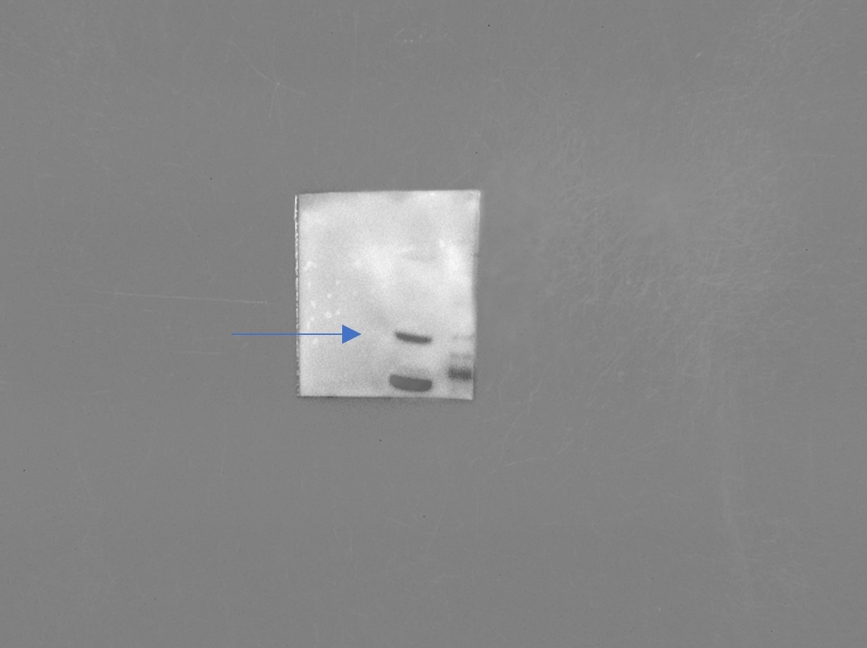
FH Nin (Verified Customer) (11-07-2024) | A good antibody to detect endogenous VANGL although there are nonspecific bands.
 |
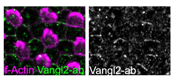
FH Stephen (Verified Customer) (05-31-2023) | works in chicken tissue fixed with 4% PFA for 2 hours, perm 0.3% tx100 for 2h primary incubated 1:200 for 24h @4 degrees,
 |
FH Louisiane (Verified Customer) (08-11-2022) | Tissues (small intestine sections) were incubated with primary antibody over the weekend at 4C and with secondary antibody overnight at 4C. The signal looks good but there is a bit of background
|

