"VDAC1/2 Antibodies" Comparison
View side-by-side comparison of VDAC1/2 antibodies from other vendors to find the one that best suits your research needs.
Tested Applications
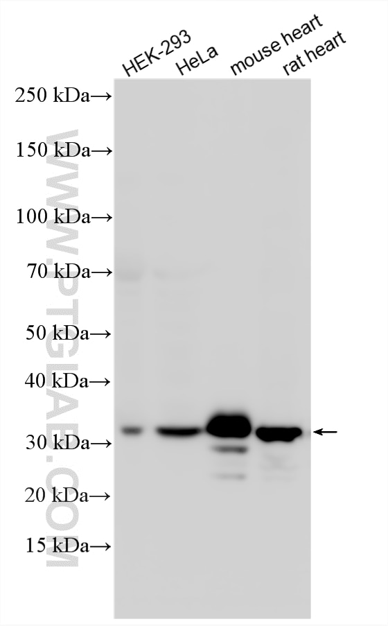
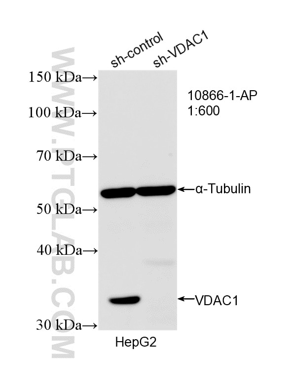
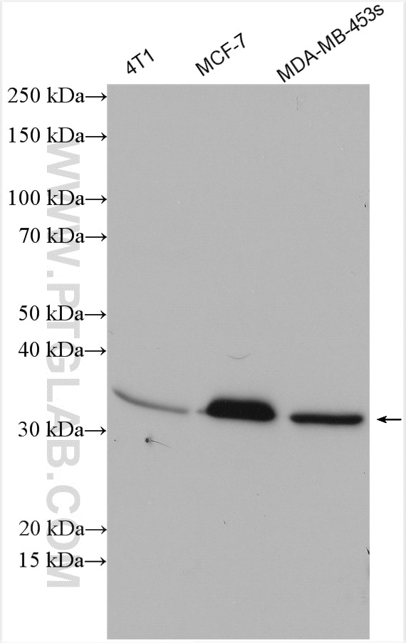
| Positive WB detected in | HEK-293 cells, 4T1 cells, HepG2 cells, MCF-7 cells, MDA-MB-453s cells, HeLa cells, mouse heart tissue, rat heart tissue |
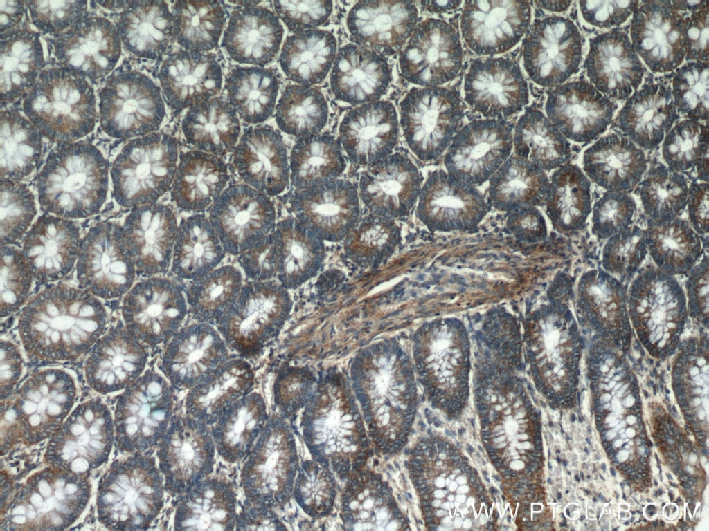
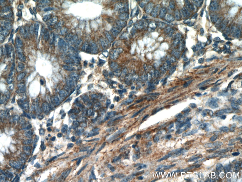
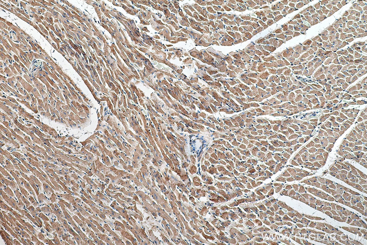
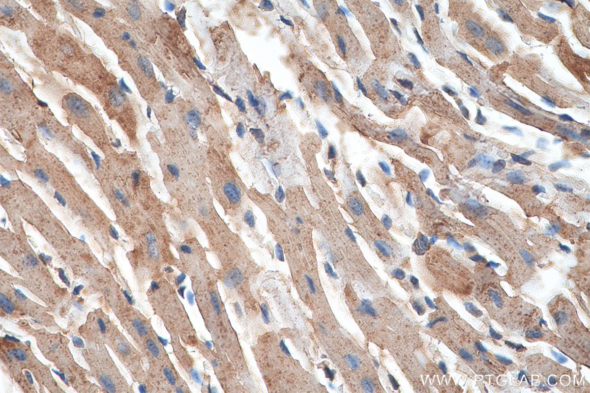
| Positive IHC detected in | human colon tissue, mouse heart tissue Note: suggested antigen retrieval with TE buffer pH 9.0; (*) Alternatively, antigen retrieval may be performed with citrate buffer pH 6.0 |
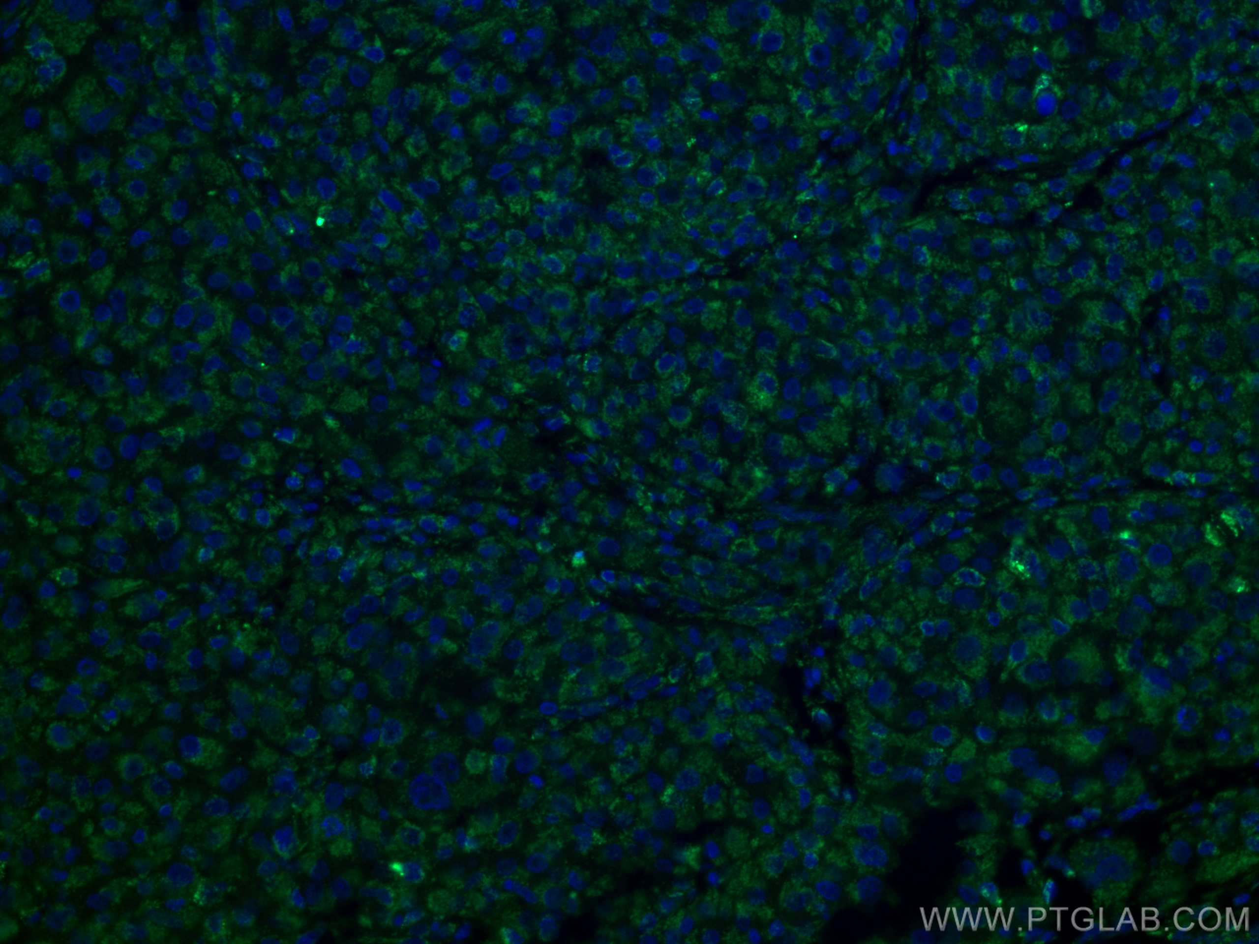
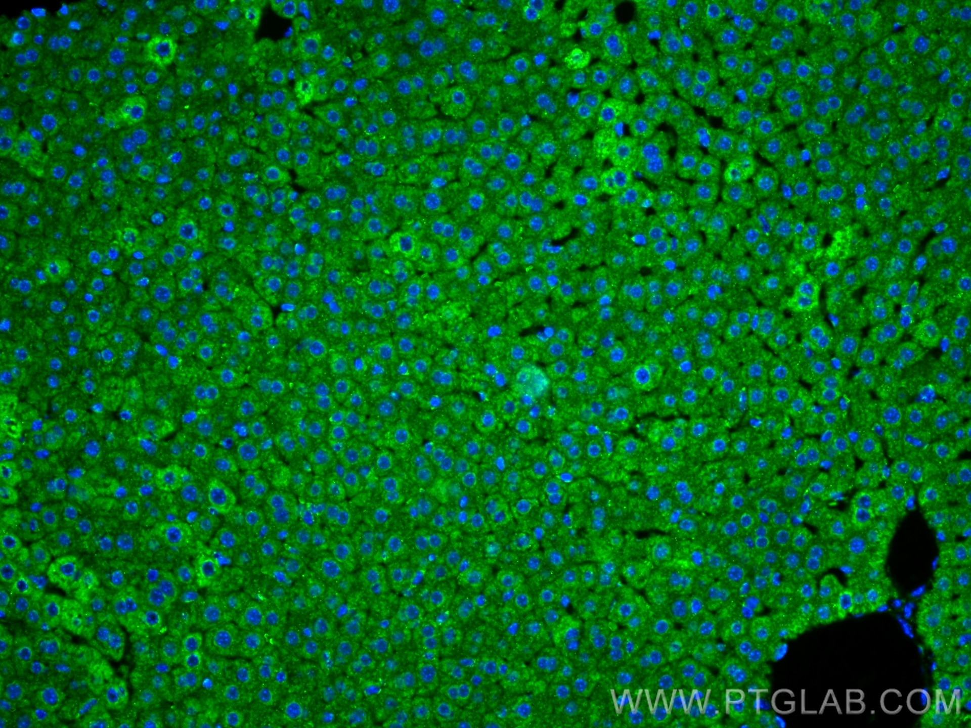
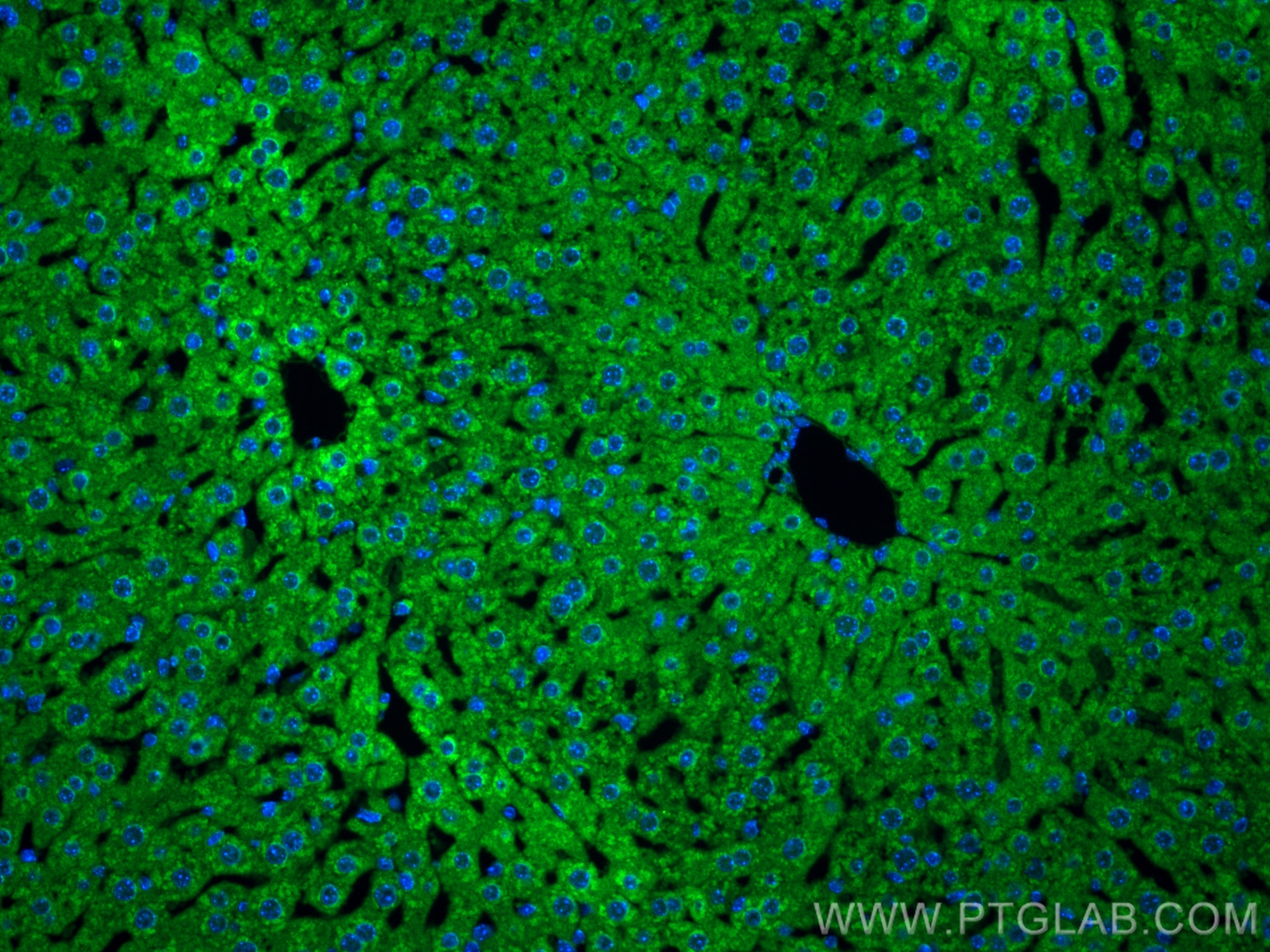
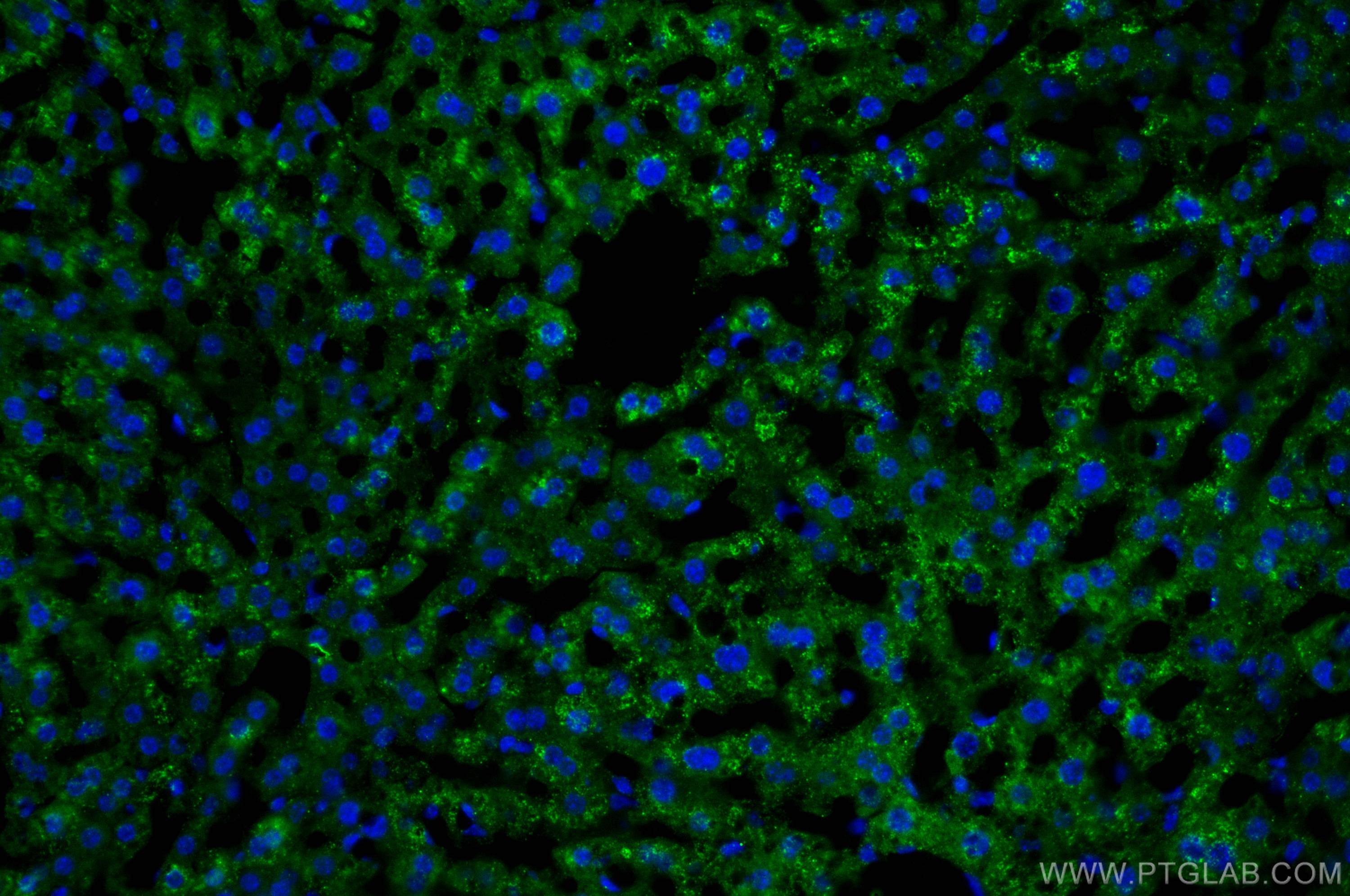
| Positive IF-P detected in | mouse liver tissue, human liver cancer tissue |
| Positive IF-Fro detected in | mouse liver tissue |
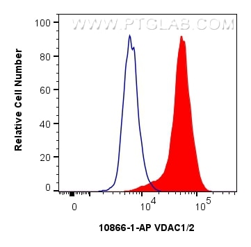
| Positive FC (Intra) detected in | HepG2 cells |
Recommended dilution
| Application | Dilution |
|---|---|
| Western Blot (WB) | WB : 1:500-1:2000 |
| Immunohistochemistry (IHC) | IHC : 1:50-1:500 |
| Immunofluorescence (IF)-P | IF-P : 1:200-1:800 |
| Immunofluorescence (IF)-FRO | IF-FRO : 1:50-1:500 |
| Flow Cytometry (FC) (INTRA) | FC (INTRA) : 0.80 ug per 10^6 cells in a 100 µl suspension |
| It is recommended that this reagent should be titrated in each testing system to obtain optimal results. | |
| Sample-dependent, Check data in validation data gallery. | |
Published Applications
| KD/KO | See 3 publications below |
| WB | See 209 publications below |
| IHC | See 5 publications below |
| IF | See 15 publications below |
| CoIP | See 5 publications below |
Product Information
10866-1-AP targets VDAC1/2 in WB, IHC, IF-P, IF-Fro, FC (Intra), CoIP, ELISA applications and shows reactivity with human, mouse, rat samples.
| Tested Reactivity | human, mouse, rat |
| Cited Reactivity | human, mouse, rat, pig, rabbit, monkey, zebrafish, pieris rapae |
| Host / Isotype | Rabbit / IgG |
| Class | Polyclonal |
| Type | Antibody |
| Immunogen |
CatNo: Ag1144 Product name: Recombinant human Porin protein Source: e coli.-derived, PGEX-4T Tag: GST Domain: 1-283 aa of BC008482 Sequence: MAVPPTYADLGKSARDVFTKGYGFGLIKLDLKTKSENGLEFTSSGSANTETTKVTGSLETKYRWTEYGLTFTEKWNTDNTLGTEITVEDQLARGLKLTFDSSFSPNTGKKNAKIKTGYKREHINLGCDMDFDIAGPSIRGALVLGYEGWLAGYQMNFETAKSRVTQSNFAVGYKTDEFQLHTNVNDGTEFGGSIYQKVNKKLETAVNLAWTAGNSNTRFGIAAKYQIDPDACFSAKVNNSSLIGLGYTQTLKPGIKLTLSALLDGKNVNAGGHKLGLGLEFQA Predict reactive species |
| Full Name | voltage-dependent anion channel 1 |
| Calculated Molecular Weight | 31 kDa |
| Observed Molecular Weight | 31 kDa |
| GenBank Accession Number | BC008482 |
| Gene Symbol | Porin |
| Gene ID (NCBI) | 7416 |
| RRID | AB_2257153 |
| Conjugate | Unconjugated |
| Form | Liquid |
| Purification Method | Antigen affinity purification |
| UNIPROT ID | P21796 |
| Storage Buffer | PBS with 0.02% sodium azide and 50% glycerol, pH 7.3. |
| Storage Conditions | Store at -20°C. Stable for one year after shipment. Aliquoting is unnecessary for -20oC storage. 20ul sizes contain 0.1% BSA. |
Background Information
VDAC1, also named as VDAC, porin 31HM, porin 31HL and plasmalemmal porin, belongs to the eukaryotic mitochondrial porin family. It adopts an open conformation at low or zero membrane potential and a closed conformation at potentials above 30-40 mV, to form a channel through the mitochondrial outer membrane and also the plasma membrane. Unlike other membrane transport proteins, porins are large enough to allow passive diffusion. Studies have shown that VDAC1 is subject to both phosphorylation and acetylation (PMID: 23233904). The apparent molecular weight of VDAC1 is 30-37 kDa (PMID: 14573604; 23754752; 25681439). Hypoxic conditions were found to trigger cleavage of the VDAC1 C-terminal to yield a 26-kDa truncated but active form (PMID: 22389449; 23233904). This polyclonal antibody raised against full-length human VDAC1 protein can cross react with VDAC2.
Protocols
| Product Specific Protocols | |
|---|---|
| FC protocol for VDAC1/2 antibody 10866-1-AP | Download protocol |
| IF protocol for VDAC1/2 antibody 10866-1-AP | Download protocol |
| IHC protocol for VDAC1/2 antibody 10866-1-AP | Download protocol |
| WB protocol for VDAC1/2 antibody 10866-1-AP | Download protocol |
| Standard Protocols | |
|---|---|
| Click here to view our Standard Protocols |
Publications
| Species | Application | Title |
|---|---|---|
Signal Transduct Target Ther Targeting CRL4 suppresses chemoresistant ovarian cancer growth by inducing mitophagy | ||
Nat Cell Biol AIDA directly connects sympathetic innervation to adaptive thermogenesis by UCP1. | ||
Cell Rep Med Urolithin A improves muscle strength, exercise performance, and biomarkers of mitochondrial health in a randomized trial in middle-aged adults. | ||
Cell Res DNA damage triggers tubular endoplasmic reticulum extension to promote apoptosis by facilitating ER-mitochondria signaling. | ||
Acta Pharm Sin B Histone deacetylase inhibitors inhibit cervical cancer growth through Parkin acetylation-mediated mitophagy. | ||
Reviews
The reviews below have been submitted by verified Proteintech customers who received an incentive for providing their feedback.
FH lu (Verified Customer) (10-02-2025) | Good antibody for western
|
FH Macarena (Verified Customer) (10-07-2022) | Good results for both IP and WB
|
FH Alberto (Verified Customer) (01-09-2020) | Clear sharp band at the expected molecular weight.
 |
FH Wanxia (Verified Customer) (10-22-2019) | Excellent WB results and very low background.
|