Tested Applications
| Positive WB detected in | HepG2 cells, COLO 320 cells, mouse skin tissue |
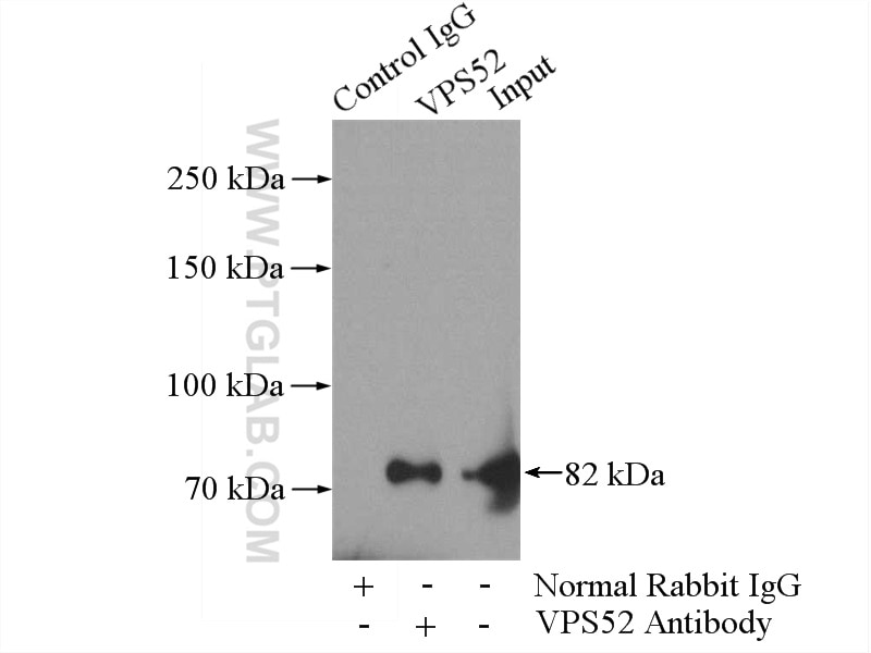
| Positive IP detected in | mouse skin tissue |
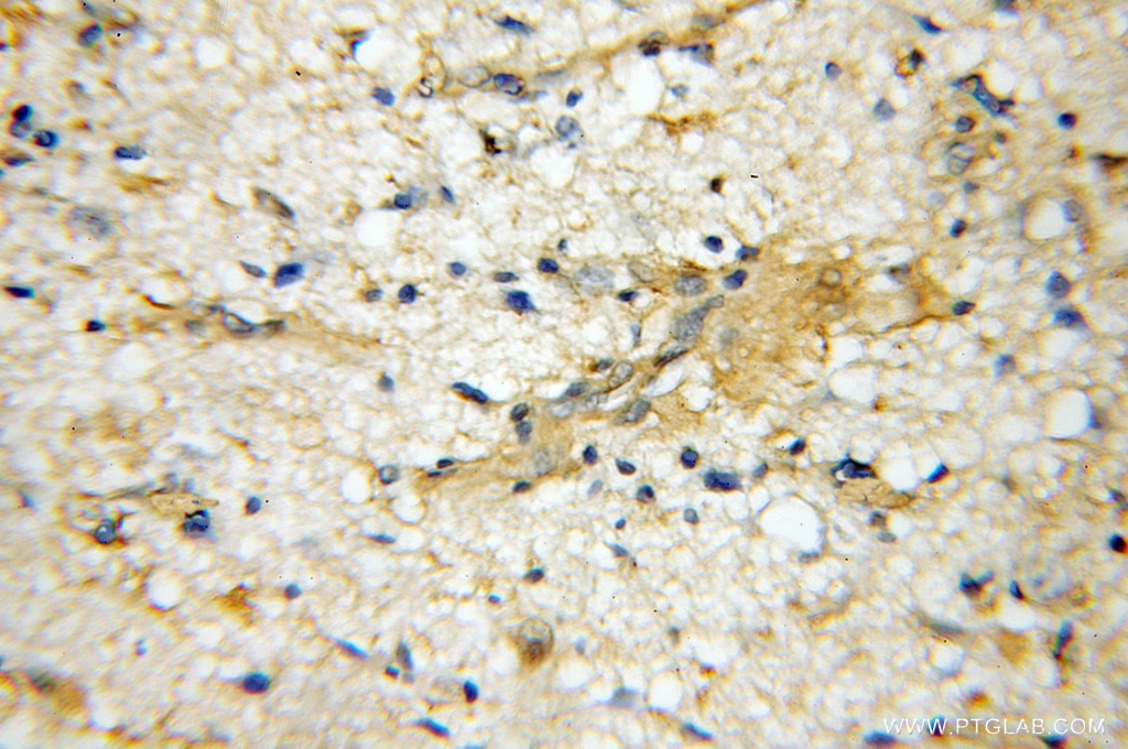
| Positive IHC detected in | human gliomas tissue Note: suggested antigen retrieval with TE buffer pH 9.0; (*) Alternatively, antigen retrieval may be performed with citrate buffer pH 6.0 |
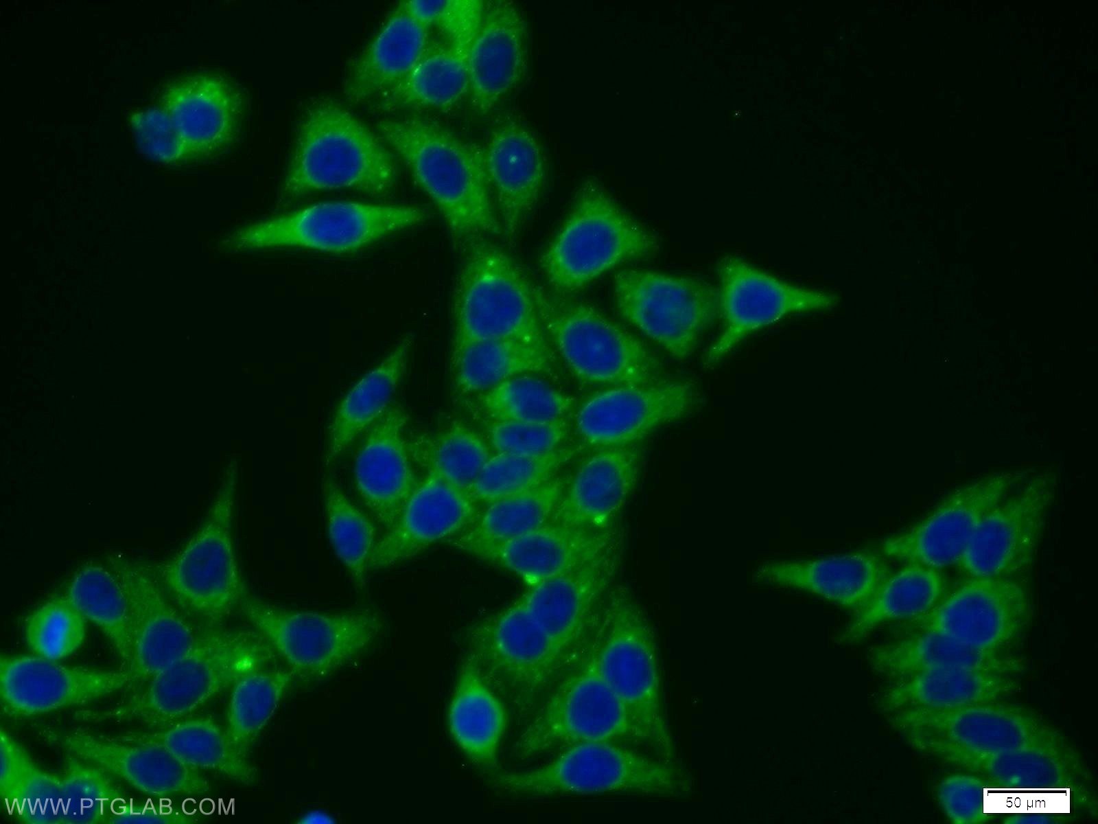
| Positive IF/ICC detected in | HepG2 cells |
Recommended dilution
| Application | Dilution |
|---|---|
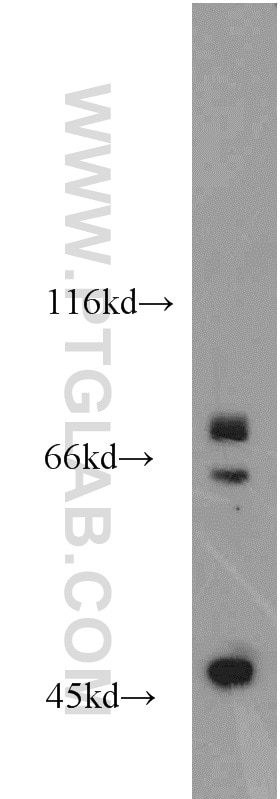
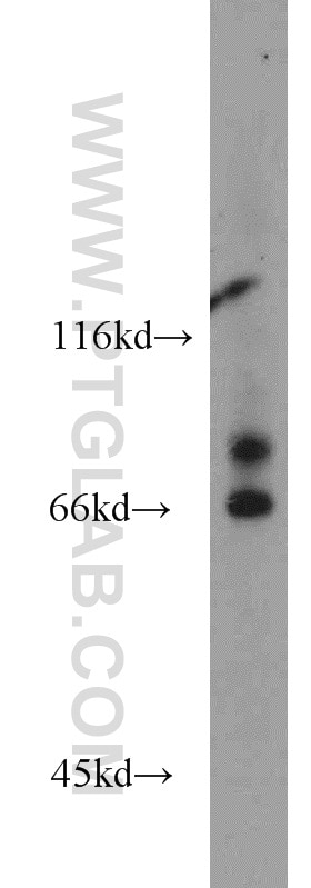
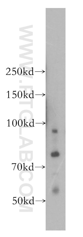
| Western Blot (WB) | WB : 1:500-1:1000 |
| Immunoprecipitation (IP) | IP : 0.5-4.0 ug for 1.0-3.0 mg of total protein lysate |
| Immunohistochemistry (IHC) | IHC : 1:20-1:200 |
| Immunofluorescence (IF)/ICC | IF/ICC : 1:10-1:100 |
| It is recommended that this reagent should be titrated in each testing system to obtain optimal results. | |
| Sample-dependent, Check data in validation data gallery. | |
Published Applications
| WB | See 3 publications below |
| IHC | See 1 publications below |
| IF | See 1 publications below |
Product Information
11662-2-AP targets VPS52 in WB, IHC, IF/ICC, IP, ELISA applications and shows reactivity with human, mouse, rat samples.
| Tested Reactivity | human, mouse, rat |
| Cited Reactivity | human, mouse |
| Host / Isotype | Rabbit / IgG |
| Class | Polyclonal |
| Type | Antibody |
| Immunogen |
CatNo: Ag2180 Product name: Recombinant human VPS52 protein Source: e coli.-derived, PGEX-4T Tag: GST Domain: 373-723 aa of BC032108 Sequence: EQRYPFEALFRSQHYALLDNSCREYLFICEFFVVSGPAAHDLFHAVMGRTLSMTLKHLDSYLADCYDAIAVFLCIHIVLRFRNIAAKRDVPALDRYWEQVLALLWPRFELILEMNVQSVRSTDPQRLGGLDTRPHYITRRYAEFSSALVSINQTIPNERTMQLLGQLQVEVENFVLRVAAEFSSRKEQLVFLINNYDMMLGVLMERAADDSKEVESFQQLLNARTQEFIEELLSPPFGGLVAFVKEAEALIERGQAERLRGEEARVTQLIRGFGSSWKSSVESLSQDVMRSFTNFRNGTSIIQGALTQLIQLYHRFHRVLSQPQLRALPARAELINIHHLMVELKKHKPNF Predict reactive species |
| Full Name | vacuolar protein sorting 52 homolog (S. cerevisiae) |
| Calculated Molecular Weight | 723 aa, 82 kDa |
| Observed Molecular Weight | 82 kDa |
| GenBank Accession Number | BC032108 |
| Gene Symbol | VPS52 |
| Gene ID (NCBI) | 6293 |
| RRID | AB_2241614 |
| Conjugate | Unconjugated |
| Form | Liquid |
| Purification Method | Antigen affinity purification |
| UNIPROT ID | Q8N1B4 |
| Storage Buffer | PBS with 0.02% sodium azide and 50% glycerol, pH 7.3. |
| Storage Conditions | Store at -20°C. Stable for one year after shipment. Aliquoting is unnecessary for -20oC storage. 20ul sizes contain 0.1% BSA. |
Background Information
VPS52 is a component of the Golgi-associated retrograde protein (GARP) complex, also called VFT (VPS fifty-three) complex, composed of VPS51, VPS52, VPS53 and VPS54. GARP complex functions in traffic from endosomes the trans-Golgi network (PMID: 15878329). GARP proteins interact with RAB proteins and SNARE proteins. VPS52 may play a role in the development of a multicellular organism through cell-cell interactions (PMID: 23142660).
Protocols
| Product Specific Protocols | |
|---|---|
| IF protocol for VPS52 antibody 11662-2-AP | Download protocol |
| IHC protocol for VPS52 antibody 11662-2-AP | Download protocol |
| IP protocol for VPS52 antibody 11662-2-AP | Download protocol |
| WB protocol for VPS52 antibody 11662-2-AP | Download protocol |
| Standard Protocols | |
|---|---|
| Click here to view our Standard Protocols |
Publications
| Species | Application | Title |
|---|---|---|
Cell Rep The GARP Complex Is Involved in Intracellular Cholesterol Transport via Targeting NPC2 to Lysosomes. | ||
Antioxid Redox Signal Thioredoxin-1 Inhibits Golgi Stress Induced by Methyl-4-Phenyl-1,2,3, 6-Tetrahydropyridine. |