"YY1 Antibodies" Comparison
View side-by-side comparison of YY1 antibodies from other vendors to find the one that best suits your research needs.
Tested Applications
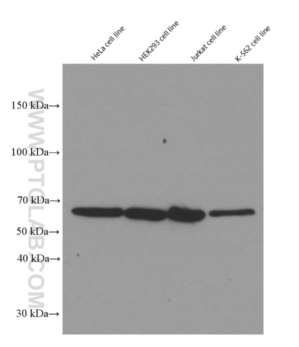
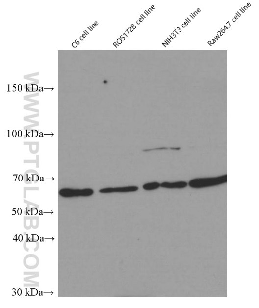
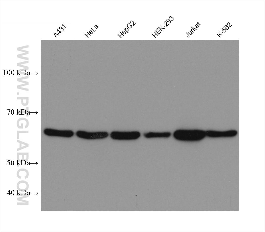
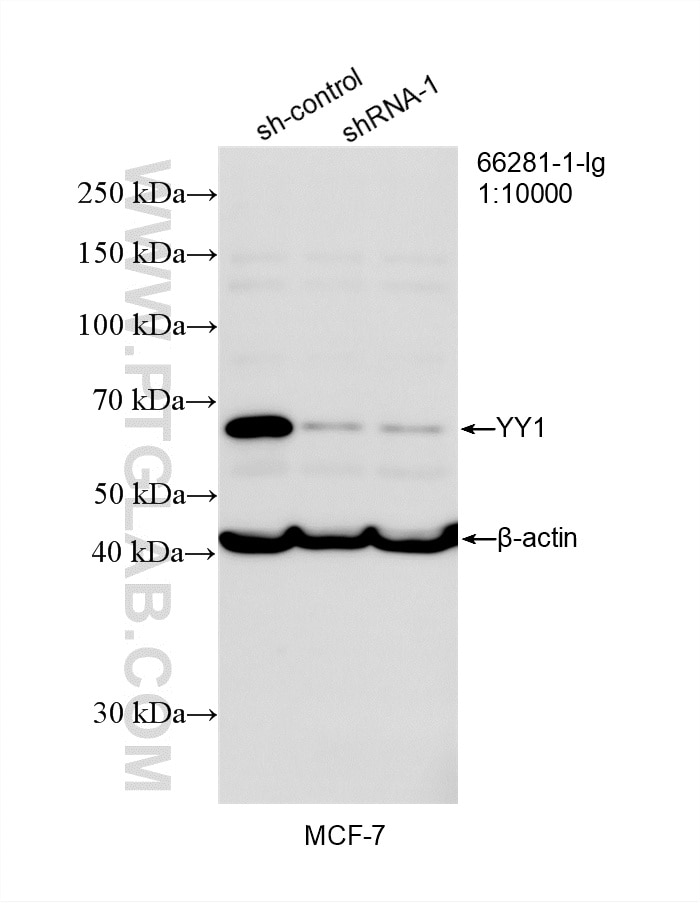
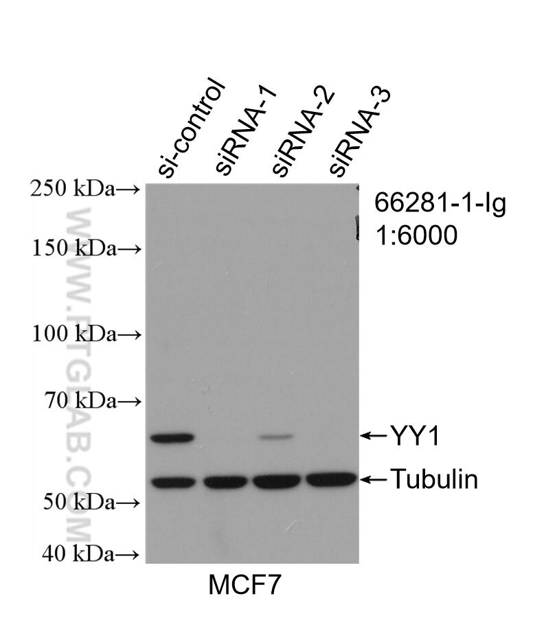
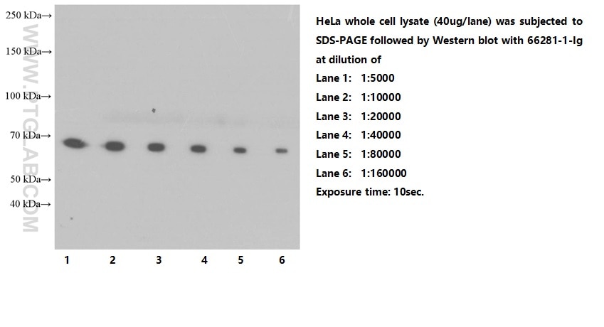
| Positive WB detected in | C6 cells, A431 cells, COS-7 cells, HeLa cells, MCF-7 cells, NIH/3T3 cells, HepG2 cells, HEK-293 cells, Jurkat cells, K-562 cells |
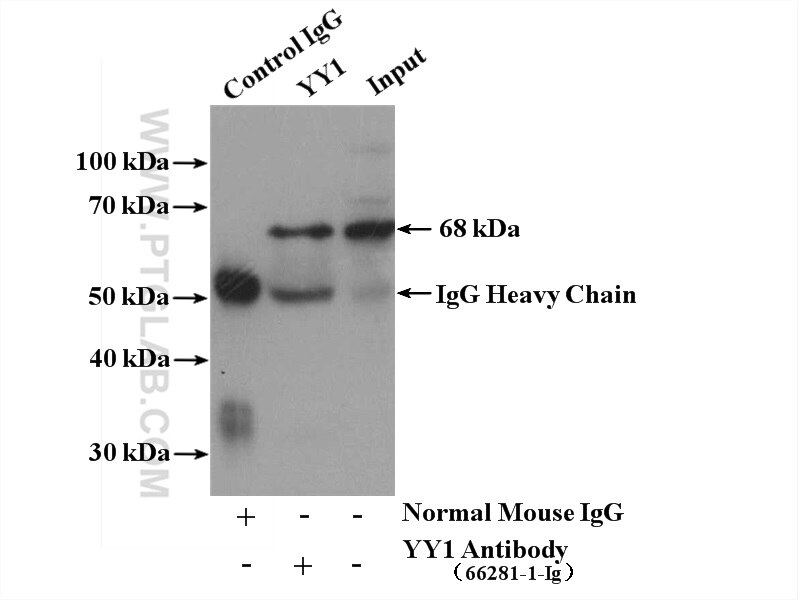
| Positive IP detected in | NIH/3T3 cells |
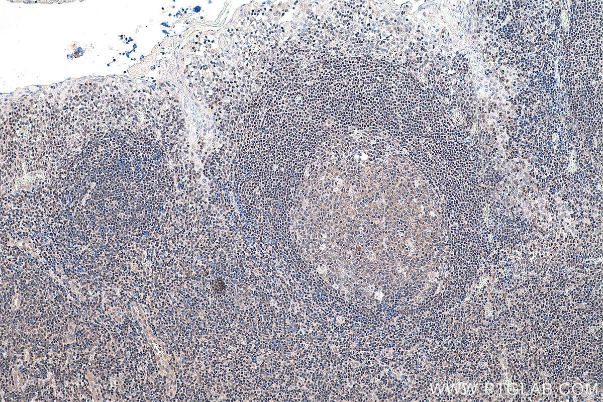
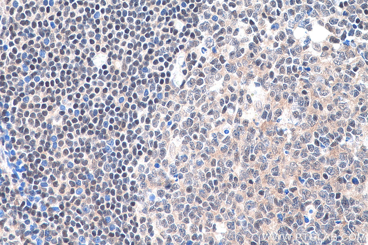
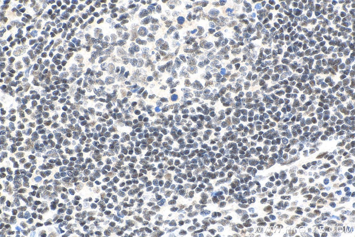
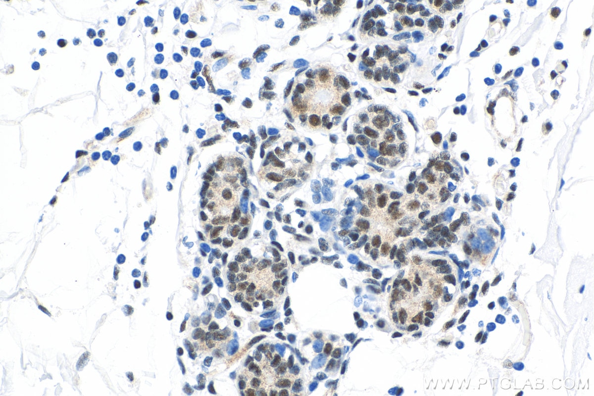
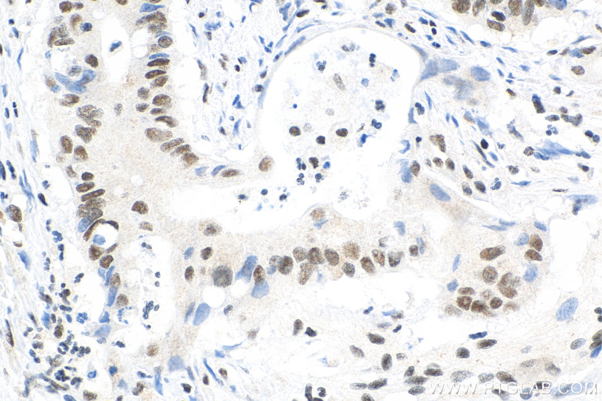
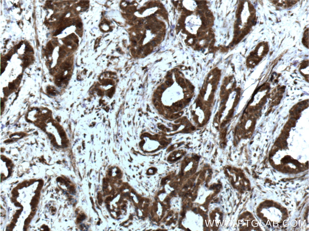
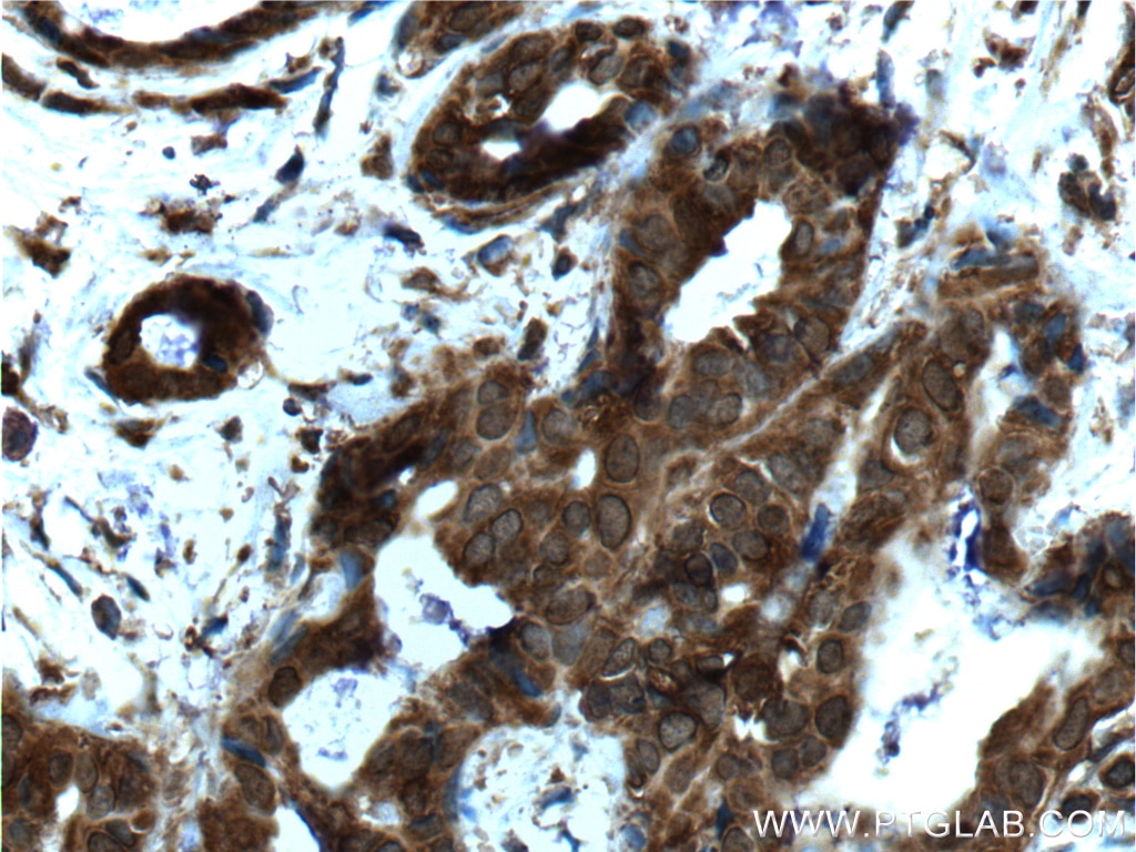
| Positive IHC detected in | human breast cancer tissue, human colon cancer tissue, human tonsillitis tissue Note: suggested antigen retrieval with TE buffer pH 9.0; (*) Alternatively, antigen retrieval may be performed with citrate buffer pH 6.0 |
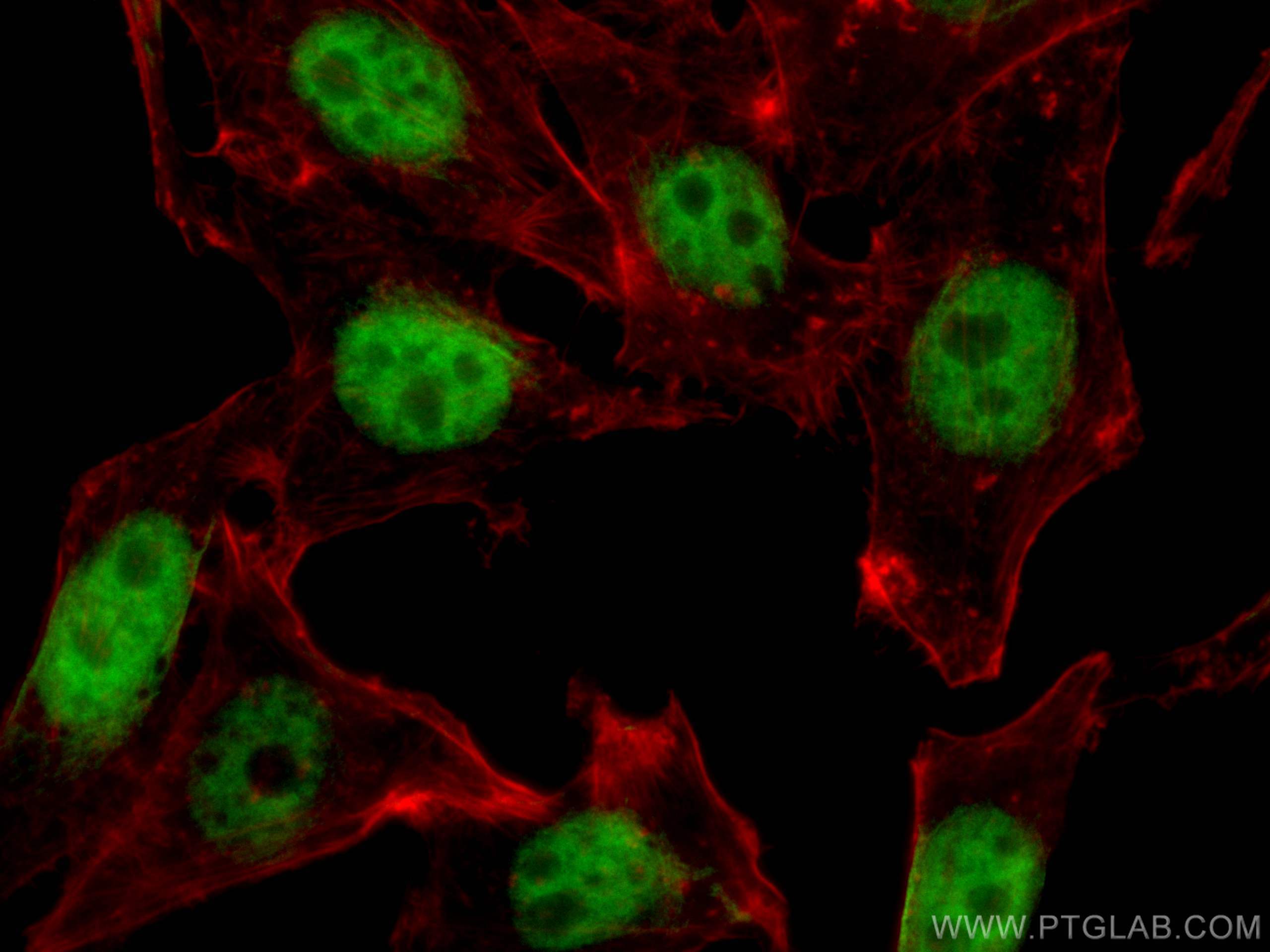
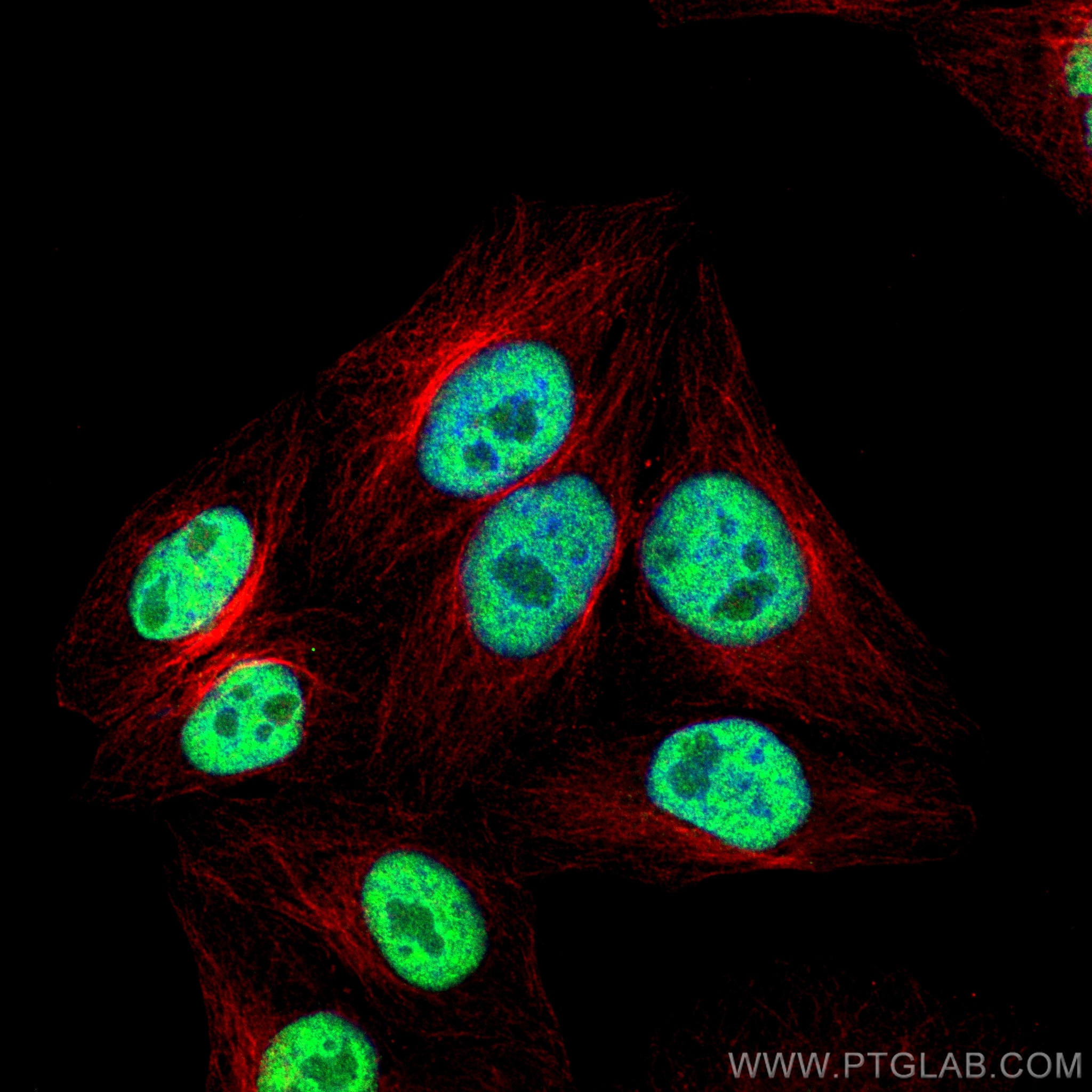
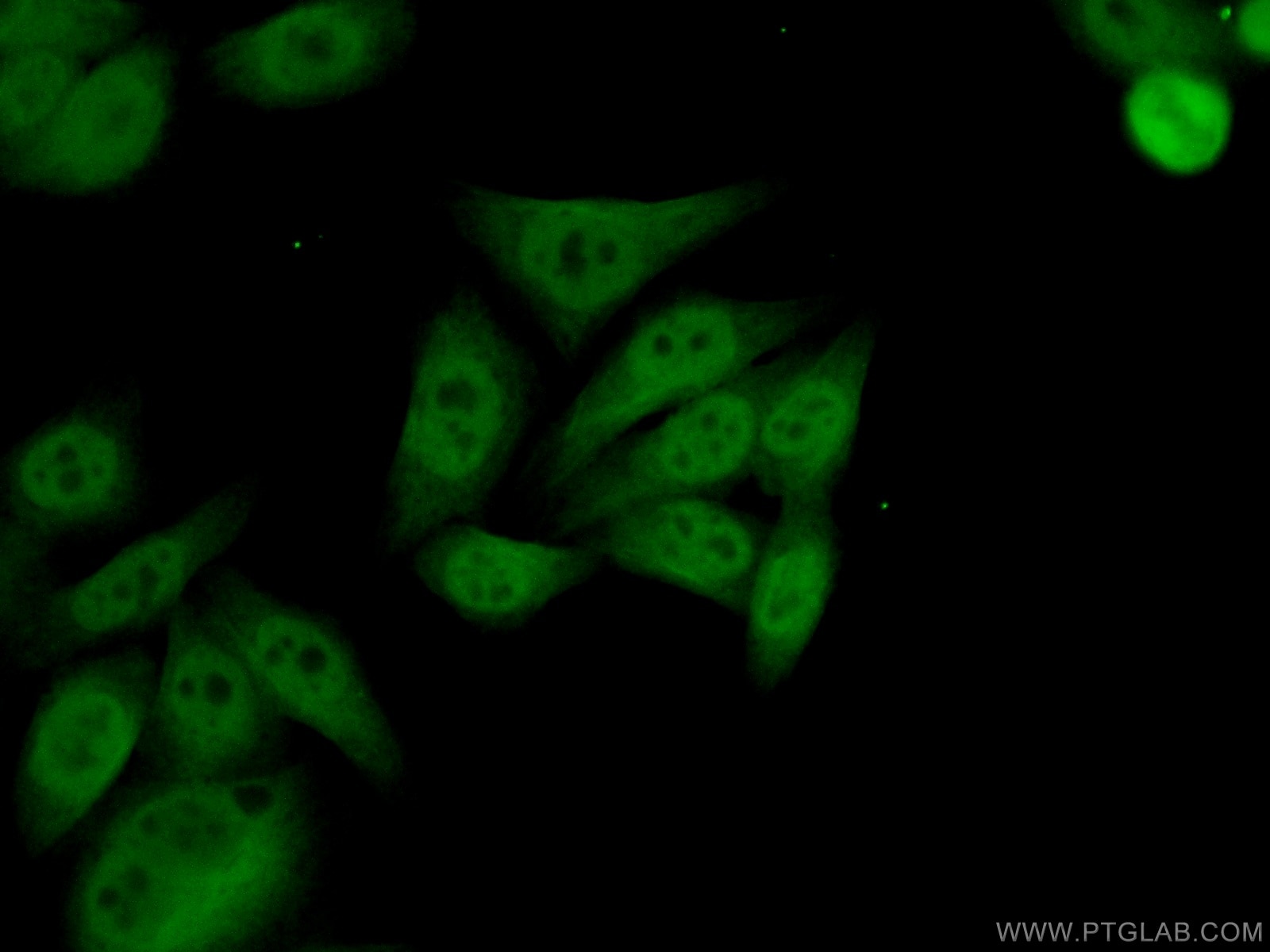
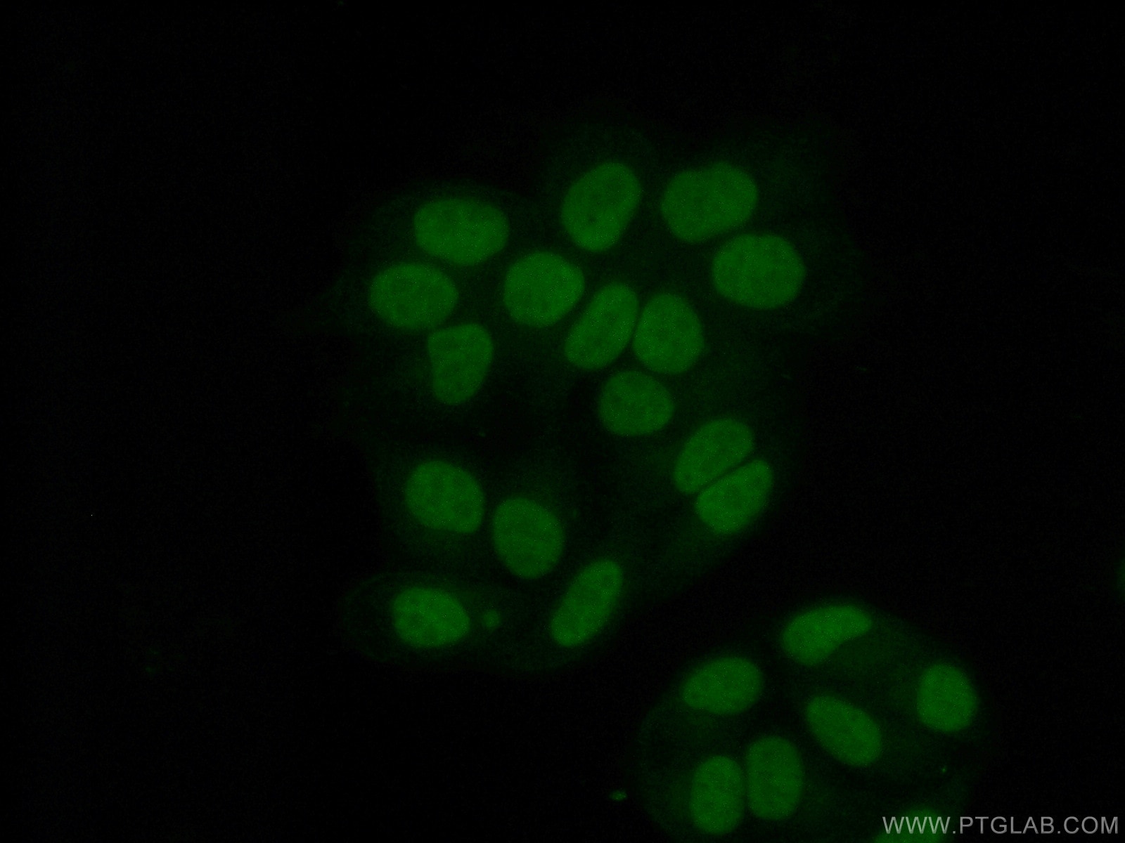
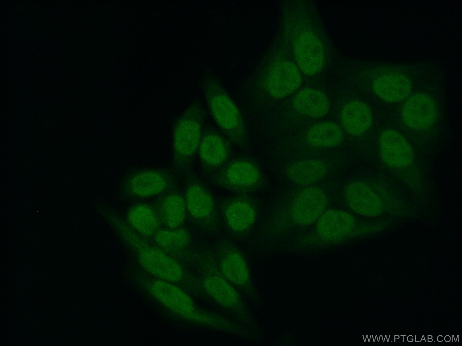
| Positive IF/ICC detected in | HepG2 cells |
Recommended dilution
| Application | Dilution |
|---|---|
| Western Blot (WB) | WB : 1:5000-1:50000 |
| Immunoprecipitation (IP) | IP : 0.5-4.0 ug for 1.0-3.0 mg of total protein lysate |
| Immunohistochemistry (IHC) | IHC : 1:5000-1:20000 |
| Immunofluorescence (IF)/ICC | IF/ICC : 1:200-1:800 |
| It is recommended that this reagent should be titrated in each testing system to obtain optimal results. | |
| Sample-dependent, Check data in validation data gallery. | |
Product Information
66281-1-Ig targets YY1 in WB, IHC, IF/ICC, IP, CoIP, ChIP, RIP, ELISA applications and shows reactivity with human, mouse, rat, monkey samples.
| Tested Reactivity | human, mouse, rat, monkey |
| Cited Reactivity | human, mouse, rat, pig |
| Host / Isotype | Mouse / IgG2a |
| Class | Monoclonal |
| Type | Antibody |
| Immunogen |
CatNo: Ag17732 Product name: Recombinant human YY1 protein Source: e coli.-derived, PET28a Tag: 6*His Domain: 1-414 aa of BC037308 Sequence: MASGDTLYIATDGSEMPAEIVELHEIEVETIPVETIETTVVGEEEEEDDDDEDGGGGDHGGGGGHGHAGHHHHHHHHHHHPPMIALQPLVTDDPTQVHHHQEVILVQTREEVVGGDDSDGLRAEDGFEDQILIPVPAPAGGDDDYIEQTLVTVAAAGKSGGGGSSSSGGGRVKKGGGKKSGKKSYLSGGAGAAGGGGADPGNKKWEQKQVQIKTLEGEFSVTMWSSDEKKDIDHETVVEEQIIGENSPPDYSEYMTGKKLPPGGIPGIDLSDPKQLAEFARMKPRKIKEDDAPRTIACPHKGCTKMFRDNSAMRKHLHTHGPRVHVCAECGKAFVESSKLKRHQLVHTGEKPFQCTFEGCGKRFSLDFNLRTHVRIHTGDRPYVCPFDGCNKKFAQSTNLKSHILTHAKAKNNQ Predict reactive species |
| Full Name | YY1 transcription factor |
| Calculated Molecular Weight | 414 aa, 45 kDa |
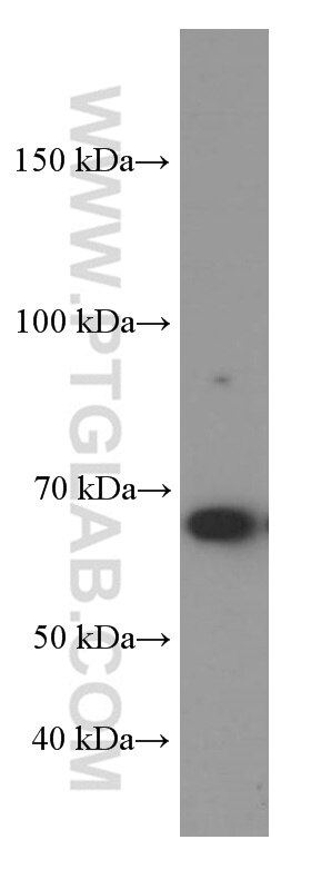
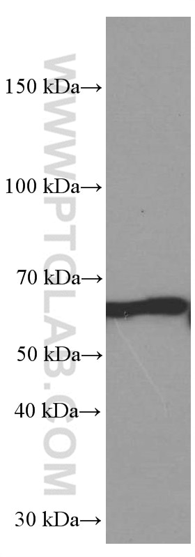
| Observed Molecular Weight | 65-70 kDa |
| GenBank Accession Number | BC037308 |
| Gene Symbol | YY1 |
| Gene ID (NCBI) | 7528 |
| RRID | AB_2881664 |
| Conjugate | Unconjugated |
| Form | Liquid |
| Purification Method | Protein A purification |
| UNIPROT ID | P25490 |
| Storage Buffer | PBS with 0.02% sodium azide and 50% glycerol, pH 7.3. |
| Storage Conditions | Store at -20°C. Stable for one year after shipment. Aliquoting is unnecessary for -20oC storage. 20ul sizes contain 0.1% BSA. |
Background Information
YY1, also named as DELTA, INO80S and NF-E1, contains four C2H2-type zinc fingers and belongs to the YY transcription factor family. YY1 is a multifunctional transcription factor that exhibits positive and negative control on a large number of cellular and viral genes by binding to sites overlapping the transcription start site. YY1 may direct histone deacetylases and histone acetyltransferases to a promoter in order to activate or repress the promoter, thus implicating histone modification in the YY1. The open reading frame of the human YY1 cDNA encodes a protein of 414 amino acids with a predicted molecular weight of 44 kDa. However, YY1 migrates on SDS gels as a 65-70 kDa protein, probably due to the structure of the protein. It is a ubiquitously expressed transcription factor with fundamental roles in embryogenesis, differentiation, replication and proliferation.
Protocols
| Product Specific Protocols | |
|---|---|
| IF protocol for YY1 antibody 66281-1-Ig | Download protocol |
| IHC protocol for YY1 antibody 66281-1-Ig | Download protocol |
| IP protocol for YY1 antibody 66281-1-Ig | Download protocol |
| WB protocol for YY1 antibody 66281-1-Ig | Download protocol |
| Standard Protocols | |
|---|---|
| Click here to view our Standard Protocols |
Publications
| Species | Application | Title |
|---|---|---|
Cell Pervasive Chromatin-RNA Binding Protein Interactions Enable RNA-Based Regulation of Transcription. | ||
Mol Ther Nucleic Acids Angiotensin II-induced muscle atrophy via PPARγ suppression is mediated by miR-29b. | ||
Cell Death Dis CircHIPK3 promotes colorectal cancer growth and metastasis by sponging miR-7. | ||
Cancers (Basel) MiR-302b as a Combinatorial Therapeutic Approach to Improve Cisplatin Chemotherapy Efficacy in Human Triple-Negative Breast Cancer. |