
Cell contamination
A guide to identifying common cell culture contaminants, including mycoplasma, bacteria, fungi and viruses.
| Cell culture contamination | |
| Bacterial contamination | Fungal (yeast/mold) contamination |
| Mycoplasma contamination | Virus contamination |
Cell contamination
Contamination of cell cultures is a serious risk to your samples. Consequently, safety measures, including aseptic techniques, should be put in place to actively prevent it and your samples should be regularly checked for any sign of infections. Cell contamination may severely affect their metabolism, growth, and viability, as well as decrease transfection efficiency and threaten the reproducibility of cell-based experiments.
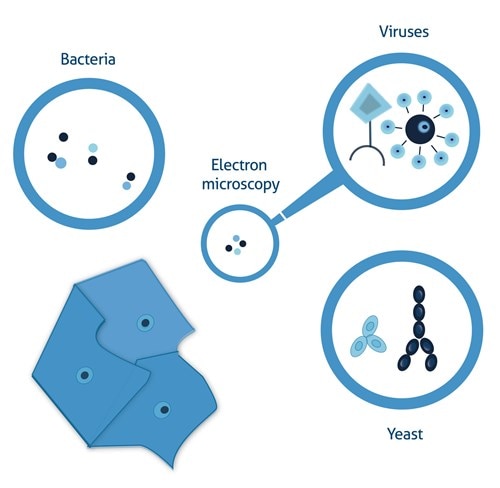
Contamination in cell culture may be due to chemical and biological agents. Chemical contamination, such as the presence of endotoxins, detergents, or impurities of used media, sera, or other media supplements can be controlled by using trusted suppliers that guarantee rigorous batch testing. Biological contamination can arise from improper aseptic techniques and most often comes from the direct environment in the cell culture area. The most common biological contaminants are bacteria, fungi (yeast and mold), viruses, and mycoplasma (summary Figure 6).
Bacterial contamination
Many bacterial strains can grow in commonly used media in mammalian cell culture. Due to rapid logarithmic growth, the signs of contamination are easily detectable within a few days of the initial contamination.
-
Loss of medium clarity.
-
Faster acidification of medium (medium turns orange/yellow if phenol red is present).
-
A thin bacterial film appears on the surface of cell culture vessels/flasks.
Bacteria can be identified by a brightfield microscope check (20–60x magnification) as slowly moving, dark, round- or rod-shaped objects that are significantly smaller than mammalian cells.
Mycoplasma contamination
Mycoplasma are very small (0.2um) bacteria characterized by their lack of a cell wall. Unlike standard bacterial contamination, mycoplasma often grow at a slow rate in infected cultures and can be present with no visible signs of infection such as loss of clarity or change of pH.
Mycoplasma do not contain cell walls, so they cannot be seen with a standard brightfield microscope.
-
Due to their slow growth rate, mycoplasma infection may develop for a long time without any signs.
-
Mycoplasma infection often affects cell growth rates, metabolism, and can even lead to chromosome aberrations of samples.
-
Routine testing using immunofluorescence with DNA stain, ELISA, or PCR is recommended to monitor possible infections.
Fungal (yeast/mold) contamination
Similar to bacterial contamination, yeasts can grow rapidly in mammalian culture media, leading to a loss of medium clarity, albeit at a slower rate.
-
Relatively rapid progression from initial infection to a serious threat.
-
pH is unchanged or slightly increases at later stages of the contamination, which can affect your experiments.
-
Brightfield microscope check (20–60x magnification): rod-shaped or branched filamentous objects that become visible to the naked eye at later stages of contamination.
Many fungi can produce spores capable of surviving for a long time in a dormant state in hostile environments and, therefore, in the event of fungal contamination it is particularly important to decontaminate all working surfaces and incubators to prevent repeated contaminations.
Virus contamination
Viral contaminations are not very common but can potentially pose a serious health threat to cell culture personnel.
-
Viruses are hard to detect because they do not affect cell culture growth and cannot be seen with a brightfield microscope.
-
Dedicated assays (e.g., ELISA, PCR) can be used to test for infections.
Cell culture contamination. Cells can be contaminated with bacteria (small, dark, rod-shaped that can form strings), yeast (elongated, branched structures, light with darker edges), or viruses (visible only with electron microscopy).
Able™