CD206 Polyklonaler Antikörper
CD206 Polyklonal Antikörper für WB, IHC, IF-P, ELISA
Wirt / Isotyp
Kaninchen / IgG
Getestete Reaktivität
human, Ratte und mehr (4)
Anwendung
WB, IHC, IF-P, ELISA, Cell treatment
Konjugation
Unkonjugiert
Kat-Nr. : 18704-1-AP
Synonyme
 at dilution of 1:1000 incubated at room temperature for 1.5 hours.")
 at dilution of 1:4000 (under 10x lens). Heat mediated antigen retrieval with Tris-EDTA buffer (pH 9.0).")
 at dilution of 1:4000 (under 40x lens). Heat mediated antigen retrieval with Tris-EDTA buffer (pH 9.0).")
 fixed paraffin-embedded human placenta tissue using CD206 antibody (18704-1-AP) at dilution of 1:200 and CoraLite®488-Conjugated Goat Anti-Rabbit IgG(H+L) (SA00013-2). Heat mediated antigen retrieval with Tris-EDTA buffer (pH 9.0).")
 fixed paraffin-embedded human placenta tissue using CD206 antibody (18704-1-AP) at dilution of 1:200 and CoraLite®488-Conjugated Goat Anti-Rabbit IgG(H+L) (SA00013-2). Heat mediated antigen retrieval with Tris-EDTA buffer (pH 9.0).")
Geprüfte Anwendungen
| Erfolgreiche Detektion in WB | humanes Plazenta-Gewebe, Rattenlebergewebe |
| Erfolgreiche Detektion in IHC | humanes Plazenta-Gewebe Hinweis: Antigendemaskierung mit TE-Puffer pH 9,0 empfohlen. (*) Wahlweise kann die Antigendemaskierung auch mit Citratpuffer pH 6,0 erfolgen. |
| Erfolgreiche Detektion in IF-P | humanes Plazenta-Gewebe |
Empfohlene Verdünnung
| Anwendung | Verdünnung |
|---|---|
| Western Blot (WB) | WB : 1:500-1:2000 |
| Immunhistochemie (IHC) | IHC : 1:2000-1:8000 |
| Immunfluoreszenz (IF)-P | IF-P : 1:50-1:500 |
| It is recommended that this reagent should be titrated in each testing system to obtain optimal results. | |
| Sample-dependent, check data in validation data gallery | |
Veröffentlichte Anwendungen
| WB | See 250 publications below |
| IHC | See 133 publications below |
| IF | See 425 publications below |
Produktinformation
18704-1-AP bindet in WB, IHC, IF-P, ELISA, Cell treatment CD206 und zeigt Reaktivität mit human, Ratten
| Getestete Reaktivität | human, Ratte |
| In Publikationen genannte Reaktivität | human, Hausschwein, Kaninchen, Ratte, Rind, Miesmuschel |
| Wirt / Isotyp | Kaninchen / IgG |
| Klonalität | Polyklonal |
| Typ | Antikörper |
| Immunogen | Peptid |
| Vollständiger Name | mannose receptor, C type 1 |
| Berechnetes Molekulargewicht | 166 kDa |
| Beobachtetes Molekulargewicht | 180-200 kDa |
| GenBank-Zugangsnummer | NM_002438 |
| Gene symbol | CD206 |
| Gene ID (NCBI) | 4360 |
| Konjugation | Unkonjugiert |
| Form | Liquid |
| Reinigungsmethode | Antigen-Affinitätsreinigung |
| Lagerungspuffer | PBS with 0.02% sodium azide and 50% glycerol |
| Lagerungsbedingungen | Bei -20°C lagern. Nach dem Versand ein Jahr lang stabil Aliquotieren ist bei -20oC Lagerung nicht notwendig. 20ul Größen enthalten 0,1% BSA. |
Hintergrundinformationen
Background
CD206 (macrophage mannose receptor 1) is a lectin-type endocytic receptor expressed on selected macrophages, dendritic cells, and non-vascular endothelium and plays a role in antigen processing and presentation, phagocytosis, and intracellular signaling.
1. What is the molecular weight of CD206?
The molecular size of full-length CD206 is 170-180 kDa, depending on the exact tissue-specific glycosylation pattern (PMID: 19427834). Additionally, CD206 can be cleaved off and a soluble form (sMR) lacking the tail, with a slightly lower molecular weight, can be released to the cell medium (PMID: 9722572).
2. What is the subcellular localization of CD206?
CD206 is a type I membrane protein composed of a large extracellular multidomain, a transmembrane domain, and a short cytoplasmic tail. It is present at the plasma membrane and in endosomes, as CD206 undergoes constant recycling between the plasma membrane and endosomal compartment.
3. Is CD206 post-translationally modified?
CD206 undergoes quite extensive post-translational modifications, predominantly N-linked glycosylation that affects ligand binding recognition and affinity (PMID: 22966131).
4. Can CD206 marker be used as a marker of M2 macrophages?
The activation of macrophages with various stimuli leads to their polarization into classical (M1) or alternatively activated (M2) subtypes spectrums and both subtypes differ in their regulatory and effector functions (PMID: 24669294). Pathogens and IFN-γ promote M1 polarization, while IL-4 released during parasite infections and allergen response promotes M2 polarization. Classically, the markers of M2 macrophages include CD206, as well as arginase-1 (ARG1; https://www.ptglab.com/products/ARG1-Antibody-16001-1-AP.htm), CD163 (https://www.ptglab.com/products/CD163-Antibody-16646-1-AP.htm), and thrombospondin 1 (TSP1/ THBS1; https://www.ptglab.com/products/TSP1-Antibody-18304-1-AP.htm).
5. How can you polarize macrophages into M2 direction?
One of the most commonly used methods is stimulation by the addition of IL-4 cytokine. We recommend using our animal-free human IL-4 (https://www.ptglab.com/products/recombinant-human-il-4.htm).
Protokolle
| PRODUKTSPEZIFISCHE PROTOKOLLE | |
|---|---|
| WB protocol for CD206 antibody 18704-1-AP | Protokoll herunterladen |
| IHC protocol for CD206 antibody 18704-1-AP | Protokoll herunterladenl |
| IF protocol for CD206 antibody 18704-1-AP | Protokoll herunterladen |
| STANDARD-PROTOKOLLE | |
|---|---|
| Klicken Sie hier, um unsere Standardprotokolle anzuzeigen |
Publikationen
| Species | Application | Title |
|---|---|---|
Adv Mater Osteoimmunity-Regulating Biomimetically Hierarchical Scaffold for Augmented Bone Regeneration. | ||
Bioact Mater Reprogramming macrophages via immune cell mobilized hydrogel microspheres for osteoarthritis treatments | ||
Bioact Mater Highly active probiotic hydrogels matrixed on bacterial EPS accelerate wound healing via maintaining stable skin microbiota and reducing inflammation | ||
ACS Nano Glutamine Antagonist Synergizes with Electrodynamic Therapy to Induce Tumor Regression and Systemic Antitumor Immunity. | ||
Adv Sci (Weinh) Colorectal Cancer-Derived Small Extracellular Vesicles Promote Tumor Immune Evasion by Upregulating PD-L1 Expression in Tumor-Associated Macrophages. |
Rezensionen
The reviews below have been submitted by verified Proteintech customers who received an incentive for providing their feedback.
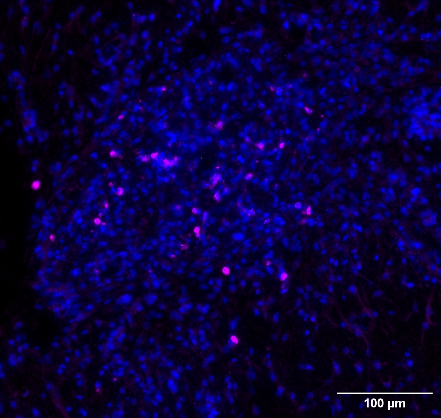
FH Marion (Verified Customer) (12-09-2024) | DAPI (blue) & CD206 (magenta) This antibody was tested to reveal CD206+/Iba-1+ cells in a murine AD model. Iba-1 staining is not presented here. The antibody works fine for IF on mouse tissue.
 |
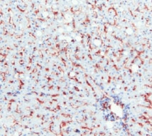
FH Murali (Verified Customer) (07-28-2022) | This amazing antibody works well for human IHC staining.
 |
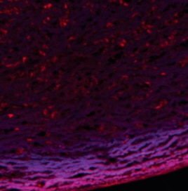
FH Uthra (Verified Customer) (10-14-2021) | The antibody works fine for IF.
 |
FH Nethaji (Verified Customer) (05-08-2019) | The antibody works fine for western and ICC.
|

