Tested Applications
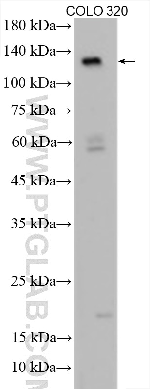
| Positive WB detected in | COLO 320 cells |
Recommended dilution
| Application | Dilution |
|---|---|
| Western Blot (WB) | WB : 1:1000-1:6000 |
| It is recommended that this reagent should be titrated in each testing system to obtain optimal results. | |
| Sample-dependent, Check data in validation data gallery. | |
Published Applications
| KD/KO | See 1 publications below |
| WB | See 8 publications below |
| IF | See 1 publications below |
Product Information
29022-1-AP targets APAF1 in WB, IF, ELISA applications and shows reactivity with Human samples.
| Tested Reactivity | Human |
| Cited Reactivity | human, mouse, rat |
| Host / Isotype | Rabbit / IgG |
| Class | Polyclonal |
| Type | Antibody |
| Immunogen |
CatNo: Ag30606 Product name: Recombinant human APAF1 protein Source: e coli.-derived, PGEX-4T Tag: GST Domain: 978-1110 aa of BC136532 Sequence: FGDENGAIEILELVNNRIFQSRFQHKKTVWHIQFTADEKTLISSSDDAEIQVWNWQLDKCIFLRGHQETVKDFRLLKNSRLLSWSFDGTVKVWNIITGNKEKDFVCHQGTVLSCDISHDATKFSSTSADKTAK Predict reactive species |
| Full Name | apoptotic peptidase activating factor 1 |
| Calculated Molecular Weight | 1248 aa, 142 kDa |
| Observed Molecular Weight | 130-140 kDa |
| GenBank Accession Number | BC136532 |
| Gene Symbol | APAF1 |
| Gene ID (NCBI) | 317 |
| RRID | AB_2923588 |
| Conjugate | Unconjugated |
| Form | Liquid |
| Purification Method | Antigen affinity purification |
| UNIPROT ID | O14727 |
| Storage Buffer | PBS with 0.02% sodium azide and 50% glycerol, pH 7.3. |
| Storage Conditions | Store at -20°C. Aliquoting is unnecessary for -20oC storage. 20ul sizes contain 0.1% BSA. |
Background Information
APAF1, also named as Apoptotic protease-activating factor 1 or KIAA0413, is a 1248 amino acid protein, which contains one CARD domain, contains one NB-ARC domain and Contains fifteen WD repeats. APAF1 localizes in the cytoplasm and is widely expressed in many tissues. Highest levels of expression is in adult spleen, peripheral blood leukocytes, in fetal brain, kidney and lung. Oligomeric APAF1 mediates the cytochrome c-dependent autocatalytic activation of pro-caspase-9 (Apaf-3), leading to the activation of caspase-3 and apoptosis.
Protocols
| Product Specific Protocols | |
|---|---|
| WB protocol for APAF1 antibody 29022-1-AP | Download protocol |
| Standard Protocols | |
|---|---|
| Click here to view our Standard Protocols |
Publications
| Species | Application | Title |
|---|---|---|
BMC Cancer LncRNA EGFR-AS1 inhibits ferroptosis to reduce radiosensitivity of cervical cancer through the m6A/IGF2BP3/APAF1 axis.
| ||
Cells Metformin Induces Apoptosis and Ferroptosis of Ovarian Cancer Cells Under Energy Stress Conditions | ||
J Agric Food Chem Polystyrene Microplastics Induced Hepatocytes Pyroptosis, Apoptosis and Ferroptosis via GSDMD-N-Mediated Mitochondrial Damage. | ||
Mol Neurobiol Oxymatrine Alleviates Cerebral Ischemia/Reperfusion Injury By Targeting HDAC1 to Regulate Mitochondria-Related Autophagy and Oxidative Stress. | ||
J Adv Res Sodium citrate targeting Ca2+/CAMKK2 pathway exhibits anti-tumor activity through inducing apoptosis and ferroptosis in ovarian cancer | ||
Nutrients Diosgenin Ameliorated Type II Diabetes-Associated Nonalcoholic Fatty Liver Disease through Inhibiting De Novo Lipogenesis and Improving Fatty Acid Oxidation and Mitochondrial Function in Rats |