- Phare
- Validé par KD/KO
Anticorps Polyclonal de lapin anti-PCNA
PCNA Polyclonal Antibody for WB, IHC, IF-P, IP, ELISA
Hôte / Isotype
Lapin / IgG
Réactivité testée
Humain, rat, souris et plus (5)
Applications
WB, IHC, IF-P, IP, CoIP, ELISA, Cell treatment
Conjugaison
Non conjugué
N° de cat : 10205-2-AP
Synonymes
 with si-control and si-PCNA transfected HEK293 cells.")
 at dilution of 1:10000 incubated at room temperature for 1.5 hours.")
 at dilution of 1:50000 incubated at room temperature for 1.5 hours.")
 at dilution of 1:30000 incubated at room temperature for 1.5 hours.")
 at dilution of 1:10000 incubated at room temperature for 1.5 hours.")
 at dilution of 1:50000 incubated at room temperature for 1.5 hours.")
 with MCF-7 cells lysate 1880 ug.")
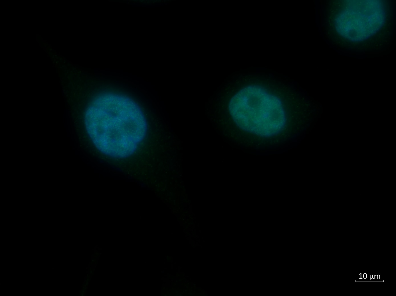
 with MCF7 cell.")
 at dilution of 1:3000 (under 10x lens). Heat mediated antigen retrieval with Tris-EDTA buffer (pH 9.0).")
 at dilution of 1:2000 (under 40x lens). Heat mediated antigen retrieval with Tris-EDTA buffer (pH 9.0).")
 at dilution of 1:3000 (under 40x lens). Heat mediated antigen retrieval with Tris-EDTA buffer (pH 9.0).")
 at dilution of 1:200 (under 10x lens)..")
 at dilution of 1:200 (under 40x lens)..")
 at dilution of 1:800 (under 10x lens). Heat mediated antigen retrieval with Tris-EDTA buffer (pH 9.0).")
 at dilution of 1:800 (under 40x lens). Heat mediated antigen retrieval with Tris-EDTA buffer (pH 9.0).")
 at dilution of 1:800 (under 40x lens). Heat mediated antigen retrieval with Tris-EDTA buffer (pH 9.0).")
 at dilution of 1:800 (under 10x lens). Heat mediated antigen retrieval with Tris-EDTA buffer (pH 9.0).")
 at dilution of 1:200 (under 10x lens). Heat mediated antigen retrieval with Tris-EDTA buffer (pH 9.0).")
 fixed mouse testis tissue using PCNA antibody (10205-2-AP) at dilution of 1:200 and CoraLite®488-Conjugated AffiniPure Goat Anti-Rabbit IgG(H+L).")
 fixed human breast cancer tissue using 10205-2-AP (PCNA antibody), at dilution of 1:100 and CoraLite®488-Conjugated AffiniPure Goat Anti-Rabbit IgG(H+L).")
"PCNA Antibodies" Comparison
View side-by-side comparison of PCNA antibodies from other vendors to find the one that best suits your research needs.
Applications testées
| Résultats positifs en WB | cellules A431, cellules C2C12, cellules HEK293, cellules HeLa, cellules Jurkat, cellules MCF-7, cellules NIH/3T3, cellules PC-12, tissu splénique de rat, tissu splénique de souris |
| Résultats positifs en IP | cellules MCF-7 |
| Résultats positifs en IHC | tissu de cancer de l'estomac humain, tissu de cancer du côlon humain, tissu de cancer du foie humain, tissu de cancer du sein humain, tissu de mélanome malin humain il est suggéré de démasquer l'antigène avec un tampon de TE buffer pH 9.0; (*) À défaut, 'le démasquage de l'antigène peut être 'effectué avec un tampon citrate pH 6,0. |
| Résultats positifs en IF-P | tissu testiculaire de souris, tissu de cancer du sein humain |
Dilution recommandée
| Application | Dilution |
|---|---|
| Western Blot (WB) | WB : 1:5000-1:50000 |
| Immunoprécipitation (IP) | IP : 0.5-4.0 ug for 1.0-3.0 mg of total protein lysate |
| Immunohistochimie (IHC) | IHC : 1:1500-1:6000 |
| Immunofluorescence (IF)-P | IF-P : 1:50-1:500 |
| It is recommended that this reagent should be titrated in each testing system to obtain optimal results. | |
| Sample-dependent, check data in validation data gallery | |
Applications publiées
| KD/KO | See 4 publications below |
| WB | See 812 publications below |
| IHC | See 283 publications below |
| IF | See 103 publications below |
| IP | See 1 publications below |
| CoIP | See 1 publications below |
Informations sur le produit
10205-2-AP cible PCNA dans les applications de WB, IHC, IF-P, IP, CoIP, ELISA, Cell treatment et montre une réactivité avec des échantillons Humain, rat, souris
| Réactivité | Humain, rat, souris |
| Réactivité citée | rat, Chèvre, Humain, Lapin, poulet, souris, embryons de médaka, fish |
| Hôte / Isotype | Lapin / IgG |
| Clonalité | Polyclonal |
| Type | Anticorps |
| Immunogène | PCNA Protéine recombinante Ag0277 |
| Nom complet | proliferating cell nuclear antigen |
| Masse moléculaire calculée | 29 kDa/31 kDa |
| Poids moléculaire observé | 36-38 kDa |
| Numéro d’acquisition GenBank | BC000491 |
| Symbole du gène | PCNA |
| Identification du gène (NCBI) | 5111 |
| Conjugaison | Non conjugué |
| Forme | Liquide |
| Méthode de purification | Purification par affinité contre l'antigène |
| Tampon de stockage | PBS with 0.02% sodium azide and 50% glycerol |
| Conditions de stockage | Stocker à -20°C. Stable pendant un an après l'expédition. L'aliquotage n'est pas nécessaire pour le stockage à -20oC Les 20ul contiennent 0,1% de BSA. |
Informations générales
Proliferating Cell Nuclear Antigen, commonly known as PCNA, is a protein that acts as a processivity factor for DNA polymerase δ in eukaryotic cells. This protein is an auxiliary protein of DNA polymerase delta and is involved in the control of eukaryotic DNA replication by increasing the polymerase's processibility during elongation of the leading strand. PCNA induces a robust stimulatory effect on the 3'-5' exonuclease and 3'-phosphodiesterase, but not apurinic-apyrimidinic (AP) endonuclease, APEX2 activities. It has to be loaded onto DNA in order to be able to stimulate APEX2. PCNA protein is highly conserved during evolution; the deduced amino acid sequences of rat and human differ by only 4 of 261 amino acids. PCNA has been used as loading control for proliferating cells. This antibody is a rabbit polyclonal antibody raised against an internal region of human PCNA. The calculated molecular weight of PCNA is 29 kDa, but modified PCNA is 36kDa (PMID: 1358458).
Protocole
| Product Specific Protocols | |
|---|---|
| WB protocol for PCNA antibody 10205-2-AP | Download protocol |
| IHC protocol for PCNA antibody 10205-2-AP | Download protocol |
| IF protocol for PCNA antibody 10205-2-AP | Download protocol |
| IP protocol for PCNA antibody 10205-2-AP | Download protocol |
| Standard Protocols | |
|---|---|
| Click here to view our Standard Protocols |
Publications
| Species | Application | Title |
|---|---|---|
J Hematol Oncol METTL16 promotes liver cancer stem cell self-renewal via controlling ribosome biogenesis and mRNA translation | ||
Mil Med Res Aspartoacylase suppresses prostate cancer progression by blocking LYN activation | ||
Protein Cell NDFIP1 limits cellular TAZ accumulation via exosomal sorting to inhibit NSCLC proliferation | ||
Brain Behav Immun An enriched environment restores hepatitis B vaccination-mediated impairments in synaptic function through IFN-γ/Arginase1 signaling. | ||
Nat Commun Selectively targeting the AdipoR2-CaM-CaMKII-NOS3 axis by SCM-198 as a rapid-acting therapy for advanced acute liver failure | ||
Nat Commun Four-dimensional hydrogel dressing adaptable to the urethral microenvironment for scarless urethral reconstruction |
Avis
The reviews below have been submitted by verified Proteintech customers who received an incentive for providing their feedback.
FH Mi (Verified Customer) (06-25-2023) | It works well in human brown adipocyte cells.
|
FH Emma (Verified Customer) (11-29-2021) | Nice antibody which worked well on PC3 cells fixed with 4% PFA. Antibody used at 1:50
 |

