
Scientist Stories: Dr Joe McKellar on Imaging Influenza A
How Dr Joe McKellar uses Proteintech antibodies in confocal microscopy to study viral infections
This story is part of #InTheLabWithProteintech, highlighting how Proteintech reagents support researchers in advancing their work.
This feature marks the first of a three-part spotlight on Joe McKellar, PhD, Scientific Director at Viroscope Imaging and winner of CiteAb Image of the Year and Nikon Small World, sharing insights from his work in advanced microscopy and his PhD research in Health Biology.
Which Proteintech products have you featured most in your workflows?
"I’ve used a wide variety of Proteintech primary antibodies over the years. However, from a practical standpoint, the trial-size format has been incredibly valuable. When you are constantly pushing into new research directions, having that trial format allows for rapid, cost-effective testing before scaling up the workflow."
Explore our portfolio of antibodies.
What’s the story behind this image?
"This image traces back to my PhD research on influenza A virus (IAV). We discovered a novel antiviral activity of the MX1 protein, actively inhibiting the later stages of viral replication. Interestingly, IAV hijacks the host's YBX1 protein to facilitate this exact stage of its lifecycle. What you are looking at here is the baseline, the distribution of YBX1 (in gold) in cells heavily infected with IAV (in blue). This served as our critical control condition to establish what an unhindered infection looks like before we introduced MX1 to study its inhibitory effects."
Find out more about the YBX1 antibody.
What are some technical aspects you considered while imaging this?
"When imaging viral infections, cell confluency is a critical variable. You have to calculate the initial seeding density and know the viral replication cycle so that the final confluency is exactly where you want it at the time of fixation. Over-confluent cells tend to pile up, which not only causes suboptimal antibody staining but also distorts the cellular morphology, making it more difficult to isolate and quantify specific events. That is exactly why you see a deliberate, sub-confluent distribution of cells in this image."
How does this work contribute to the broader landscape of the field?
"It shifts how we view the vulnerabilities of the IAV lifecycle. We now understand that the late stages of viral replication can be actively hampered by this cellular defence protein. The next step for the field will be to map the exact molecular mechanisms driving MX1’s inhibition of IAV at this step of its lifecycle. If we can decode this, it opens the door to developing new antiviral therapeutics designed to mimic that precise effect."
Read more: https://pmc.ncbi.nlm.nih.gov/articles/PMC12541310/
Featured Product
The following Proteintech reagent was used in this study:
- YBX1 Antibody (Cat No. 20339-1-AP)
Used to detect and visualise YBX1 distribution in Influenza A virus-infected A549 cells, forming the baseline for analysing host–virus interactions during late-stage replication.
View product: https://www.ptglab.com/products/YBX1-Antibody-20339-1-AP.htm
About the Scientist
Joe McKellar is a virologist and cell biologist with a PhD in Health Biology, specializing in advanced microscopy. As the Scientific Director of Viroscope Imaging, he leads an agency dedicated to highlighting biotechnologies and cutting-edge research through high-resolution visual narratives. His academic work has consistently relied on the lens of a microscope to answer complex biological questions, from understanding cellular defense mechanisms to characterizing novel forms of viral transmission. This dedication to visual science has earned him recognition in international competitions, such as the CiteAb Image of the Year and Nikon Small World. When he isn't exploring cellular landscapes under a confocal microscope, you can usually find him strategizing over a game of Magic: The Gathering.
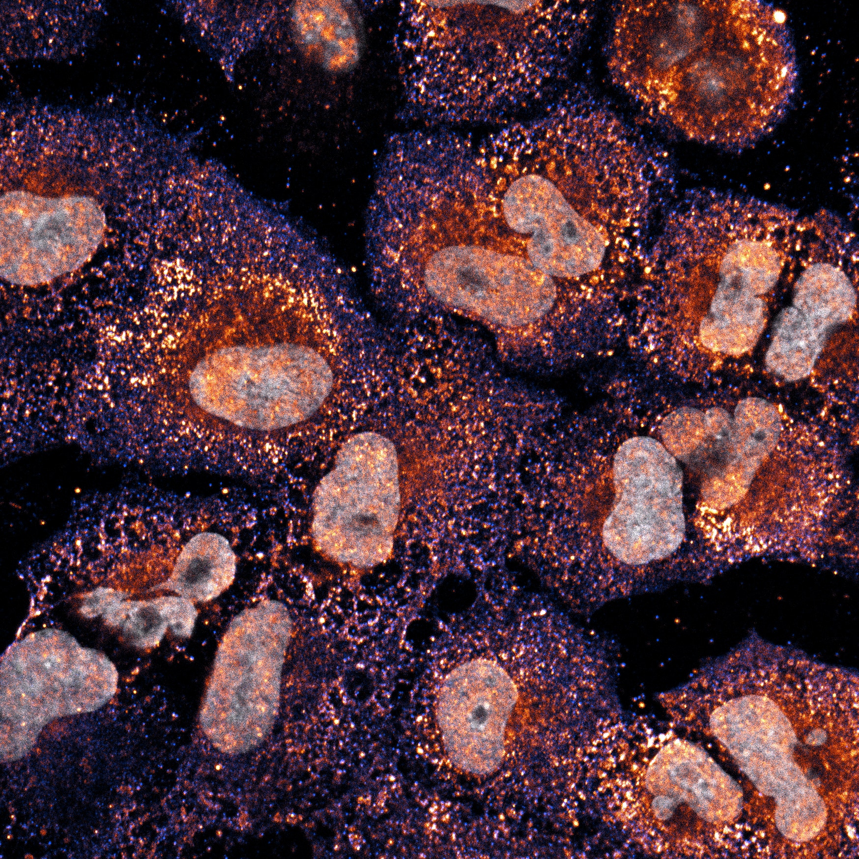
Confocal fluorescence image of A549 cells infected with Influenza A virus (IAV) for 24 hours. YBX1 was detected using a Proteintech anti-YBX1 antibody (Cat. No. 20339-1-AP) and is shown in gold. Viral nucleoprotein (IAV NP) is shown in blue, indicating infected cells, while nuclei are counterstained in grey. YBX1 expression is visibly upregulated in response to viral infection stress. Image acquired using a ZEISS LSM880 confocal microscope.
Joe McKellar, PhD