
Scientist Stories: Dr Joe McKellar on Septins, the Understudied Fourth Component of the Cytoskeleton
The fourth component of the cytoskeleton, and why it deserves more attention
This story is part of #InTheLabWithProteintech, highlighting how Proteintech reagents support researchers in advancing their work.
This feature marks the second of a three-part spotlight on Joe McKellar, PhD, Scientific Director at Viroscope Imaging and winner of CiteAb Image of the Year and Nikon Small World, sharing insights from his work in advanced microscopy and his PhD research in Health Biology.
Read Part 1 here: https://www.ptglab.com/news/blog/scientist-stories-dr-joe-mckellar-on-imaging-influenza-a/
What motivated a focus on the basal layer? What does the image reveal?
"The focus here stems from my ongoing fascination with septins, often referred to as the "fourth component" of the cytoskeleton. When you adjust your focal plane specifically to the basal layer, where the cell makes contact with the glass coverslip, the septin network is dense and magnificent. This image strips away the cellular noise to highlight the beautiful, structural complexity of a cytoskeletal element that is still somewhat understudied compared to actin or tubulin."
Explore our portfolio of antibodies.
How do you design a multiplex experiment and make sure the signals don’t bleed into each other, especially when the proteins are both from the same family?
"It all comes down to spectral distance and epitope specificity. When you are only looking at two targets, the easiest way to eliminate bleed-through is to choose fluorophores far apart on the spectrum, like a green and a far-red, which is exactly what I did here. As for the proteins belonging to the same family, it isn’t a problem provided the primary antibodies used are rigorously validated. If the antibodies target unique epitopes that don't cross-react, you can confidently image closely related proteins in the exact same sample without worrying about false overlap. Hence the importance of antibody validation."
Did anything stand out to you while you were imaging this setup?
"What stood out the most was how flawlessly the SEPT8 antibody performed right out of the box. It worked perfectly on the very first try, which is always a great feeling in the lab! Beyond that, the image clearly reveals distinct spatial patterns between the SEPT7 and SEPT8 staining. Seeing them resolved side-by-side like this is a great visual confirmation that, despite being in the same protein family, they govern both distinct and overlapping structural roles within the cell's architecture."
View the SEPT8 antibody Joe used. https://www.ptglab.com/products/SEPT8-Antibody-66188-1-Ig.htm?
How does this work contribute to the broader landscape of the field?
"While this specific image wasn't taken to answer a specific biological question, it serves a different, equally important purpose: scientific communication. I captured this purely for its aesthetic value to highlight the striking architecture of the septin basal layer. In the broader landscape, creating visuals like this is crucial. It engages the public, inspires the next generation of biologists, and reminds even seasoned researchers of the sheer beauty of the microscopic world we work in every day."
Featured Product
The following Proteintech reagent was used in this study:
- Septin 8 Monoclonal Antibody (Cat No. 66188-1-Ig)
Used to detect and visualise the distribution of Septin 8 within the basal layer of A549 cells, enabling side-by-side comparison of SEPT7 and SEPT8 spatial organisation within the septin cytoskeletal network.
View product: https://www.ptglab.com/products/SEPT8-Antibody-66188-1-Ig.htm
About the Scientist
Joe McKellar is a virologist and cell biologist with a PhD in Health Biology, specializing in advanced microscopy. As the Scientific Director of Viroscope Imaging, he leads an agency dedicated to highlighting biotechnologies and cutting-edge research through high-resolution visual narratives. His academic work has consistently relied on the lens of a microscope to answer complex biological questions, from understanding cellular defense mechanisms to characterizing novel forms of viral transmission. This dedication to visual science has earned him recognition in international competitions, such as the CiteAb Image of the Year and Nikon Small World. When he isn't exploring cellular landscapes under a confocal microscope, you can usually find him strategizing over a game of Magic: The Gathering.
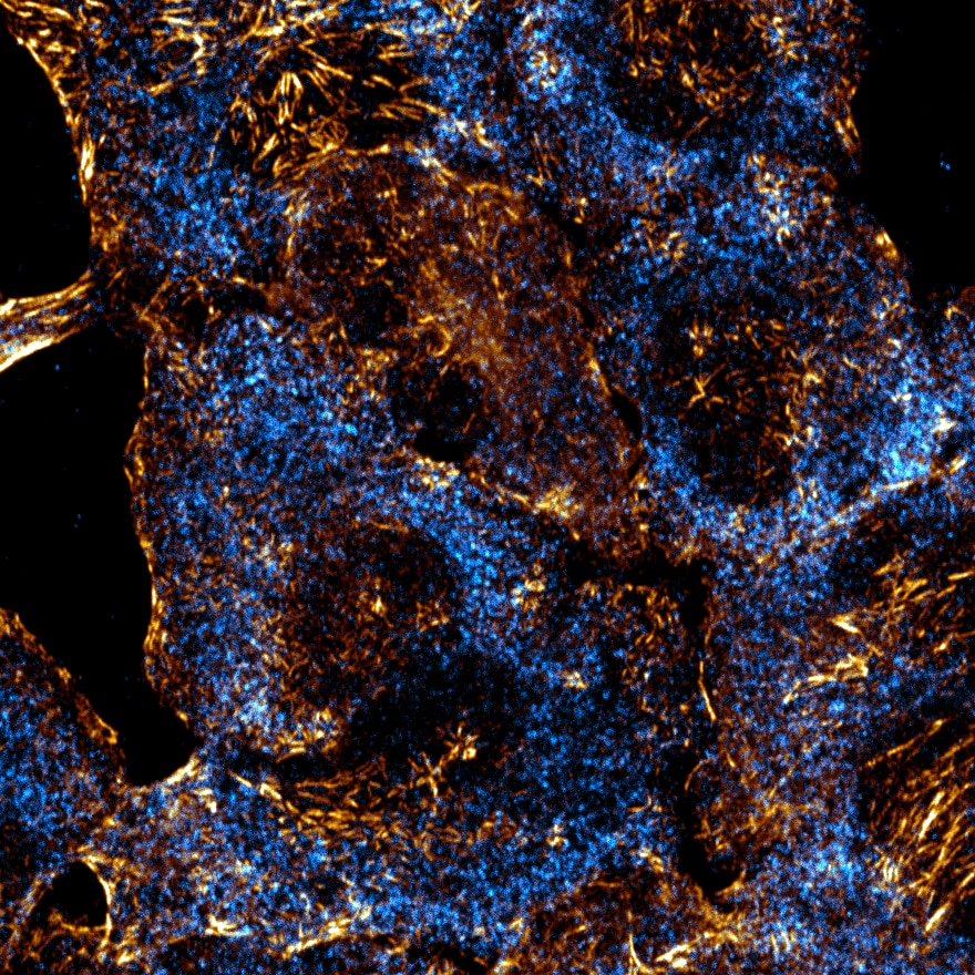
Confocal fluorescence image of the basal layer of A549 cells, imaged at the point of contact with the glass coverslip. Septin 8 (SEPT8) was detected using a Proteintech anti-SEPT8 antibody (Cat. No. 66188-1-Ig) and is shown in cyan. Septin 7 (SEPT7) is shown in gold, revealing the dense, interconnected architecture of the septin network at the cell periphery. Image acquired using a ZEISS LSM880 confocal microscope.
Joe McKellar, PhD