Tested Applications
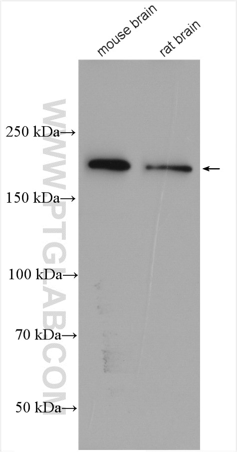
| Positive WB detected in | mouse brain tissue, rat brain tissue |
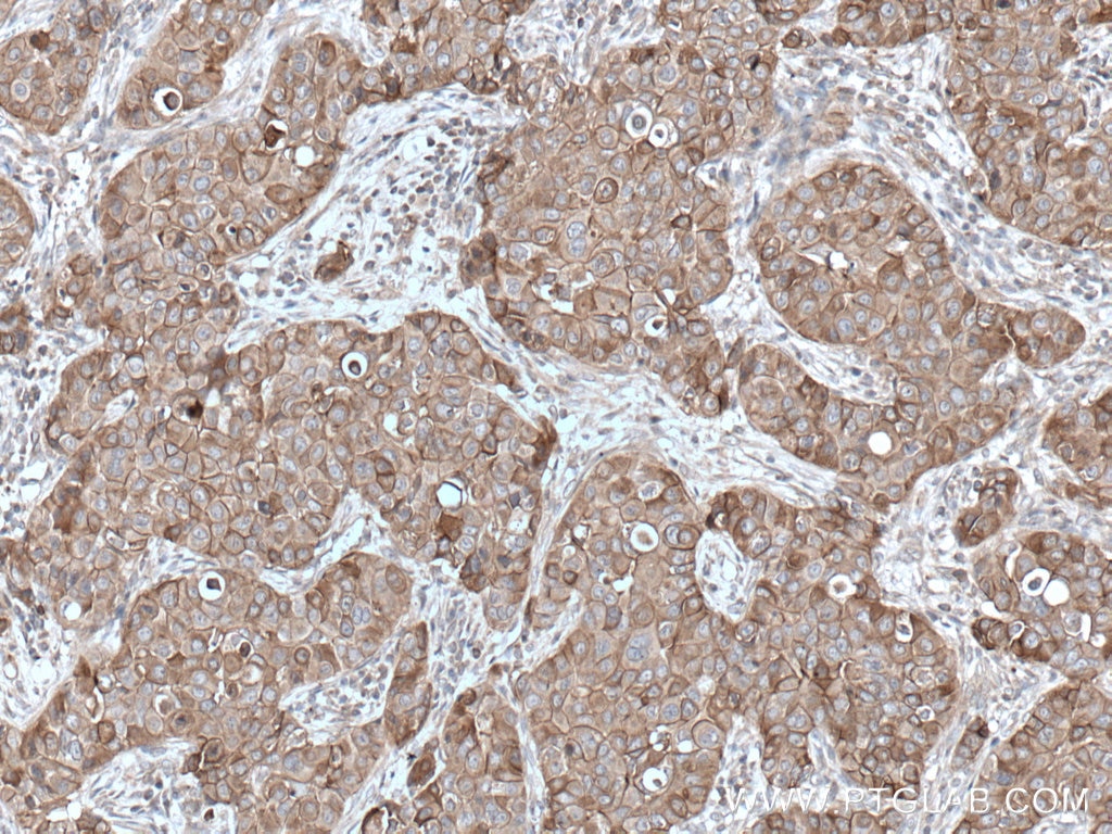
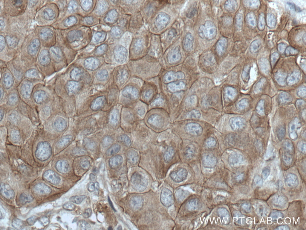
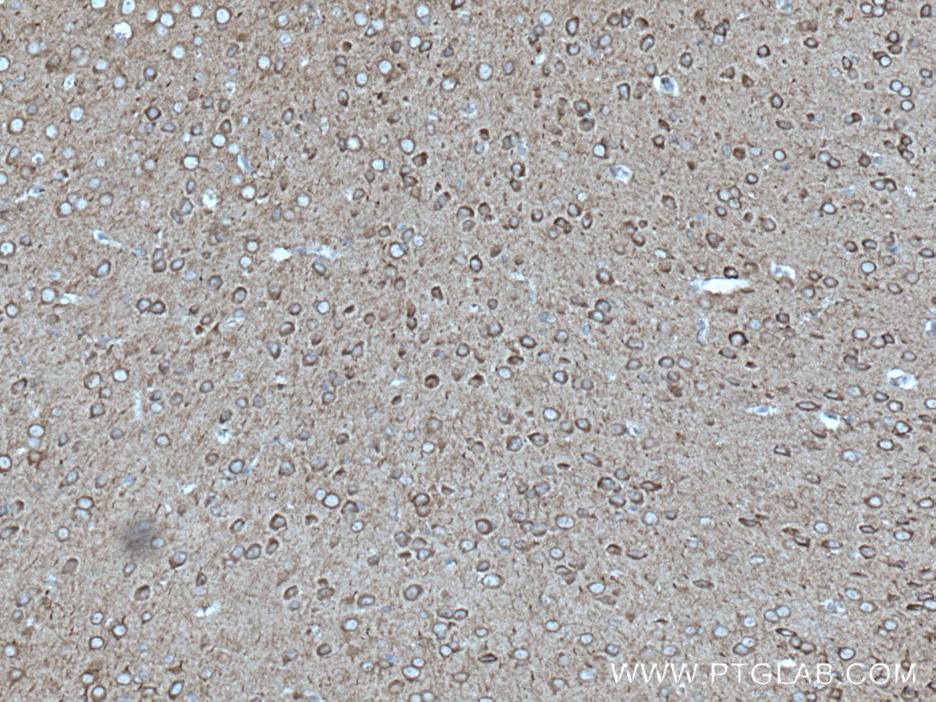
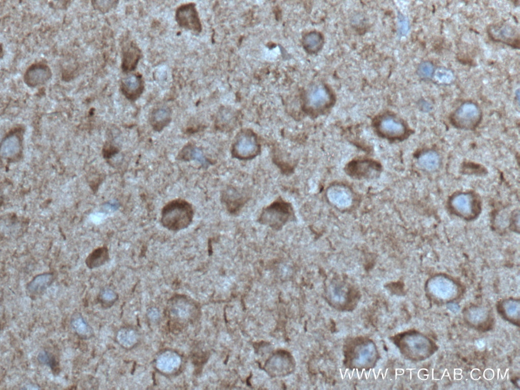
| Positive IHC detected in | human breast cancer tissue, mouse brain tissue Note: suggested antigen retrieval with TE buffer pH 9.0; (*) Alternatively, antigen retrieval may be performed with citrate buffer pH 6.0 |
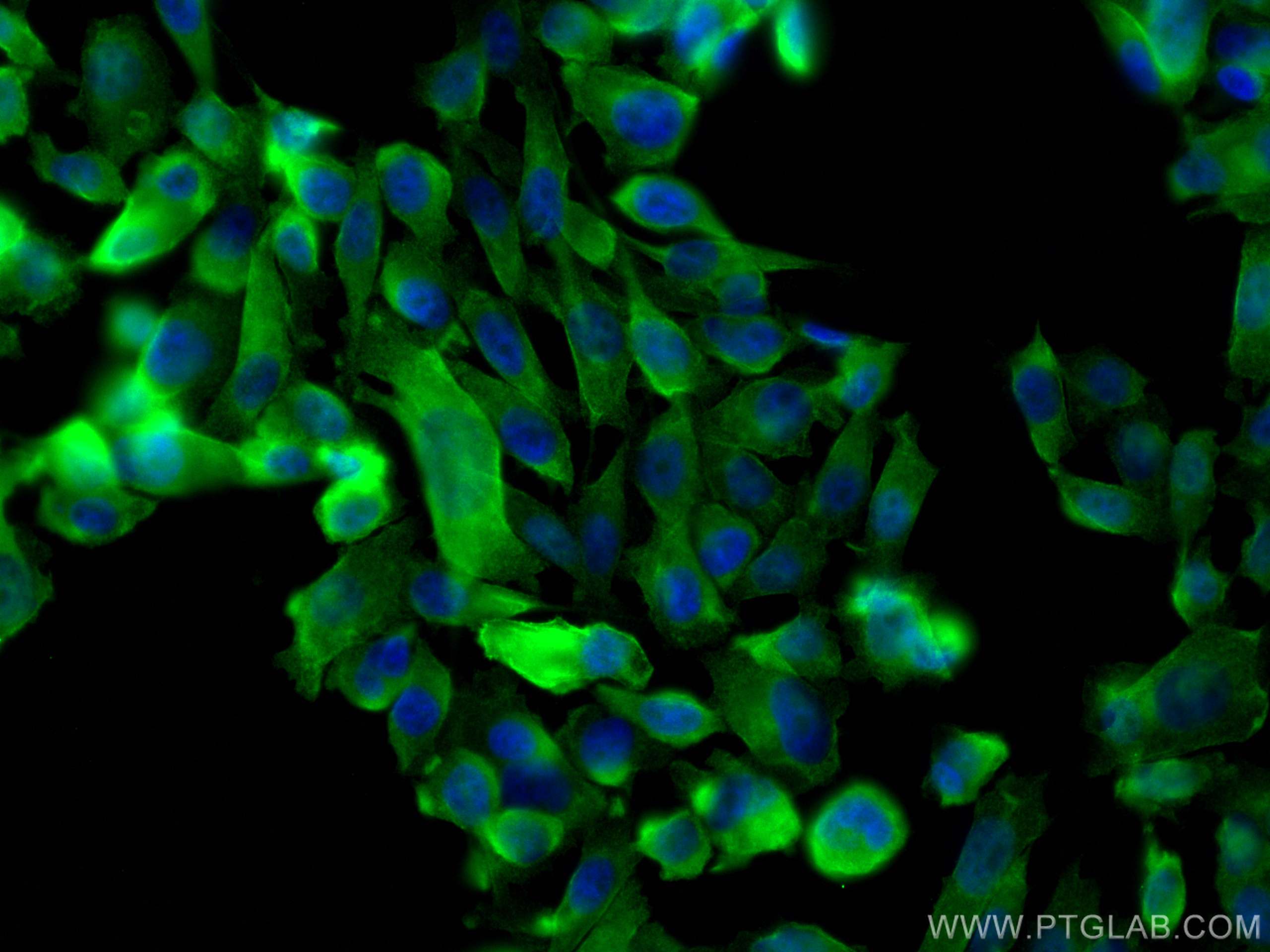
| Positive IF/ICC detected in | PC-3 cells |
Recommended dilution
| Application | Dilution |
|---|---|
| Western Blot (WB) | WB : 1:500-1:2000 |
| Immunohistochemistry (IHC) | IHC : 1:50-1:500 |
| Immunofluorescence (IF)/ICC | IF/ICC : 1:200-1:800 |
| It is recommended that this reagent should be titrated in each testing system to obtain optimal results. | |
| Sample-dependent, Check data in validation data gallery. | |
Published Applications
| WB | See 5 publications below |
| IHC | See 2 publications below |
| IF | See 2 publications below |
Product Information
27980-1-AP targets ANK3 in WB, IHC, IF/ICC, ELISA applications and shows reactivity with Human, Mouse, Rat samples.
| Tested Reactivity | Human, Mouse, Rat |
| Cited Reactivity | human, mouse, zebrafish |
| Host / Isotype | Rabbit / IgG |
| Class | Polyclonal |
| Type | Antibody |
| Immunogen |
CatNo: Ag27635 Product name: Recombinant human ANK3 protein Source: e coli.-derived, PGEX-4T Tag: GST Domain: 4221-4377 aa of NM_001149 Sequence: MGLLDRLDDSPDQCRDSITSYLKGEAGKFEANGSHTEITPEAKTKSYFPESQNDVGKQSTKETLKPKIHGSGHVEEPASPLAAYQKSLEETSKLIIEETKPCVPVSMKKMSRTSPADGKPRLSLHEEEGSSGSEQKQGEGFKVKTKKEIRHVEKKSHS Predict reactive species |
| Full Name | ankyrin 3, node of Ranvier (ankyrin G) |
| Calculated Molecular Weight | 480 kDa |
| Observed Molecular Weight | 160 kDa, 204 kDa |
| GenBank Accession Number | NM_001149 |
| Gene Symbol | ANK3 |
| Gene ID (NCBI) | 288 |
| RRID | AB_2881028 |
| Conjugate | Unconjugated |
| Form | Liquid |
| Purification Method | Antigen affinity purification |
| UNIPROT ID | Q12955 |
| Storage Buffer | PBS with 0.02% sodium azide and 50% glycerol, pH 7.3. |
| Storage Conditions | Store at -20°C. Stable for one year after shipment. Aliquoting is unnecessary for -20oC storage. 20ul sizes contain 0.1% BSA. |
Background Information
ANK3 is associated with neurodevelopment and neuronal function. It has been reported that ANK3 plays a key role in bipolar disorder. ANK3 is expressed in brain at a high level. There are several isoforms of ANK3 associated with different tissue expression and function. The immune region we selected can recognize 480 kDa, 204 kDa, 202 kDa and 111 kDa protein and 27980-1-AP detects 204 kDa isoform in SDS-PAGE. (PMID: 27217151, 28687526, 30297702, 28109561, 30046097)
Protocols
| Product Specific Protocols | |
|---|---|
| IF protocol for ANK3 antibody 27980-1-AP | Download protocol |
| IHC protocol for ANK3 antibody 27980-1-AP | Download protocol |
| WB protocol for ANK3 antibody 27980-1-AP | Download protocol |
| Standard Protocols | |
|---|---|
| Click here to view our Standard Protocols |
Publications
| Species | Application | Title |
|---|---|---|
Front Immunol A comprehensive cuproptosis score and associated gene signatures reveal prognostic and immunological features of idiopathic pulmonary fibrosis | ||
Front Biosci (Landmark Ed) ANK3 Is Regulated by Recursive Splicing and Inhibits Hepatocellular Carcinoma Metastasis by Inhibiting E-Cadherin Protein Degradation. | ||
J Ovarian Res Deciphering the molecular landscape of NMDAR-E associated ovarian teratomas: a Multi-Omics approach. | ||
Epilepsia kcnb1 loss of function in zebrafish causes neurodevelopmental and epileptic disorders associated with γ-aminobutyric acid dysregulation | ||
J Proteomics Hippocampal proteome comparison of infant and adult Fmr1 deficiency mice reveals adult-related changes associated with postsynaptic density. | ||
Bioact Mater A continuous adhesion-enhanced osteogenic pathway in artificial scaffold drives cellular infiltration and condensed mineralization for rapid bone regeneration. |