Tested Applications
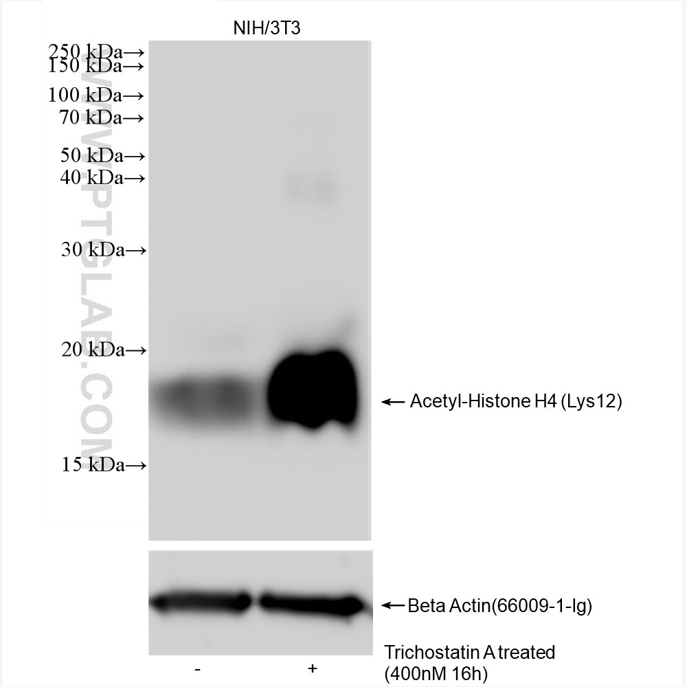
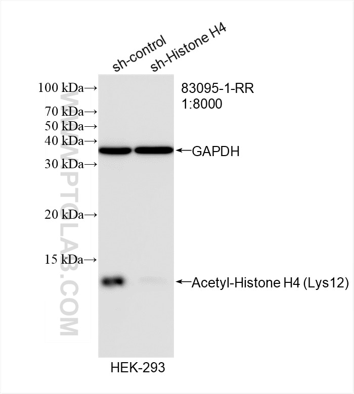
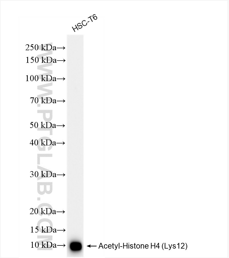
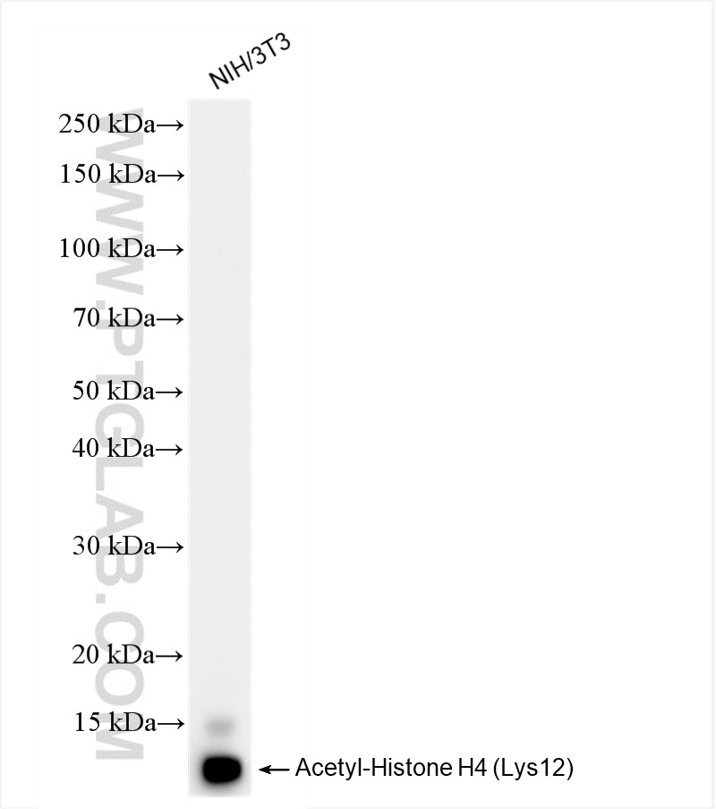
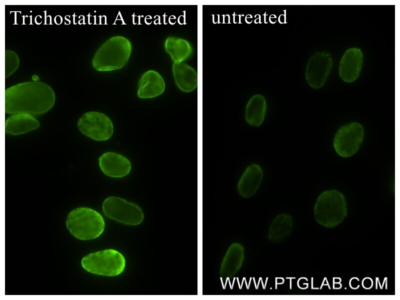
| Positive WB detected in | Trichostatin A treated NIH/3T3 cells, HEK-293 cells, HSC-T6 cells, NIH/3T3 cells |
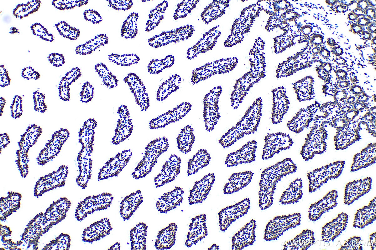
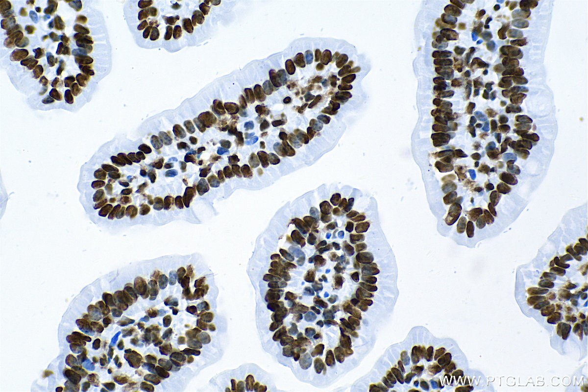
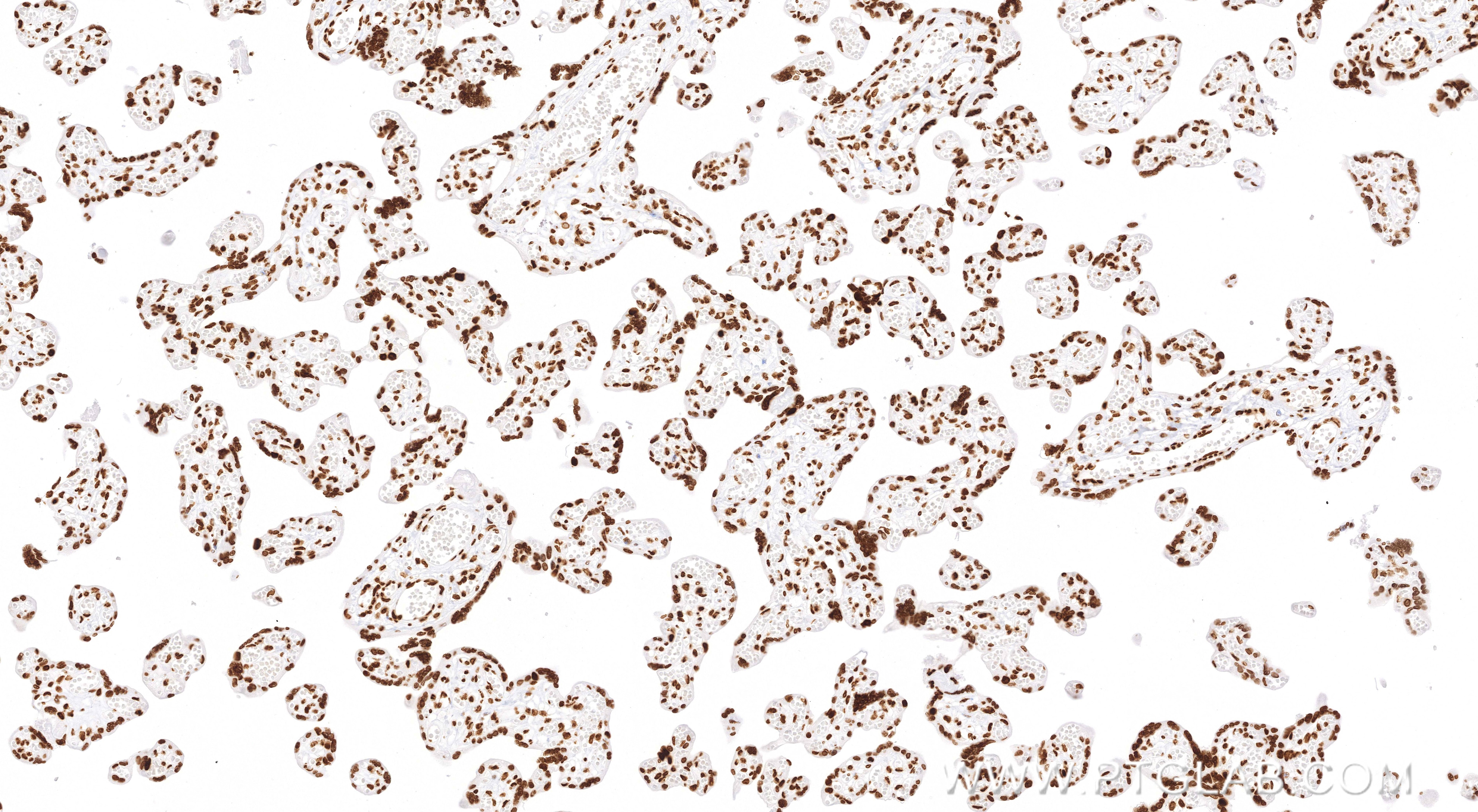
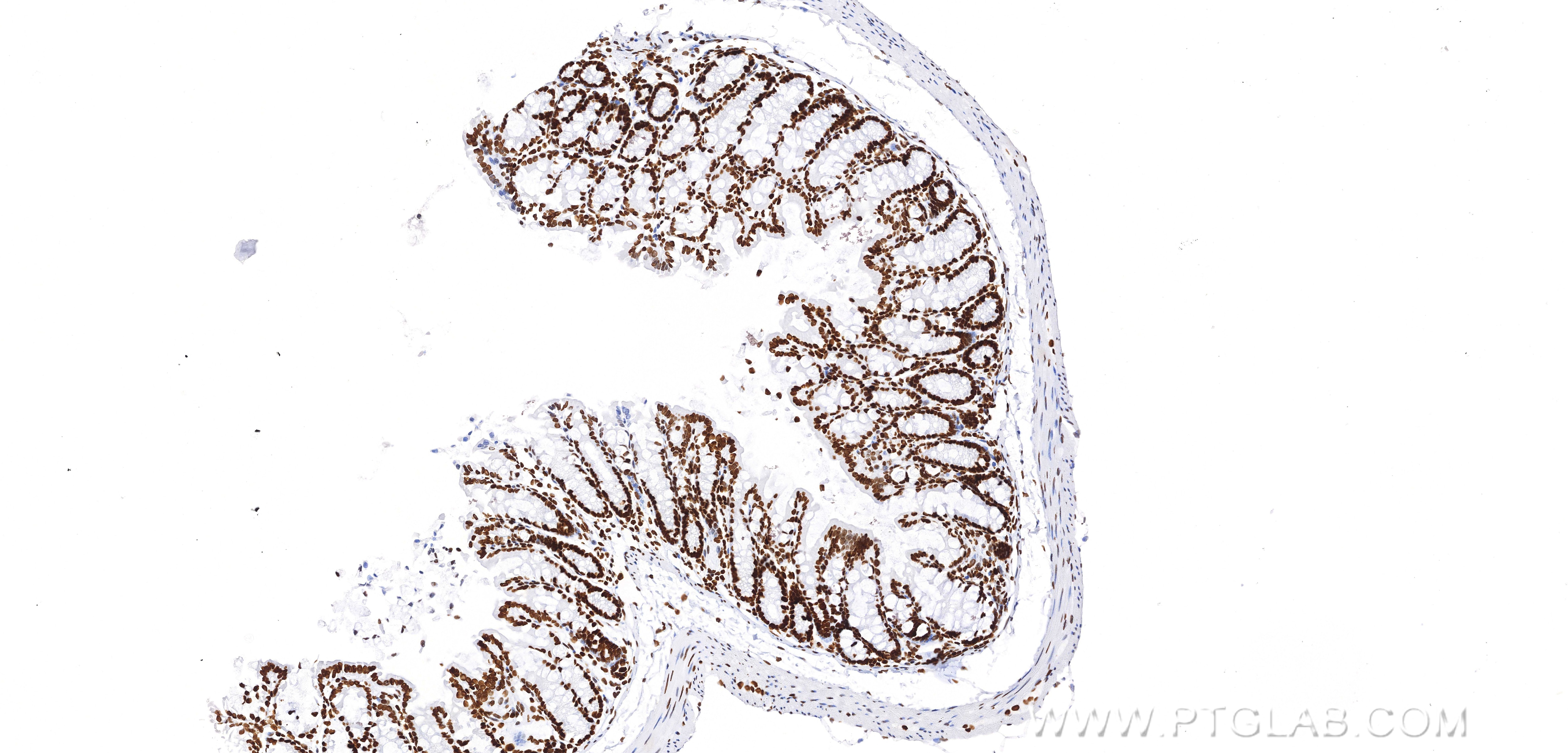
| Positive IHC detected in | mouse small intestine tissue, human placenta tissue, mouse colon tissue Note: suggested antigen retrieval with TE buffer pH 9.0; (*) Alternatively, antigen retrieval may be performed with citrate buffer pH 6.0 |
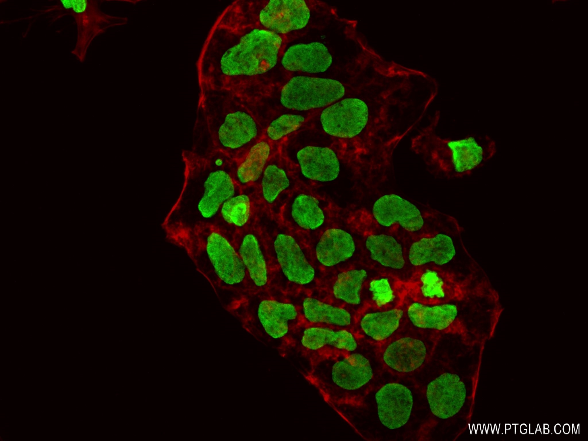
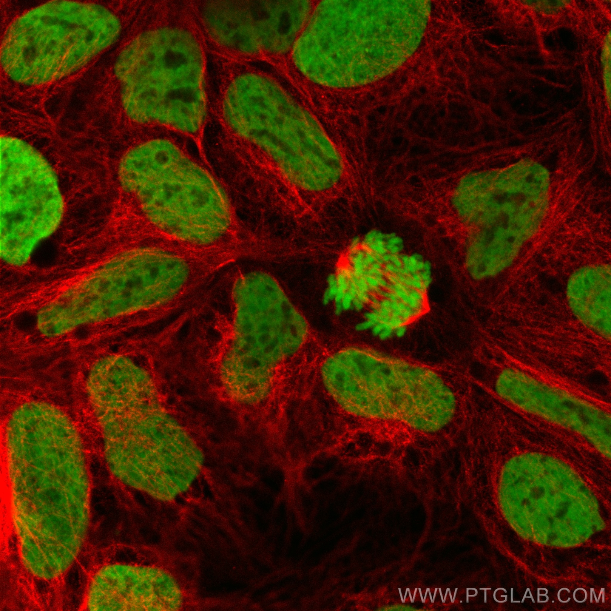
| Positive IF/ICC detected in | Caco-2 cells, Trichostatin A treated NIH/3T3 cells |
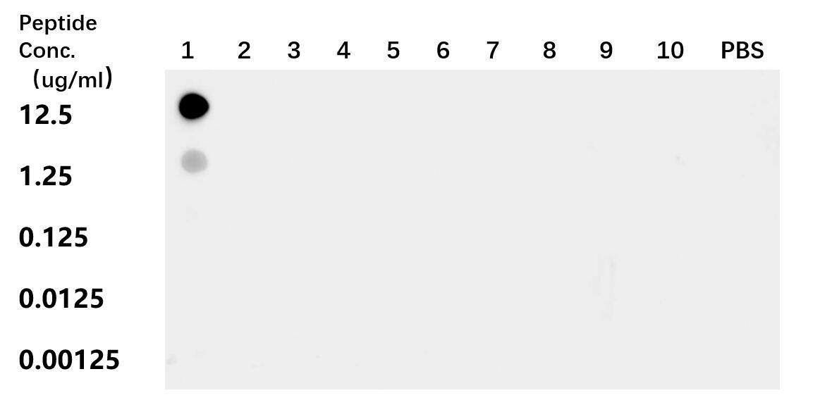
| Positive Dot Blot detected in | peptide |
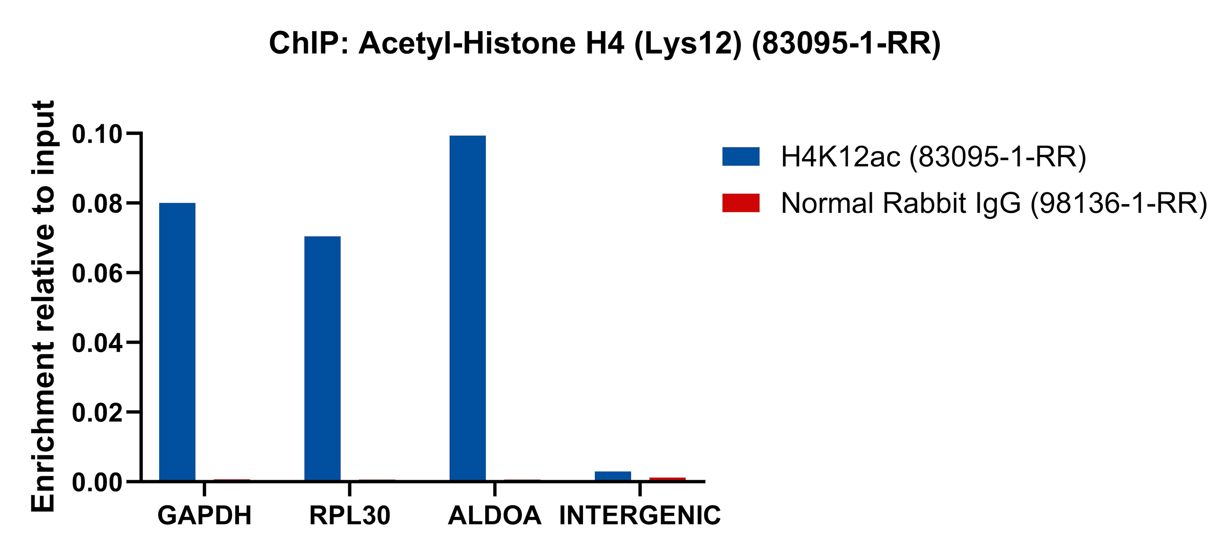
| Positive ChIP-qPCR detected in | HeLa cells |
Recommended dilution
| Application | Dilution |
|---|---|
| Western Blot (WB) | WB : 1:5000-1:50000 |
| Immunohistochemistry (IHC) | IHC : 1:1000-1:4000 |
| Immunofluorescence (IF)/ICC | IF/ICC : 1:150-1:600 |
| DOT BLOT | DOT BLOT : 1:10-1:100 |
| CHIP-QPCR | CHIP-QPCR : 1:10-1:100 |
| It is recommended that this reagent should be titrated in each testing system to obtain optimal results. | |
| Sample-dependent, Check data in validation data gallery. | |
Published Applications
| WB | See 1 publications below |
| ChIP | See 2 publications below |
Product Information
83095-1-RR targets Acetyl-Histone H4 (Lys12) in WB, IHC, IF/ICC, Dot Blot, ELISA, ChIP-qPCR applications and shows reactivity with human, mouse, rat samples.
| Tested Reactivity | human, mouse, rat |
| Cited Reactivity | human |
| Host / Isotype | Rabbit / IgG |
| Class | Recombinant |
| Type | Antibody |
| Immunogen |
Peptide Predict reactive species |
| Full Name | histone cluster 1, H4a |
| Observed Molecular Weight | 12 kDa |
| GenBank Accession Number | BC069654 |
| Gene Symbol | HIST1H4A |
| Gene ID (NCBI) | 8359 |
| RRID | AB_3670809 |
| Conjugate | Unconjugated |
| Form | Liquid |
| Purification Method | Protein A purification |
| UNIPROT ID | P62805 |
| Storage Buffer | PBS with 0.02% sodium azide and 50% glycerol, pH 7.3. |
| Storage Conditions | Store at -20°C. Stable for one year after shipment. Aliquoting is unnecessary for -20oC storage. 20ul sizes contain 0.1% BSA. |
Background Information
Histone H4 is a 103 amino acid protein, which belongs to the histone H4 family. Histone H4 localizes in the nucleus and is a core component of nucleosome. Nucleosomes wrap and compact DNA into chromatin, limiting DNA accessibility to the cellular machineries which require DNA as a template. Histones thereby play a central role in transcription regulation, DNA repair, DNA replication and chromosomal stability. DNA accessibility is regulated via a complex set of post-translational modifications of histones, also called histone code, and nucleosome remodeling. Acetylation of histone H4 is necessary for chromatin decompaction during DNA replication.
Protocols
| Product Specific Protocols | |
|---|---|
| IF protocol for Acetyl-Histone H4 (Lys12) antibody 83095-1-RR | Download protocol |
| IHC protocol for Acetyl-Histone H4 (Lys12) antibody 83095-1-RR | Download protocol |
| WB protocol for Acetyl-Histone H4 (Lys12) antibody 83095-1-RR | Download protocol |
| Standard Protocols | |
|---|---|
| Click here to view our Standard Protocols |
Publications
| Species | Application | Title |
|---|---|---|
Adv Sci (Weinh) ACSS2-Mediated Histone H4 Lysine 12 Crotonylation (H4K12cr) Alleviates Colitis via Enhancing Transcription of CLDN7 | ||
Cell Signal N-α-Acetyltransferase 40 promotes oral squamous cell carcinoma progression by enhancing FEN1 transcription and suppressing CD8(+) T cell antitumor immunity. |
Reviews
The reviews below have been submitted by verified Proteintech customers who received an incentive for providing their feedback.
FH Sree Rama Chaitanya (Verified Customer) (07-21-2025) | I have tested this antibody to evaluate H4K12ac levels in mammalian cells, and it works well for WB. Highly recommend!
|