Tested Applications
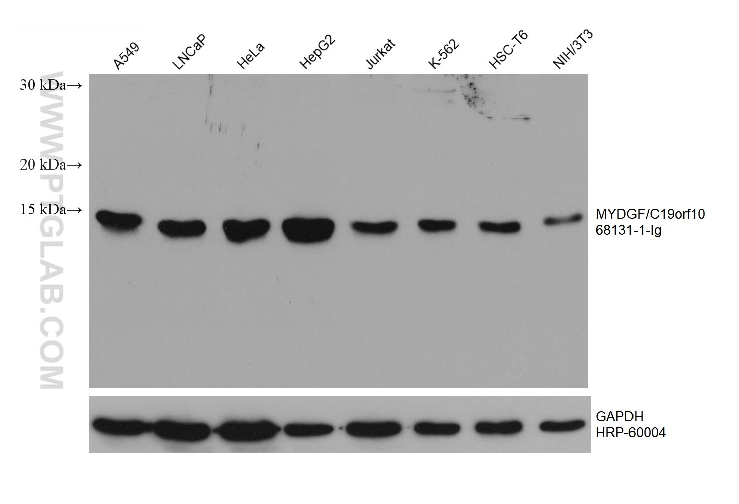
| Positive WB detected in | A549 cells, LNCaP cells, HeLa cells, HepG2 cells, Jurkat cells, K-562 cells, HSC-T6 cells, NIH/3T3 cells |
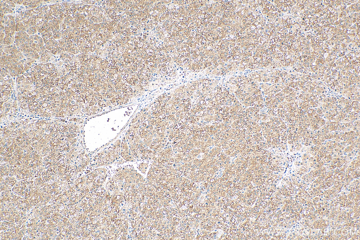
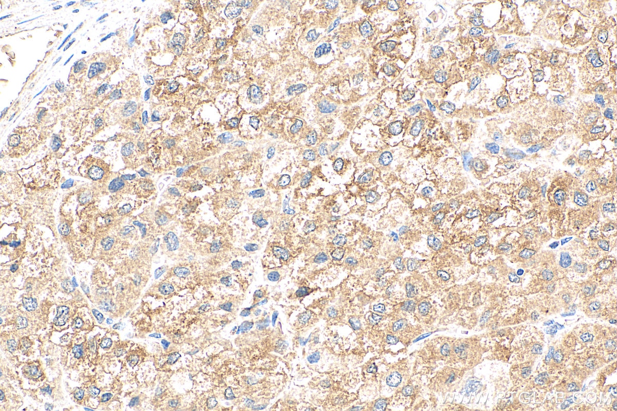
| Positive IHC detected in | human liver cancer tissue Note: suggested antigen retrieval with TE buffer pH 9.0; (*) Alternatively, antigen retrieval may be performed with citrate buffer pH 6.0 |
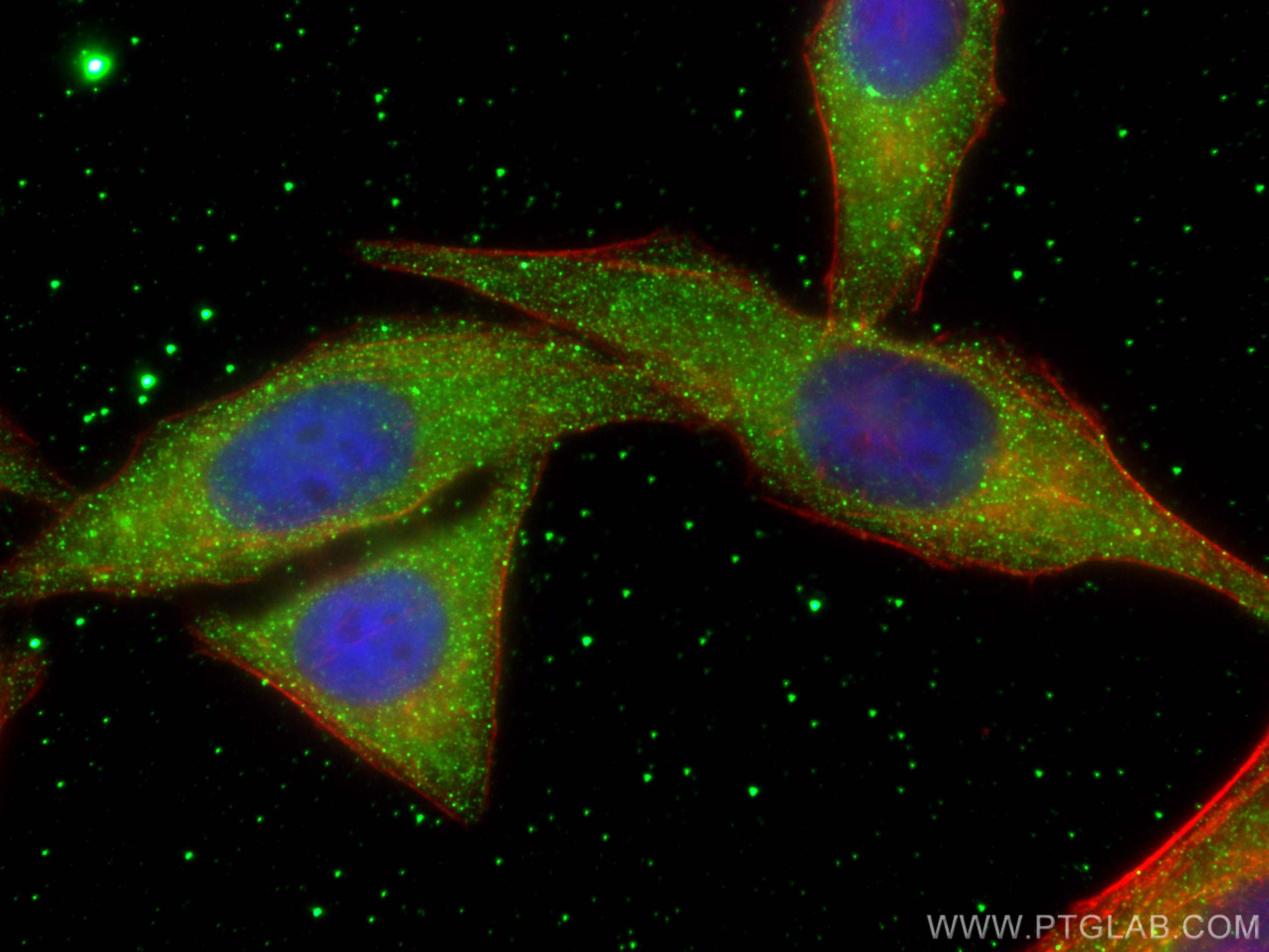
| Positive IF/ICC detected in | HepG2 cells |
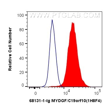
| Positive FC (Intra) detected in | HepG2 cells |
Recommended dilution
| Application | Dilution |
|---|---|
| Western Blot (WB) | WB : 1:5000-1:50000 |
| Immunohistochemistry (IHC) | IHC : 1:500-1:2000 |
| Immunofluorescence (IF)/ICC | IF/ICC : 1:400-1:1600 |
| Flow Cytometry (FC) (INTRA) | FC (INTRA) : 0.40 ug per 10^6 cells in a 100 µl suspension |
| It is recommended that this reagent should be titrated in each testing system to obtain optimal results. | |
| Sample-dependent, Check data in validation data gallery. | |
Product Information
68131-1-Ig targets MYDGF/C19orf10 in WB, IHC, IF/ICC, FC (Intra), ELISA applications and shows reactivity with human, mouse, rat samples.
| Tested Reactivity | human, mouse, rat |
| Host / Isotype | Mouse / IgG1 |
| Class | Monoclonal |
| Type | Antibody |
| Immunogen |
CatNo: Ag13438 Product name: Recombinant human C19orf10 protein Source: e coli.-derived, PET28a Tag: 6*His Domain: 1-173 aa of BC010129 Sequence: MAAPSGGWNGVGASLWAALLLGAVALRPAEAVSEPTTVAFDVRPGGVVHSFSHNVGPGDKYTCMFTYASQGGTNEQWQMSLGTSEDHQHFTCTIWRPQGKSYLYFTQFKAEVRGAEIEYAMAYSKAAFERESDVPLKTEEFEVTKTAVAHRPGAFKAELSKLVIVAKASRTEL Predict reactive species |
| Full Name | chromosome 19 open reading frame 10 |
| Calculated Molecular Weight | 173 aa, 19 kDa |
| Observed Molecular Weight | 15-19 kDa |
| GenBank Accession Number | BC010129 |
| Gene Symbol | MYDGF/C19orf10 |
| Gene ID (NCBI) | 56005 |
| RRID | AB_2923658 |
| Conjugate | Unconjugated |
| Form | Liquid |
| Purification Method | Protein G purification |
| UNIPROT ID | Q969H8 |
| Storage Buffer | PBS with 0.02% sodium azide and 50% glycerol, pH 7.3. |
| Storage Conditions | Store at -20°C. Stable for one year after shipment. Aliquoting is unnecessary for -20oC storage. 20ul sizes contain 0.1% BSA. |
Background Information
Myeloid-derived growth factor (MYDGF), also named as chromosome 19 open reading frame 10 (C19orf10), is an FLS-derived protein that is secreted into the synovial fluid. MYDGF as a paracrine-acting protein secreted from monocytes/macrophages that improves tissue repair and heart function after myocardial infarction. MYDGF has been reported to regulates neutrophil motility in interstitial tissue damage(PMID: 34047769).
Protocols
| Product Specific Protocols | |
|---|---|
| FC protocol for MYDGF/C19orf10 antibody 68131-1-Ig | Download protocol |
| IF protocol for MYDGF/C19orf10 antibody 68131-1-Ig | Download protocol |
| IHC protocol for MYDGF/C19orf10 antibody 68131-1-Ig | Download protocol |
| WB protocol for MYDGF/C19orf10 antibody 68131-1-Ig | Download protocol |
| Standard Protocols | |
|---|---|
| Click here to view our Standard Protocols |