Tested Applications
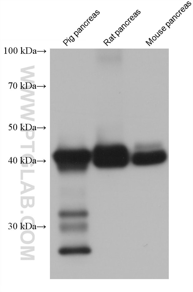
| Positive WB detected in | pig pancreas tissue, rat pancreas tissue, mouse pancreas tissue |
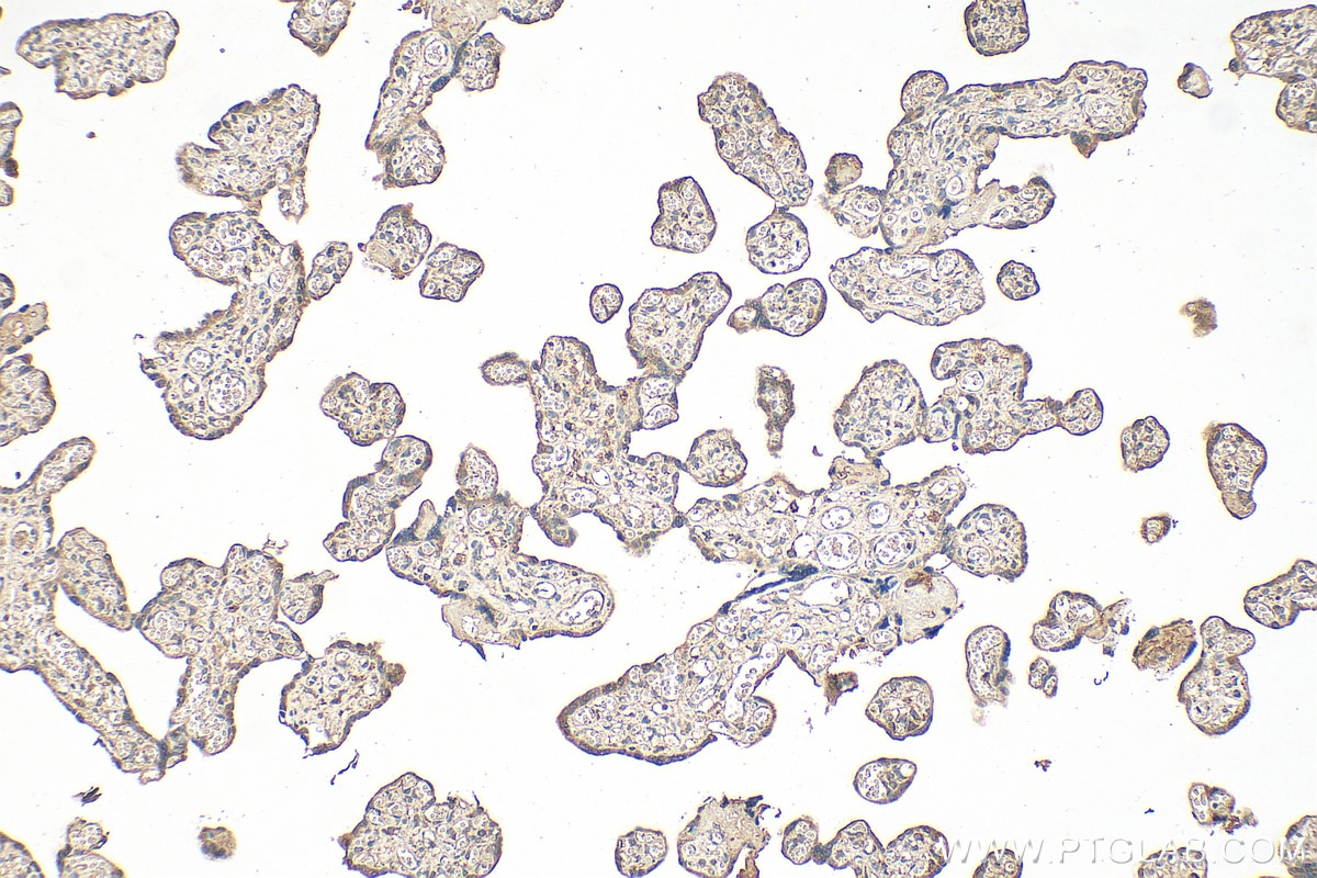
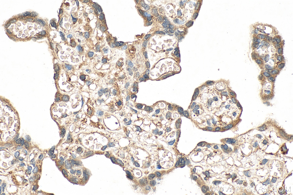
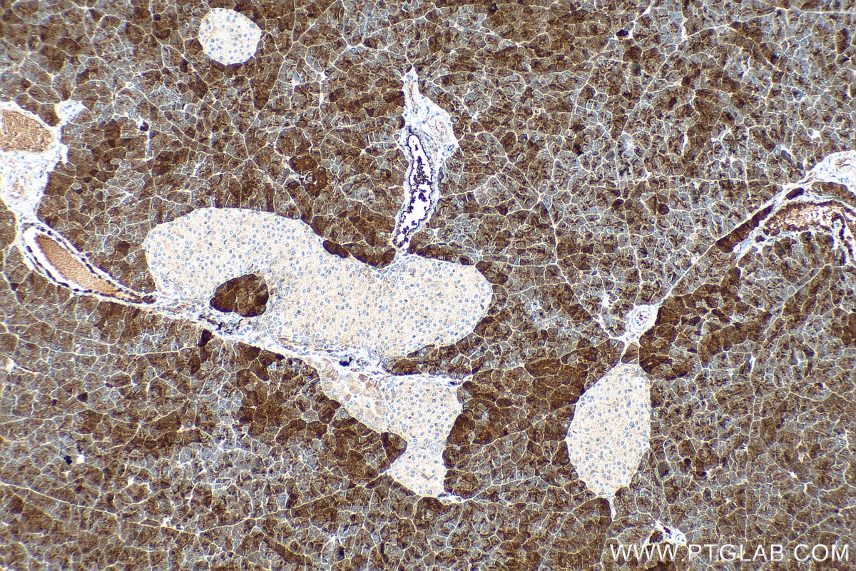
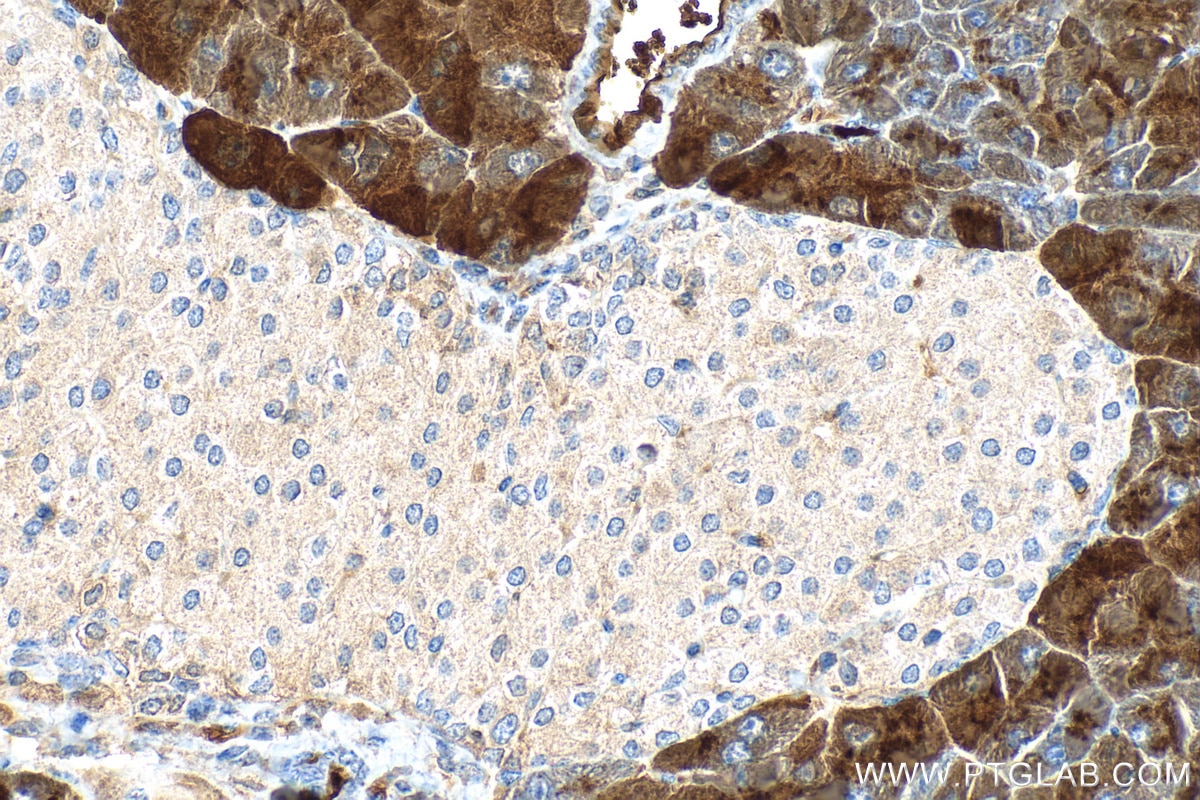
| Positive IHC detected in | mouse pancreas tissue, human placenta tissue Note: suggested antigen retrieval with TE buffer pH 9.0; (*) Alternatively, antigen retrieval may be performed with citrate buffer pH 6.0 |
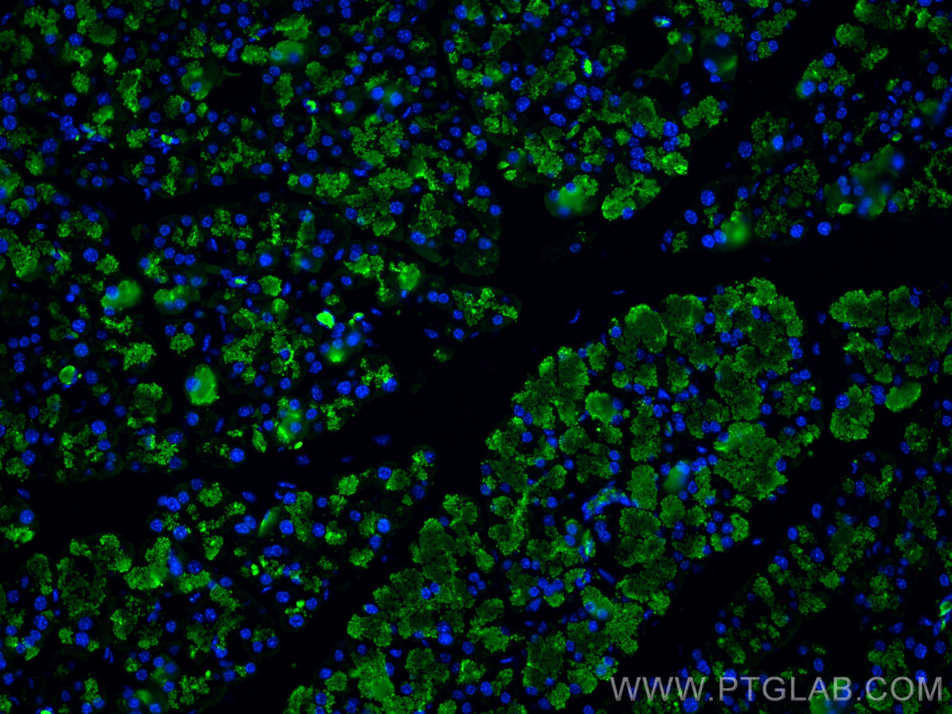
| Positive IF-P detected in | mouse pancreas tissue |
Recommended dilution
| Application | Dilution |
|---|---|
| Western Blot (WB) | WB : 1:5000-1:50000 |
| Immunohistochemistry (IHC) | IHC : 1:300-1:1200 |
| Immunofluorescence (IF)-P | IF-P : 1:200-1:800 |
| It is recommended that this reagent should be titrated in each testing system to obtain optimal results. | |
| Sample-dependent, Check data in validation data gallery. | |
Product Information
68826-1-Ig targets CPA1 in WB, IHC, IF-P, ELISA applications and shows reactivity with human, mouse, rat, pig samples.
| Tested Reactivity | human, mouse, rat, pig |
| Host / Isotype | Mouse / IgG1 |
| Class | Monoclonal |
| Type | Antibody |
| Immunogen |
CatNo: Ag8579 Product name: Recombinant human CPA1 protein Source: e coli.-derived, PET28a Tag: 6*His Domain: 71-419 aa of BC005279 Sequence: SIQAVKIFLESHGISYETMIEDVQSLLDEEQEQMFAFRSRARSTDTFNYATYHTLEEIYDFLDLLVAENPHLVSKIQIGNTYEGRPIYVLKFSTGGSKRPAIWIDTGIHSREWVTQASGVWFAKKITQDYGQDAAFTAILDTLDIFLEIVTNPDGFAFTHSTNRMWRKTRSHTAGSLCIGVDPNRNWDAGFGLSGASSNPCSETYRGKFANSEVEVKSIVDFVKDHGNIKAFISIHSYSQLLMYPYGYKTEPVPDQDELDQLSKAAVTALASLYGTKFNYGSIIKAIYQASGSTIDWTYSQGIKYSFTFELRDTGRYGFLLPASQIIPTAKETWLALLTIMEHTLNHPY Predict reactive species |
| Full Name | carboxypeptidase A1 (pancreatic) |
| Calculated Molecular Weight | 419 aa, 47 kDa |
| Observed Molecular Weight | 47 kDa |
| GenBank Accession Number | BC005279 |
| Gene Symbol | Carboxypeptidase A1 |
| Gene ID (NCBI) | 1357 |
| RRID | AB_3670437 |
| Conjugate | Unconjugated |
| Form | Liquid |
| Purification Method | Protein G purification |
| UNIPROT ID | P15085 |
| Storage Buffer | PBS with 0.02% sodium azide and 50% glycerol, pH 7.3. |
| Storage Conditions | Store at -20°C. Stable for one year after shipment. Aliquoting is unnecessary for -20oC storage. 20ul sizes contain 0.1% BSA. |
Background Information
CPA1(Carboxypeptidase A1) is also named as CPA and belongs to the the peptidase M14 family. CPA1 is one of the genes whose expression in the pancreas and the protein which it encodes is synthesized as an inactive precursor, proCPA1, which is processed to the active enzyme by the proteolytic removal of the 95-amino acid N-terminal prodomain. The mature CPA1 is not secreted and is trapped and degraded intracellularly(PMID:8639628). This antibody is speicific to CPA1.
Protocols
| Product Specific Protocols | |
|---|---|
| IF protocol for CPA1 antibody 68826-1-Ig | Download protocol |
| IHC protocol for CPA1 antibody 68826-1-Ig | Download protocol |
| WB protocol for CPA1 antibody 68826-1-Ig | Download protocol |
| Standard Protocols | |
|---|---|
| Click here to view our Standard Protocols |