


Tested Applications
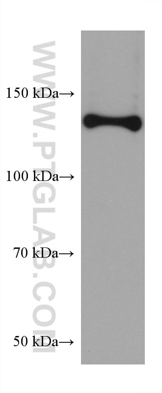
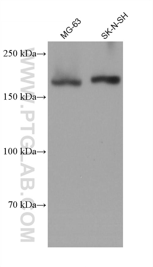
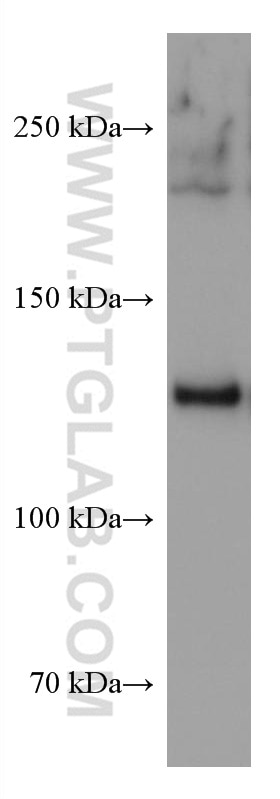
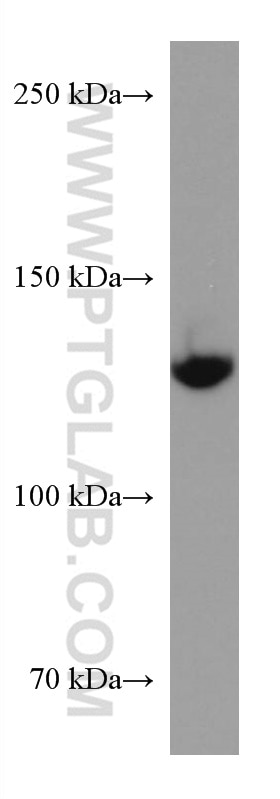
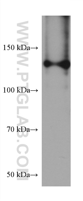
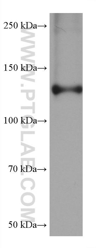
| Positive WB detected in | pig colon tissue, human cervical cancer tissue, human placenta tissue, MG-63 cells, pig lung tissue, pig skin tissue |
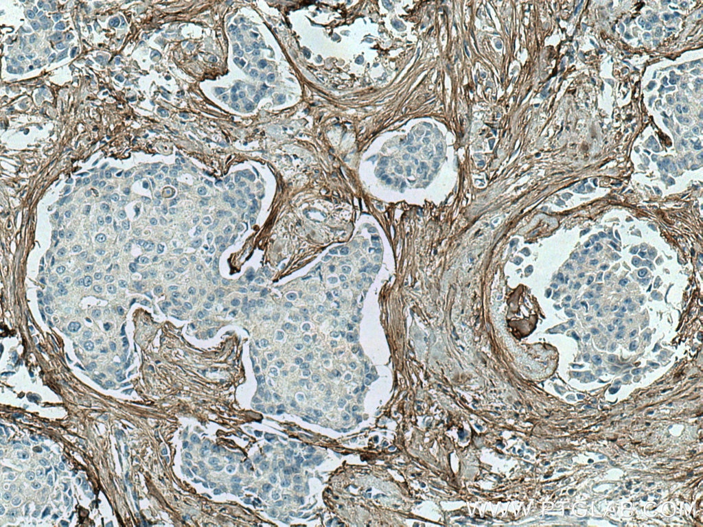
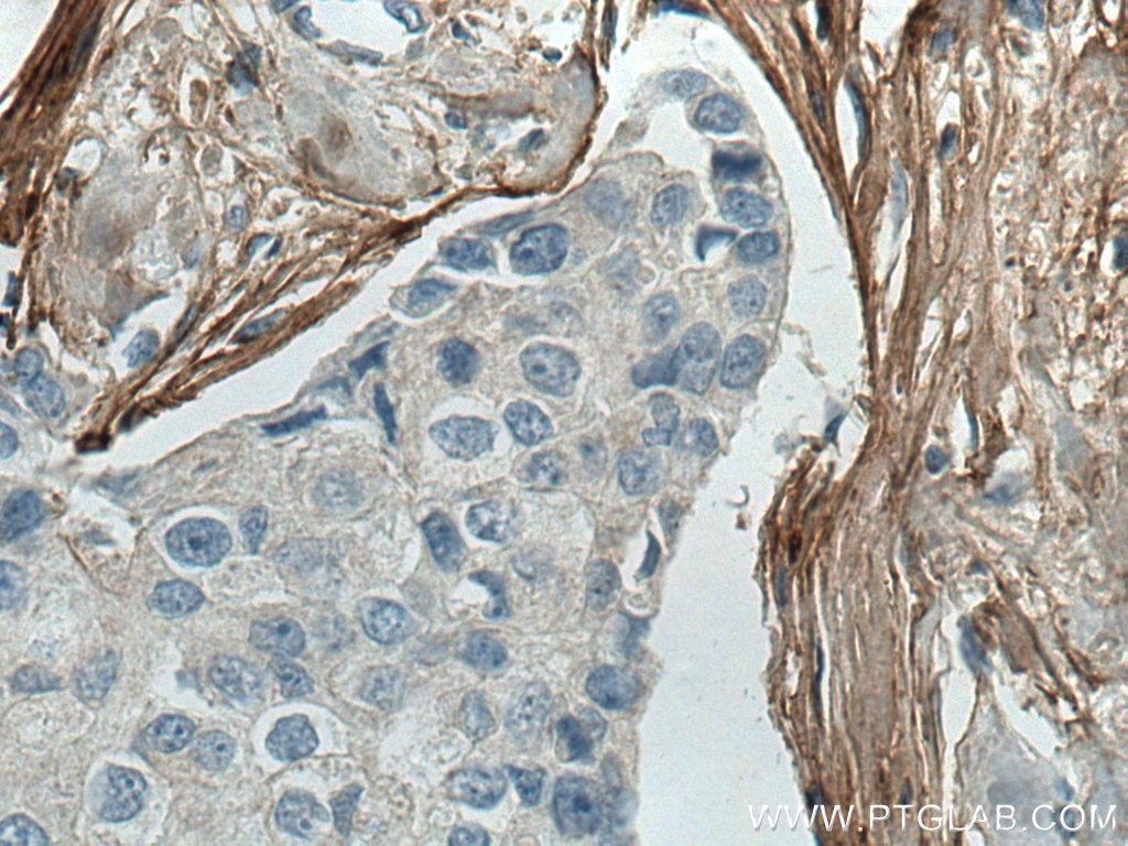
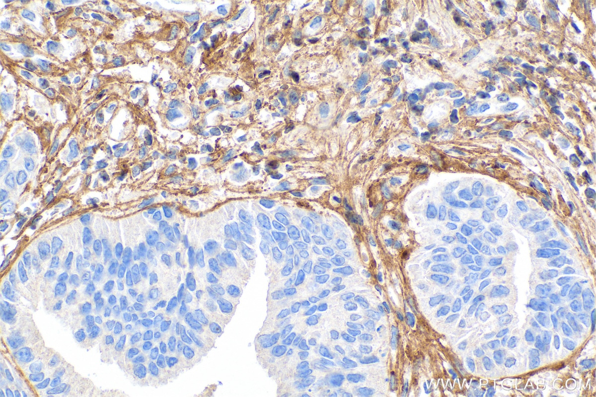
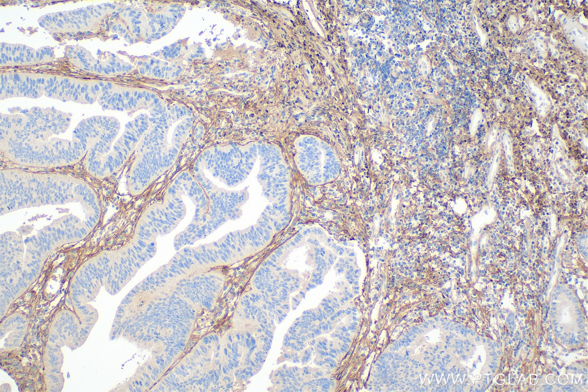
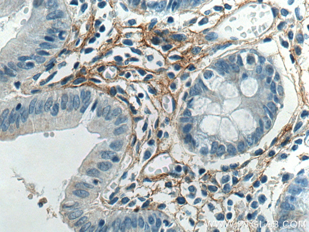
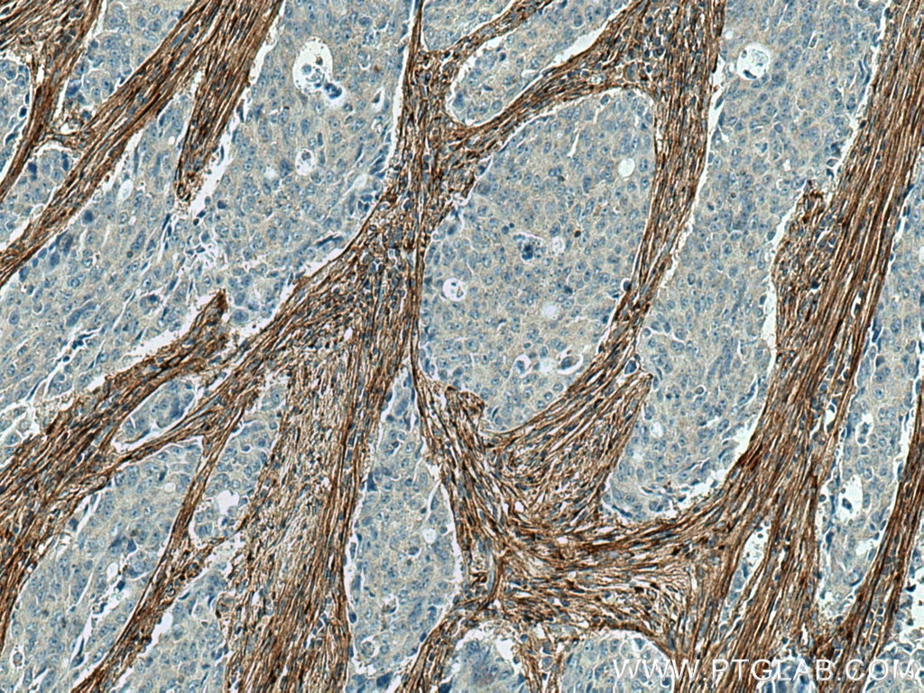
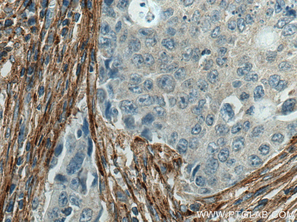
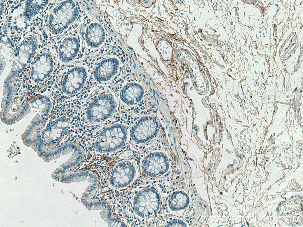
| Positive IHC detected in | human breast cancer tissue, human colon tissue, human colon cancer tissue Note: suggested antigen retrieval with TE buffer pH 9.0; (*) Alternatively, antigen retrieval may be performed with citrate buffer pH 6.0 |
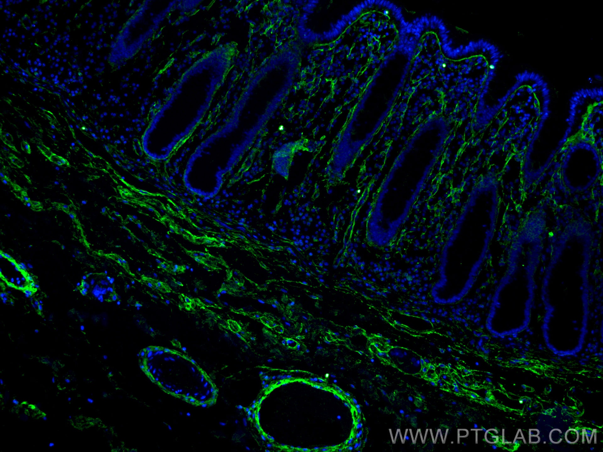
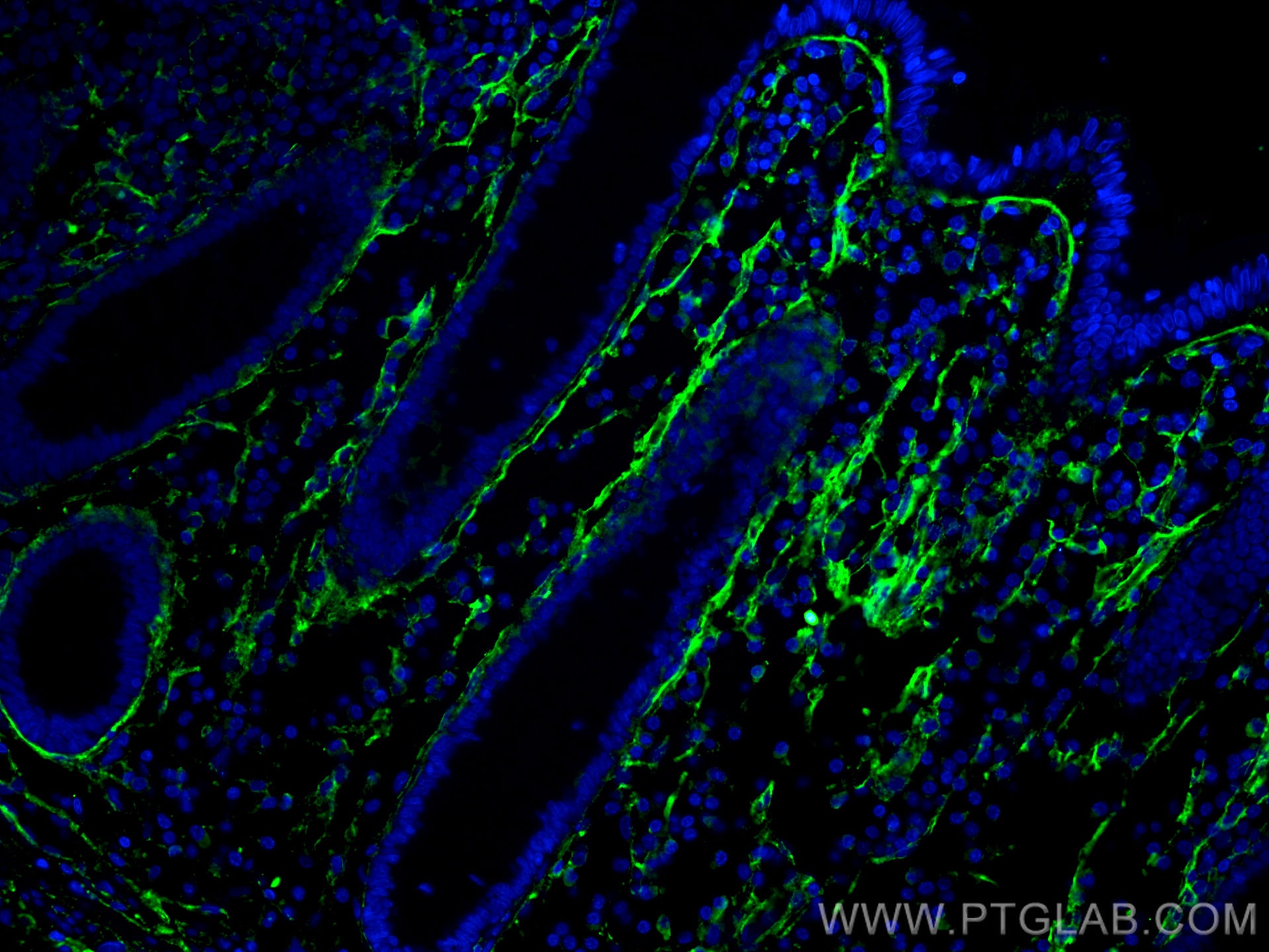
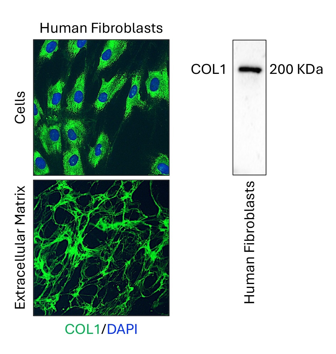
| Positive IF-P detected in | human colon cancer tissue, Human Fibroblasts |
| Positive IF/ICC detected in | U-251 cells |
Recommended dilution
| Application | Dilution |
|---|---|
| Western Blot (WB) | WB : 1:5000-1:50000 |
| Immunohistochemistry (IHC) | IHC : 1:2500-1:10000 |
| Immunofluorescence (IF)-P | IF-P : 1:200-1:800 |
| Immunofluorescence (IF)/ICC | IF/ICC : 1:400-1:1600 |
| It is recommended that this reagent should be titrated in each testing system to obtain optimal results. | |
| Sample-dependent, Check data in validation data gallery. | |
Product Information
67288-1-Ig targets Collagen Type I in WB, IHC, IF/ICC, IF-P, CoIP, IP-MS, ELISA applications and shows reactivity with human, pig samples.
| Tested Reactivity | human, pig |
| Cited Reactivity | human, pig, rabbit, chicken, zebrafish |
| Host / Isotype | Mouse / IgG1 |
| Class | Monoclonal |
| Type | Antibody |
| Immunogen |
Peptide Predict reactive species |
| Full Name | collagen, type I, alpha 1 |
| Calculated Molecular Weight | 139 kDa |
| Observed Molecular Weight | 100-140 kDa,200kDa |
| GenBank Accession Number | NM_000088 |
| Gene Symbol | COL1A1 |
| Gene ID (NCBI) | 1277 |
| RRID | AB_2882554 |
| Conjugate | Unconjugated |
| Form | Liquid |
| Purification Method | Protein G purification |
| UNIPROT ID | P02452 |
| Storage Buffer | PBS with 0.02% sodium azide and 50% glycerol, pH 7.3. |
| Storage Conditions | Store at -20°C. Stable for one year after shipment. Aliquoting is unnecessary for -20oC storage. 20ul sizes contain 0.1% BSA. |
Background Information
Type I collagen, the major structural component of connective tissues such as skin, tendon and bone, is the most abundant and widely expressed collagen in humans (PMID: 7620364; 8645190; 9016532). Type I collagen is a heterotrimer comprising one alpha 2(I) and two alpha 1(I) chains which are encoded by the unlinked loci COL1A2 and COL1A1 respectively. Mutations in COL1A1 are associated with osteogenesis imperfecta types I-IV, Ehlers-Danlos syndrome type VIIA, Ehlers-Danlos syndrome Classical type, Caffey Disease and idiopathic osteoporosis. This antibody raised against a synthesized peptide corresponding to 1206-1218 aa of human pro-alpha 1 chain of type I collagen recognize collagen alpha-1(I) chain. The presence of unprocessed, intermediate, and mature chains of type I collagen was clearly detected only in static constructs. Indeed, in sponges cultured under perfusion the presence of type I collagen was mainly restricted to mature chains, suggesting that HACs were no longer actively producing type I collagen (PMID: 27584727). It is reported that the molecular weight is 200kDa(PMID: 28526934).
Protocols
| Product Specific Protocols | |
|---|---|
| IF protocol for Collagen Type I antibody 67288-1-Ig | Download protocol |
| IHC protocol for Collagen Type I antibody 67288-1-Ig | Download protocol |
| WB protocol for Collagen Type I antibody 67288-1-Ig | Download protocol |
| Standard Protocols | |
|---|---|
| Click here to view our Standard Protocols |
Publications
What published studies show
The anti-Collagen I antibody (catalog no. 67288-1-Ig) has 471 total citations. These appear across high-impact journals including Signal Transduction and Targeted Therapy, Cancer Cell, Cell Research, Immunity, Nature Communications, Theranostics, Advanced Science, Biomaterials, and Journal of Translational Medicine. Research fields span fibrosis (pulmonary, cardiac, renal, hepatic), cancer and tumor microenvironment, bone/cartilage regeneration, wound healing, extracellular matrix remodeling, and stem cell biology, with applications mainly in WB, IF, and IHC.
| Species | Application | Title |
|---|---|---|
Signal Transduct Target Ther Nucleosome assembly protein-like 1 degradation-dependent novel cardioprotection mechanism of Wnt2 against ischemia‒reperfusion injury | ||
Cancer Cell Lipid oxidation reprogramming in cancer-associated fibroblasts enhances CD8(+) T cell cytotoxicity and therapeutic response. | ||
J Hematol Oncol Proteogenomic insights into the biology and treatment of pancreatic ductal adenocarcinoma | ||
Cell Res α-myosin heavy chain lactylation maintains sarcomeric structure and function and alleviates the development of heart failure | ||
Cell Res Increased PRSS56 expression is a causal factor and therapeutic target for human axial high myopia. | ||
Immunity Alarmin-loaded extracellular lipid droplets induce airway neutrophil infiltration during type 2 inflammation |
Reviews
What customers say
Most reviewers report strong performance for both Western Blot and Immunofluorescence, with clear, specific signals and no unspecific bands. The antibody works well at dilutions ranging from 1:400 to 1:5000, detecting both intracellular and extracellular collagen in fibroblasts and BM-MSCs. Two reviewers noted limitations: one found it ineffective on pepsin-digested human collagen I, and another reported it did not work for their specific tissue type (endometrial lesion). Overall, the antibody is well-received for standard WB and IF applications on intact cells and tissues.
The reviews below have been submitted by verified Proteintech customers who received an incentive for providing their feedback.
Sai Sindhura (Verified Customer) (01-08-2026) | Collagen type 1 antibody is very good.
|
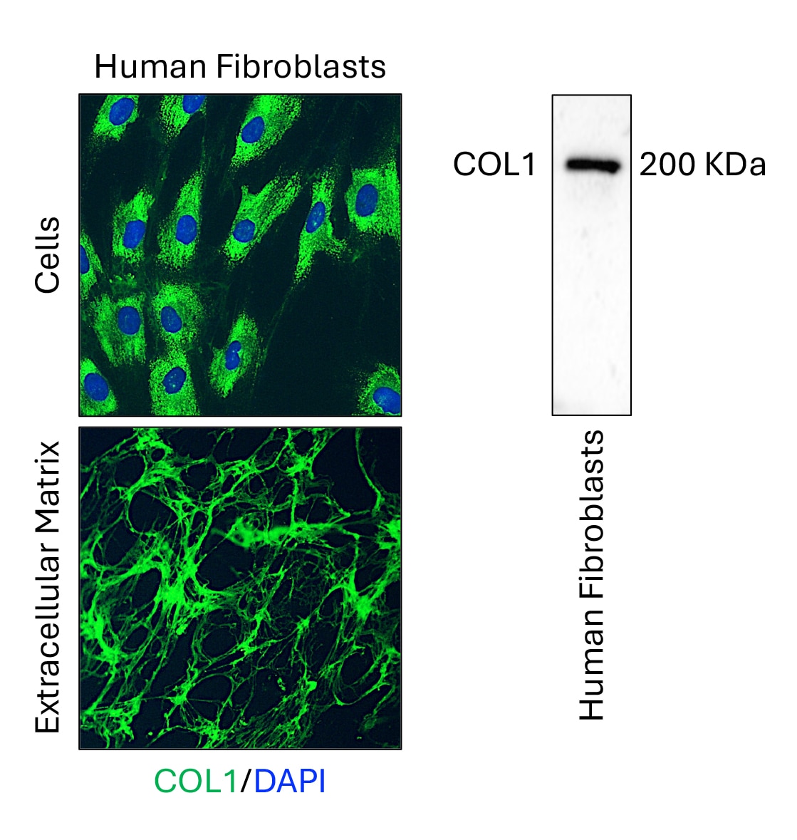
Ludovica (Verified Customer) (12-19-2025) | The antibody work nicely for both IF and WB. For IF, the antibody detects nicely both intracellular and extracellular collagen (although only after decellularization). For WB the signal is clear and without detection of other unspecific bands.
 |
Niky (Verified Customer) (02-26-2024) | Arrived very quickly on ice. However, didnt work for my tissue type
|
Udesh (Verified Customer) (12-22-2023) | Worked well for IF at 1:400
|
Niklas (Verified Customer) (11-08-2022) | great staining with even a high dilution factor
|
Ren Jie (Verified Customer) (06-22-2022) | Does not work for pepsin digested human collagen I
|
Hala (Verified Customer) (02-26-2021) | very good
|
Hala (Verified Customer) (02-23-2021) | very good
|



















