Tested Applications
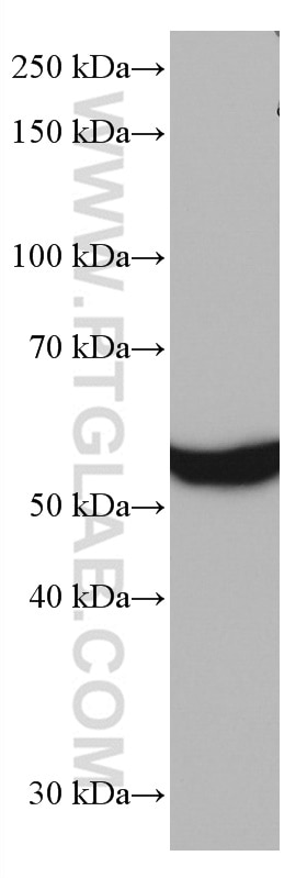
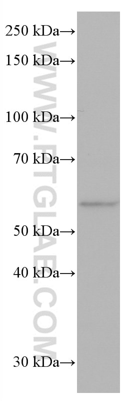
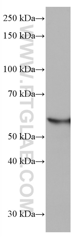
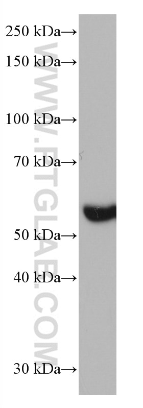
| Positive WB detected in | mouse liver tissue, pig kidney tissue, pig liver tissue, rat liver tissue |
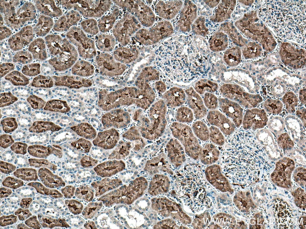
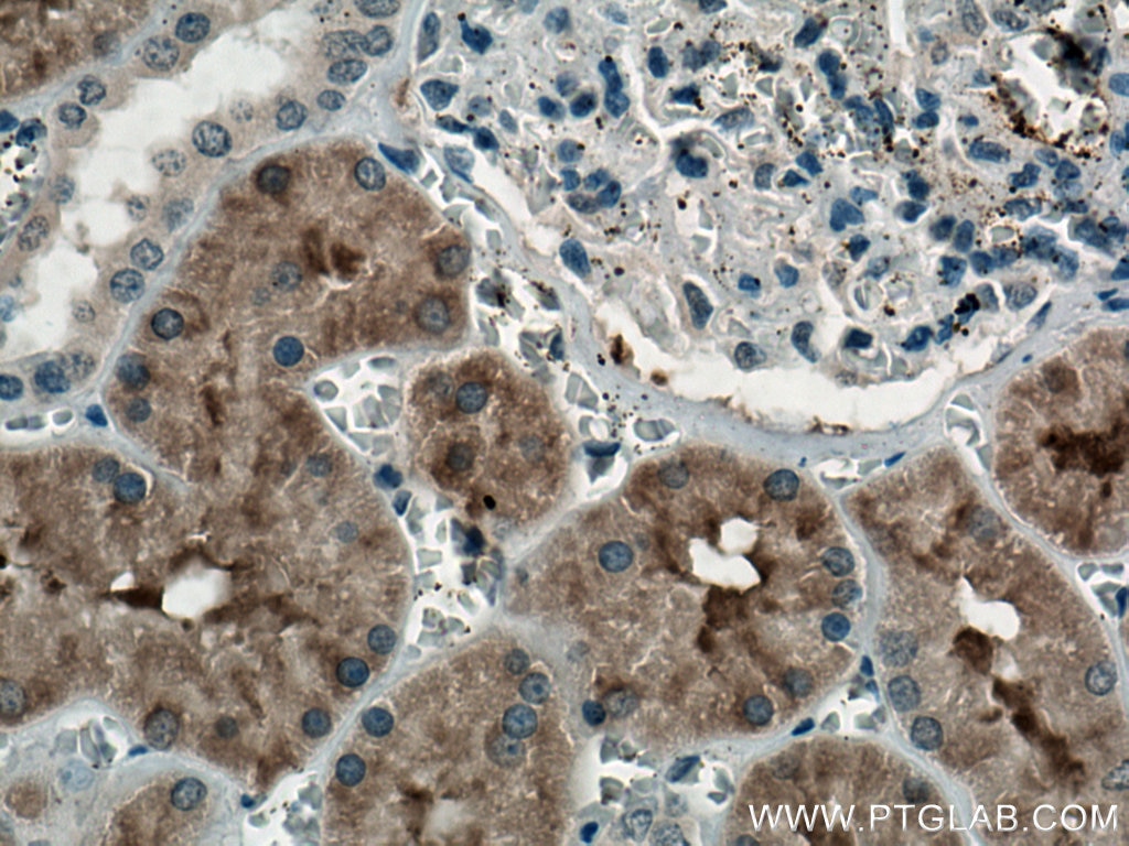
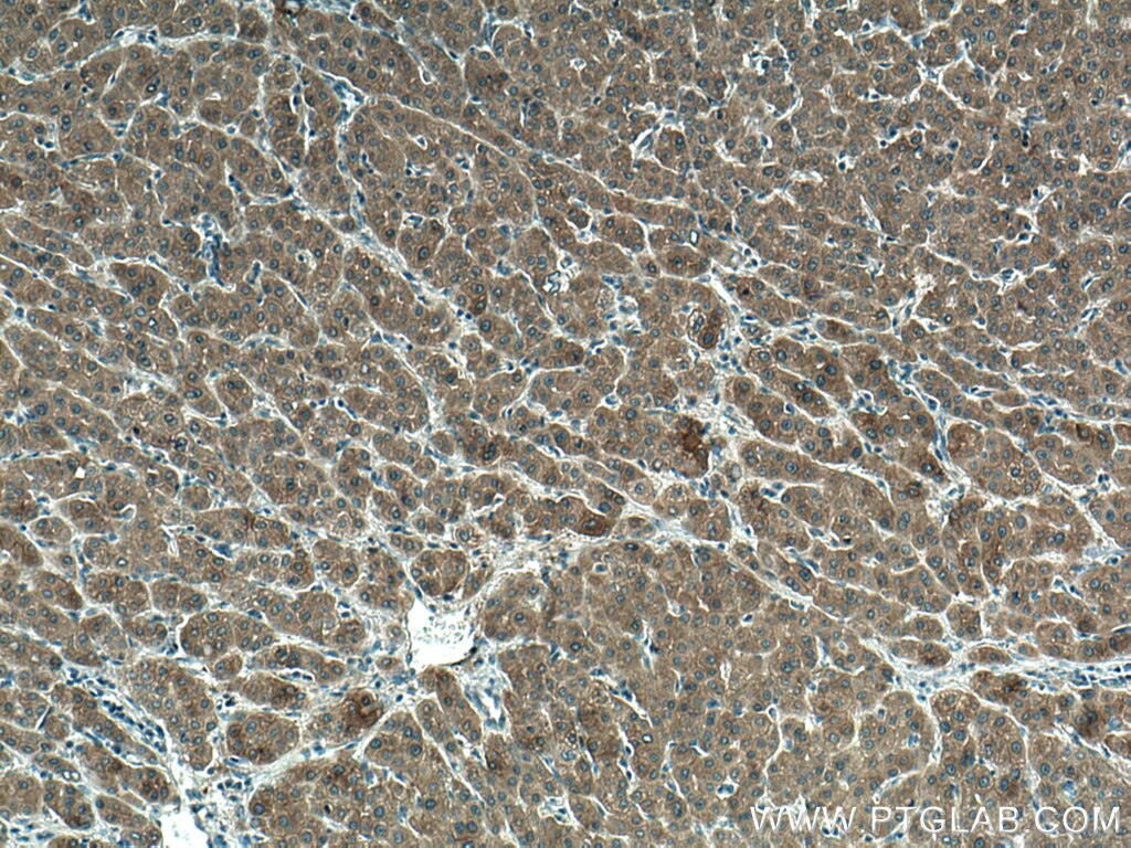
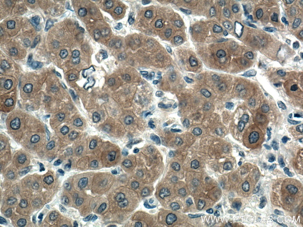
| Positive IHC detected in | human kidney tissue, human liver cancer tissue Note: suggested antigen retrieval with TE buffer pH 9.0; (*) Alternatively, antigen retrieval may be performed with citrate buffer pH 6.0 |
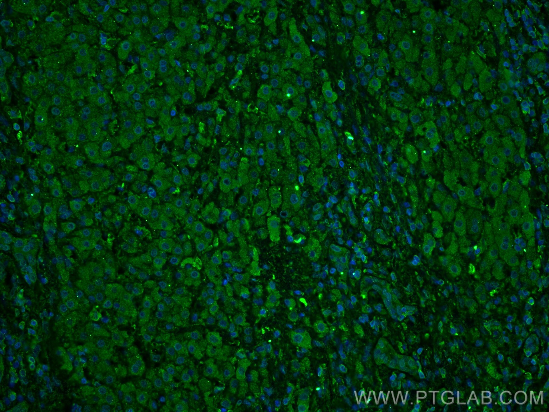
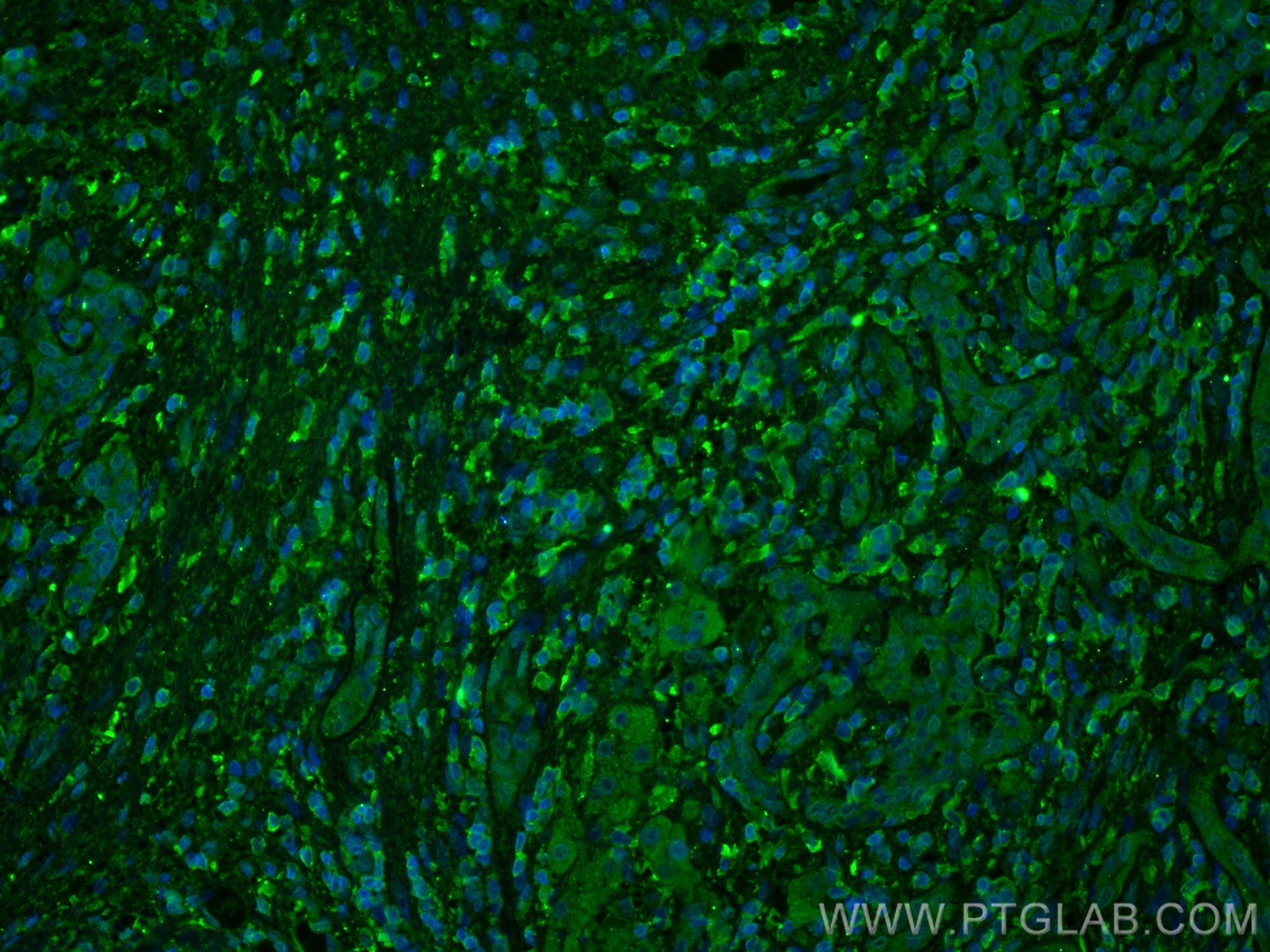
| Positive IF-P detected in | human liver cancer tissue, mouse kidney tissue |
Recommended dilution
| Application | Dilution |
|---|---|
| Western Blot (WB) | WB : 1:3000-1:10000 |
| Immunohistochemistry (IHC) | IHC : 1:150-1:600 |
| Immunofluorescence (IF)-P | IF-P : 1:200-1:800 |
| It is recommended that this reagent should be titrated in each testing system to obtain optimal results. | |
| Sample-dependent, Check data in validation data gallery. | |
Published Applications
| KD/KO | See 1 publications below |
| WB | See 1 publications below |
Product Information
66979-1-Ig targets FTCD in WB, IHC, IF-P, ELISA applications and shows reactivity with human, mouse, rat, pig samples.
| Tested Reactivity | human, mouse, rat, pig |
| Cited Reactivity | mouse |
| Host / Isotype | Mouse / IgG2b |
| Class | Monoclonal |
| Type | Antibody |
| Immunogen |
CatNo: Ag17429 Product name: Recombinant human FTCD protein Source: e coli.-derived, PET28a Tag: 6*His Domain: 192-541 aa of BC136395 Sequence: TKEQAHRIALNLREQGRGKDQPGRLKKVQGIGWYLDEKNLAQVSTNLLDFEVTALHTVYEETCREAQELSLPVVGSQLVGLVPLKALLDAAAFYCEKENLFILEEEQRIRLVVSRLGLDSLCPFSPKERIIEYLVPERGPERGLGSKSLRAFVGEVGARSAAPGGGSVAAAAAAMGAALGSMVGLMTYGRRQFQSLDTTMRRLIPPFREASAKLTTLVDADAEAFTAYLEAMRLPKNTPEEKDRRTAALQEGLRRAVSVPLTLAETVASLWPALQELARCGNLACRSDLQVAAKALEMGVFGAYFNVLINLRDITDEAFKDQIHHRVSSLLQEAKTQAALVLDCLETRQE Predict reactive species |
| Full Name | formiminotransferase cyclodeaminase |
| Calculated Molecular Weight | 541 aa, 59 kDa |
| Observed Molecular Weight | 53-62 kDa |
| GenBank Accession Number | BC136395 |
| Gene Symbol | FTCD |
| Gene ID (NCBI) | 10841 |
| RRID | AB_2882299 |
| Conjugate | Unconjugated |
| Form | Liquid |
| Purification Method | Protein A purification |
| UNIPROT ID | O95954 |
| Storage Buffer | PBS with 0.02% sodium azide and 50% glycerol, pH 7.3. |
| Storage Conditions | Store at -20°C. Stable for one year after shipment. Aliquoting is unnecessary for -20oC storage. 20ul sizes contain 0.1% BSA. |
Background Information
FTCD (formimidoyltransferase cyclodeaminase) is critical for the catabolism of histidine. This process is one-carbon units that can enter the one-carbon/folate cycle, which provides methyl groups for arsenic metabolism (PMID: 30893314). In addition, FTCD could serve as a predictive and prognostic marker for patients with HCC (PMID: 37675273).
Protocols
| Product Specific Protocols | |
|---|---|
| IF protocol for FTCD antibody 66979-1-Ig | Download protocol |
| IHC protocol for FTCD antibody 66979-1-Ig | Download protocol |
| WB protocol for FTCD antibody 66979-1-Ig | Download protocol |
| Standard Protocols | |
|---|---|
| Click here to view our Standard Protocols |