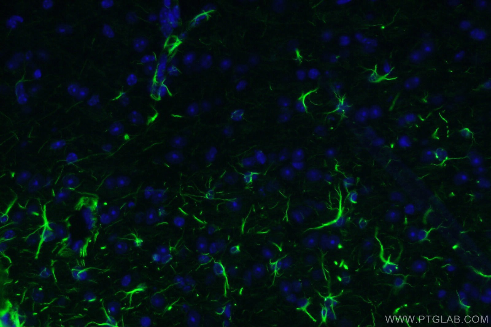
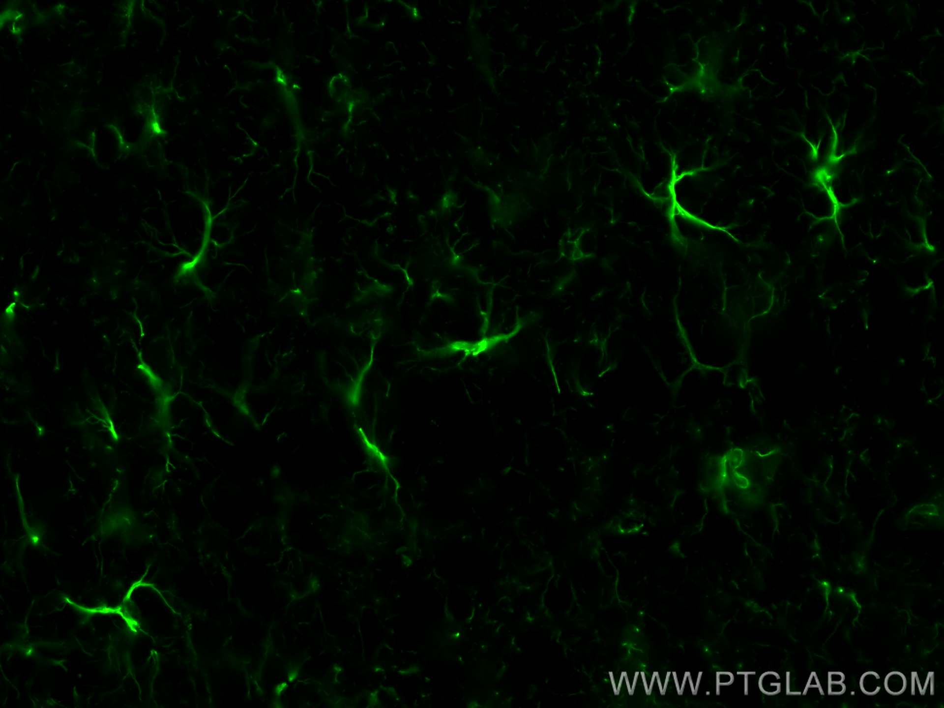
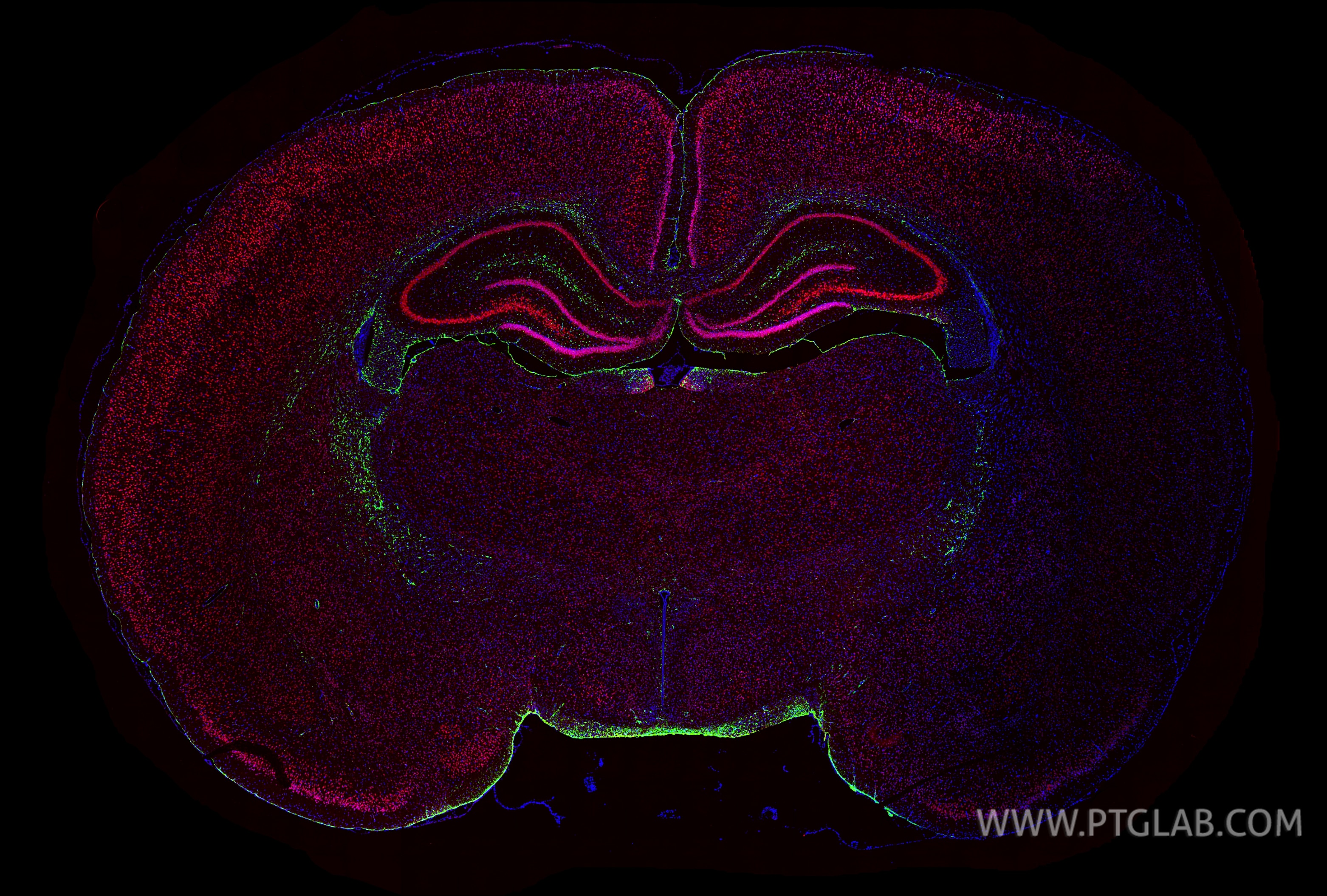
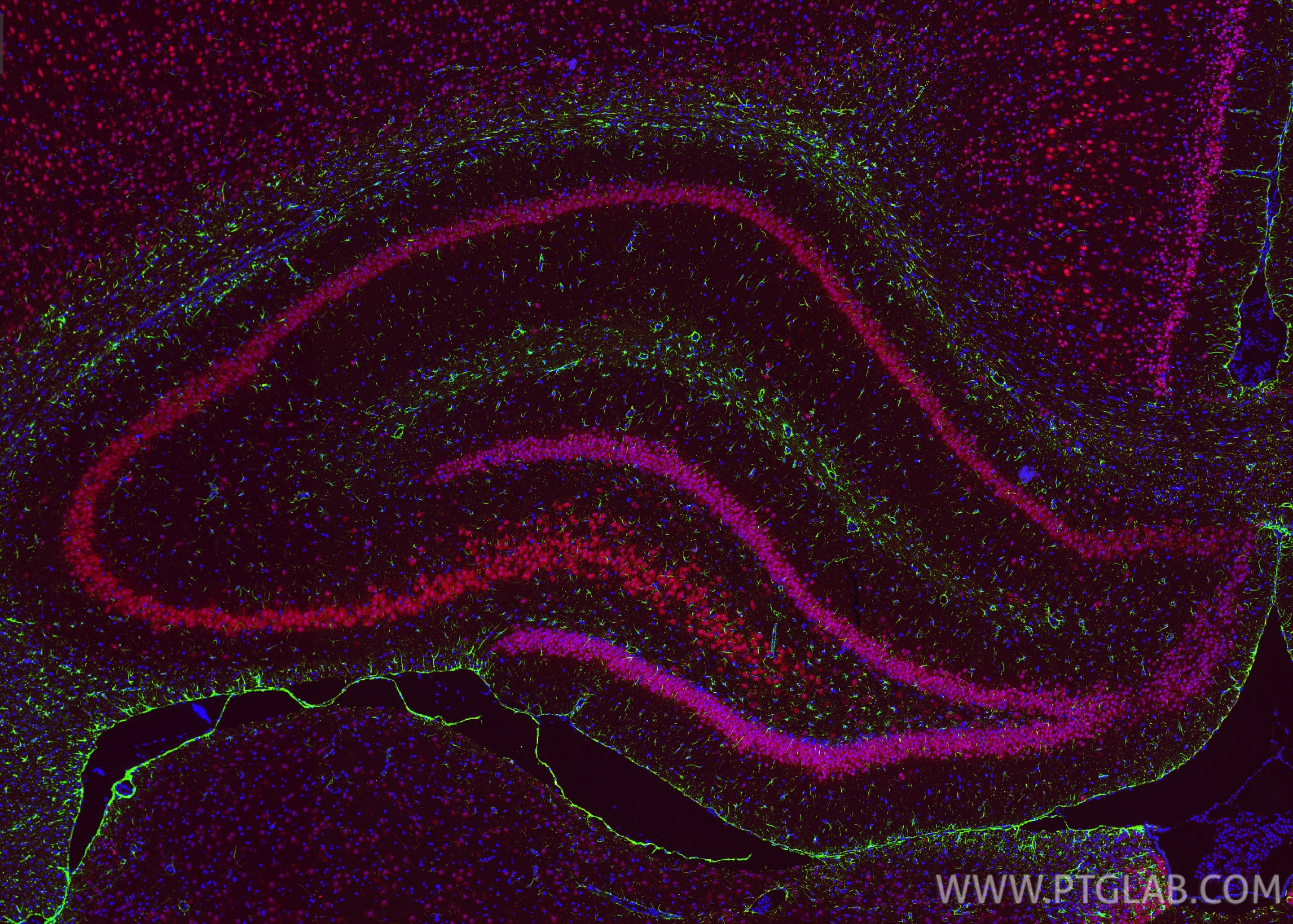
Tested Applications
| Positive IF-P detected in | rat brain tissue, mouse brain tissue |
This antibody is not recommended for immunocytofluorescent assays. It is not suitable for frozen sections.
Recommended dilution
| Application | Dilution |
|---|---|
| Immunofluorescence (IF)-P | IF-P : 1:50-1:500 |
| It is recommended that this reagent should be titrated in each testing system to obtain optimal results. | |
| Sample-dependent, Check data in validation data gallery. | |
This antibody is not recommended for immunocytofluorescent assays. It is not suitable for frozen sections.
Published Applications
| IF | See 8 publications below |
Product Information
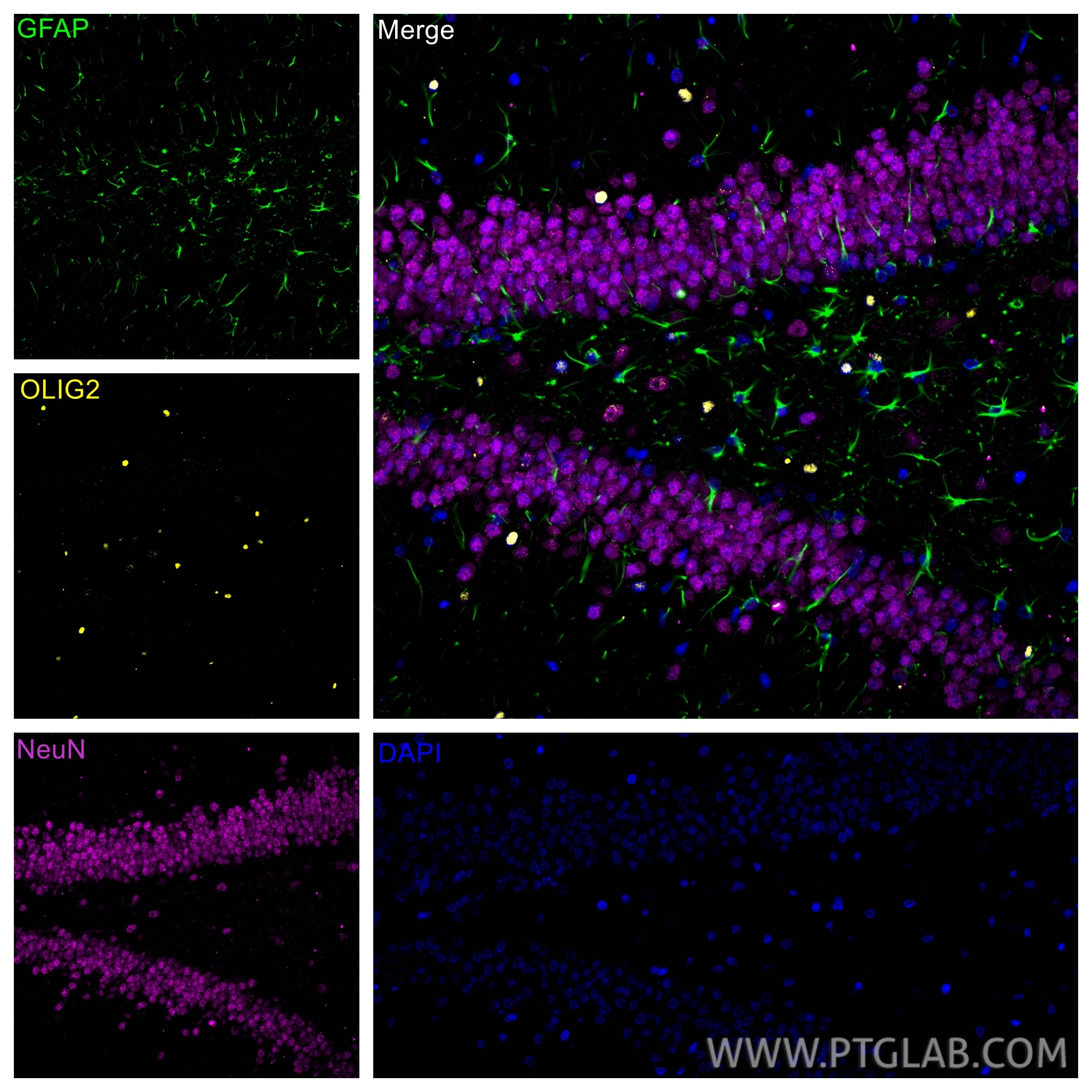
CL488-60190 targets GFAP in IF-P applications and shows reactivity with human, mouse, rat, pig samples.
| Tested Reactivity | human, mouse, rat, pig |
| Cited Reactivity | mouse, rat |
| Host / Isotype | Mouse / IgG2a |
| Class | Monoclonal |
| Type | Antibody |
| Immunogen |
CatNo: Ag10452 Product name: Recombinant human GFAP protein Source: e coli.-derived, PET28a Tag: 6*His Domain: 83-432 aa of BC013596 Sequence: YIEKVRFLEQQNKALAAELNQLRAKEPTKLADVYQAELRELRLRLDQLTANSARLEVERDNLAQDLATVRQKLQDETNLRLEAENNLAAYRQEADEATLARLDLERKIESLEEEIRFLRKIHEEEVRELQEQLARQQVHVELDVAKPDLTAALKEIRTQYEAMASSNMHEAEEWYRSKFADLTDAAARNAELLRQAKHEANDYRRQLQSLTCDLESLRGTNESLERQMREQEERHVREAASYQEALARLEEEGQSLKDEMARHLQEYQDLLNVKLALDIEIATYRKLLEGEENRITIPVQTFSNLQIRETSLDTKSVSEGHLKRNIVVKTVEMRDGEVIKESKQEHKDVM Predict reactive species |
| Full Name | glial fibrillary acidic protein |
| Calculated Molecular Weight | 432 aa, 50 kDa |
| GenBank Accession Number | BC013596 |
| Gene Symbol | GFAP |
| Gene ID (NCBI) | 2670 |
| RRID | AB_2883116 |
| Conjugate | CoraLite® Plus 488 Fluorescent Dye |
| Excitation/Emission Maxima Wavelengths | 493 nm / 522 nm |
| Excitation Laser | Blue laser (488 nm) |
| Form | Liquid |
| Purification Method | Protein A purification |
| UNIPROT ID | P14136 |
| Storage Buffer | PBS with 50% glycerol, 0.05% Proclin300, 0.5% BSA, pH 7.3. |
| Storage Conditions | Store at -20°C. Avoid exposure to light. Stable for one year after shipment. Aliquoting is unnecessary for -20oC storage. |
Background Information
Function
GFAP (Glial fibrillary acidic protein) is a type III intermediate filament (IF) protein specific to the central nervous system (CNS). GFAP is one of the main components of the intermediate filament network in astrocytes and has been proposed as playing a role in cell migration, cell motility, maintaining mechanical strength, and in mitosis.Tissue specificity
GFAP is expressed in central nervous system cells, predominantly in astrocytes. GFAP is commonly used as an astrocyte marker. However, GFAP is also present in peripheral glia and in non-CNS cells, including fibroblasts, chondrocytes, lymphocytes, and liver stellate cells (PMID: 21219963).Involvement in disease
- Mutations in GFAP lead to Alexander disease (OMIM: 203450), an autosomal dominant CNS disorder. The mutations present in affected individuals are thought to be gain-of-function.
- Upregulation of GFAP is a hallmark of reactive astrocytes, in which GFAP is present in hypertrophic cellular processes. Reactive astrogliosis is present in many neurological disorders, such as stroke, various neurodegenerative diseases (including Alzheimer's and Parkinson's disease), and neurotrauma.
Isoforms
Astrocytes express 10 different isoforms of GFAP that differ in the rod and tail domains (PMID: 25726916), which means that they differ in molecular size. Isoform expression varies during the development and across different subtypes of astrocytes. Not all isoforms are upregulated in reactive astrocytes.Post-translational modifications
Intermediate filament proteins are regulated by phosphorylation. Six phosphorylation sites have been identified in GFAP protein, at least some of which are reported to control filament assembly (PMID: 21219963).Cellular localization
GFAP localizes to intermediate filaments and stains well in astrocyte cellular processes.
Protocols
| Product Specific Protocols | |
|---|---|
| IF protocol for CL Plus 488 GFAP antibody CL488-60190 | Download protocol |
| Standard Protocols | |
|---|---|
| Click here to view our Standard Protocols |
Publications
| Species | Application | Title |
|---|---|---|
J Neurosci Res Neuronal deficiency of hypoxia-inducible factor 2α increases hypoxic-ischemic brain injury in neonatal mice. | ||
Front Pharmacol Extracellular Vesicle-Mediated Delivery of Ultrasmall Superparamagnetic Iron Oxide Nanoparticles to Mice Brain. | ||
Front Neurosci Downregulation of m6A Methyltransferase in the Hippocampus of Tyrobp -/- Mice and Implications for Learning and Memory Deficits. | ||
Acta Biochim Biophys Sin (Shanghai) Cannabidiol alleviates the inflammatory response in rats with traumatic brain injury through the PGE 2-EP2-cAMP-PKA signaling pathway | ||
Brain Res Effects of cannabinoid (CBD) on blood brain barrier permeability after brain injury in rats. | ||
Elife The myeloid cell-driven transdifferentiation of endothelial cells into pericytes promotes the restoration of BBB function and brain self-repair after stroke |