

Tested Applications
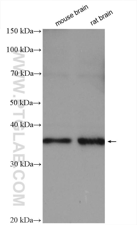
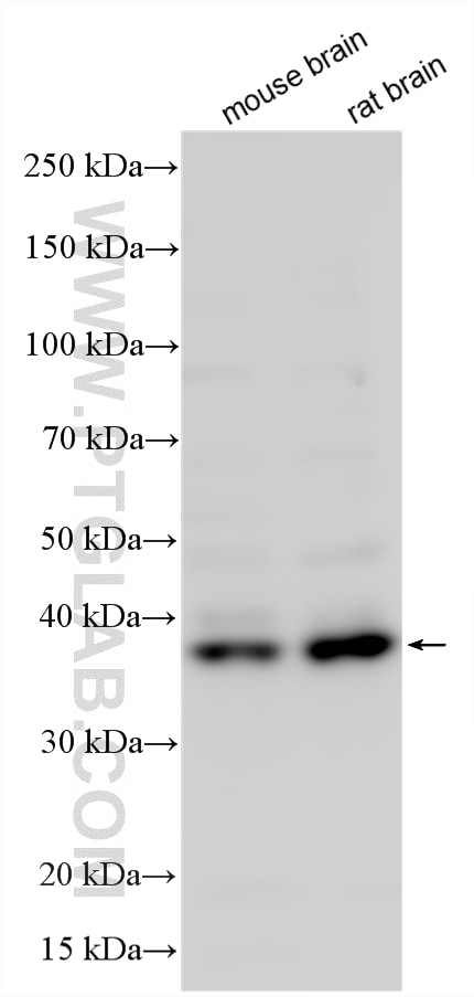
| Positive WB detected in | mouse brain tissue, rat brain |
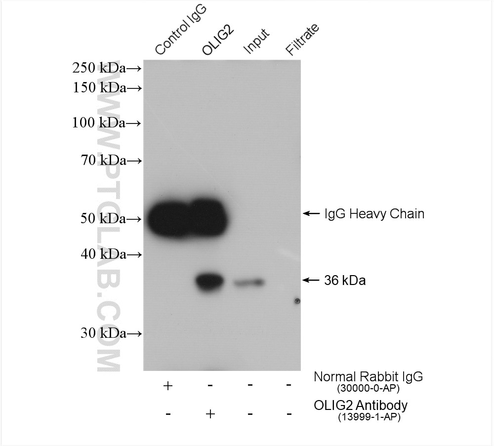
| Positive IP detected in | mouse brain tissue |
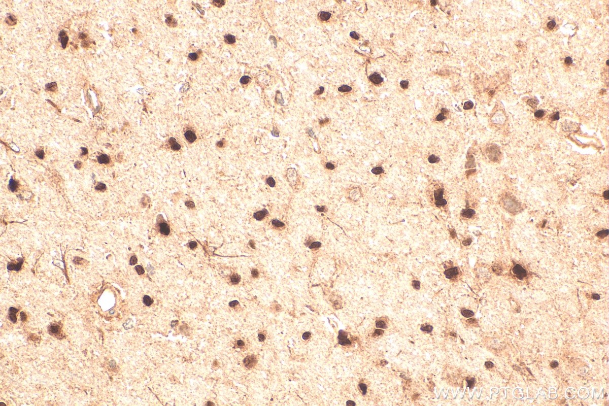
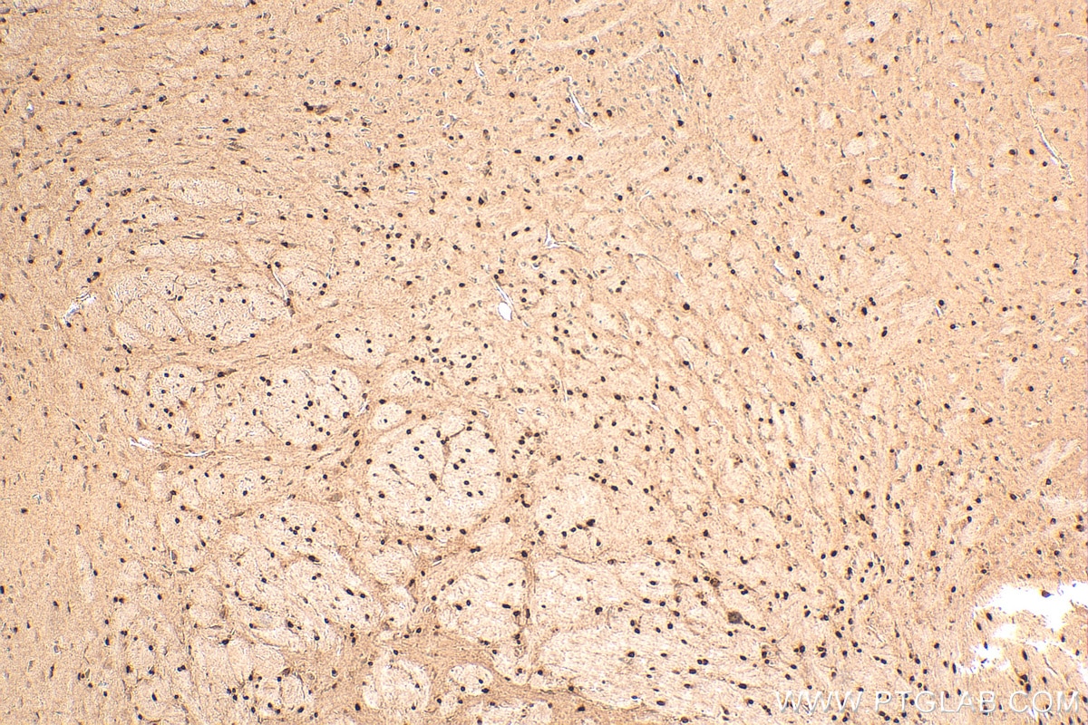
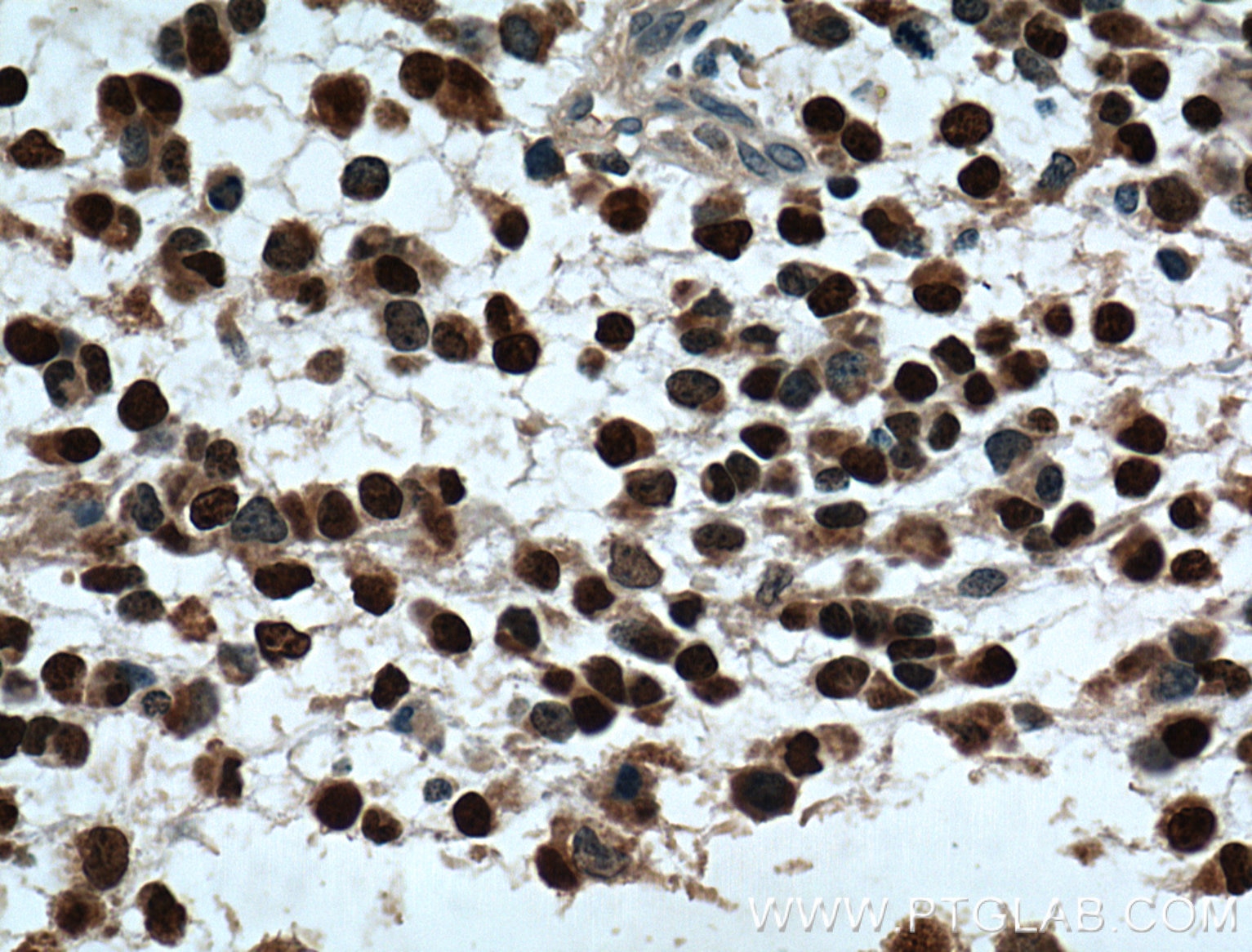
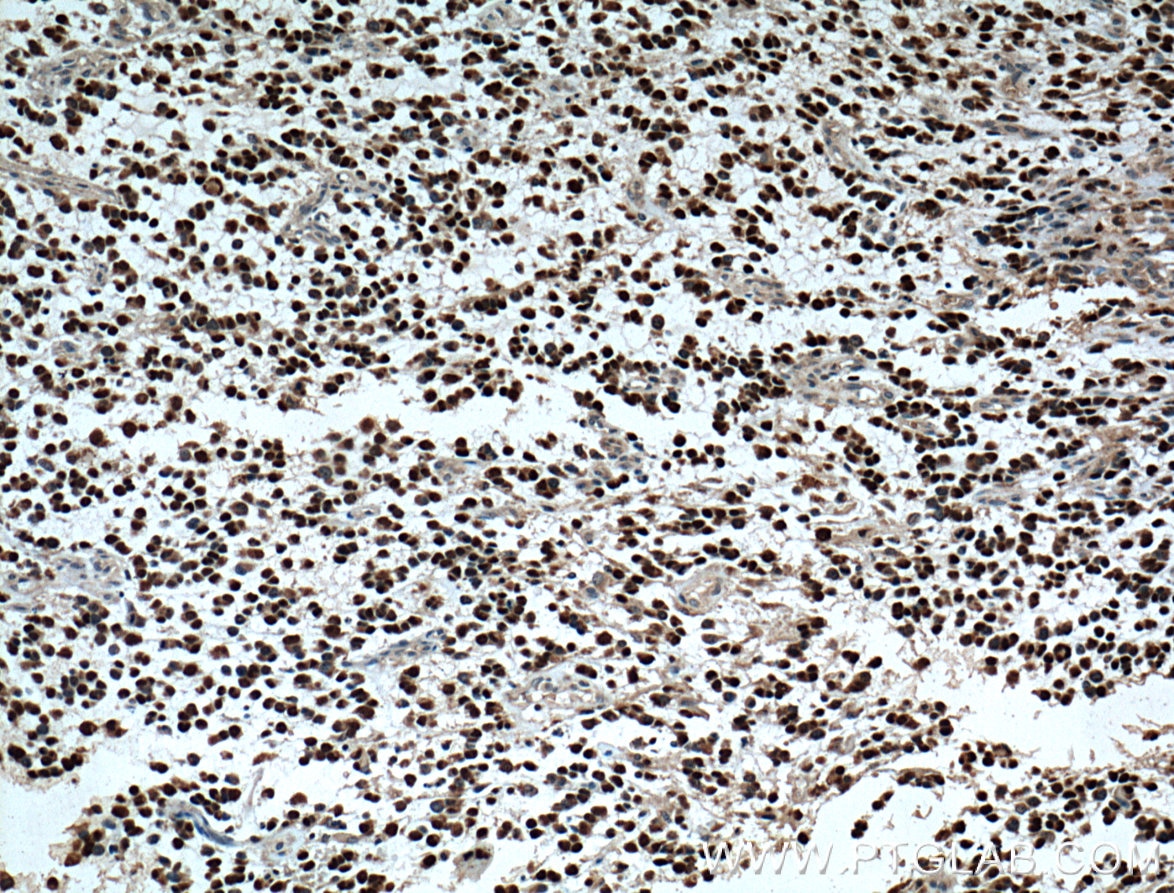
| Positive IHC detected in | mouse brain tissue, human gliomas tissue, rat brain tissue Note: suggested antigen retrieval with TE buffer pH 9.0; (*) Alternatively, antigen retrieval may be performed with citrate buffer pH 6.0 |
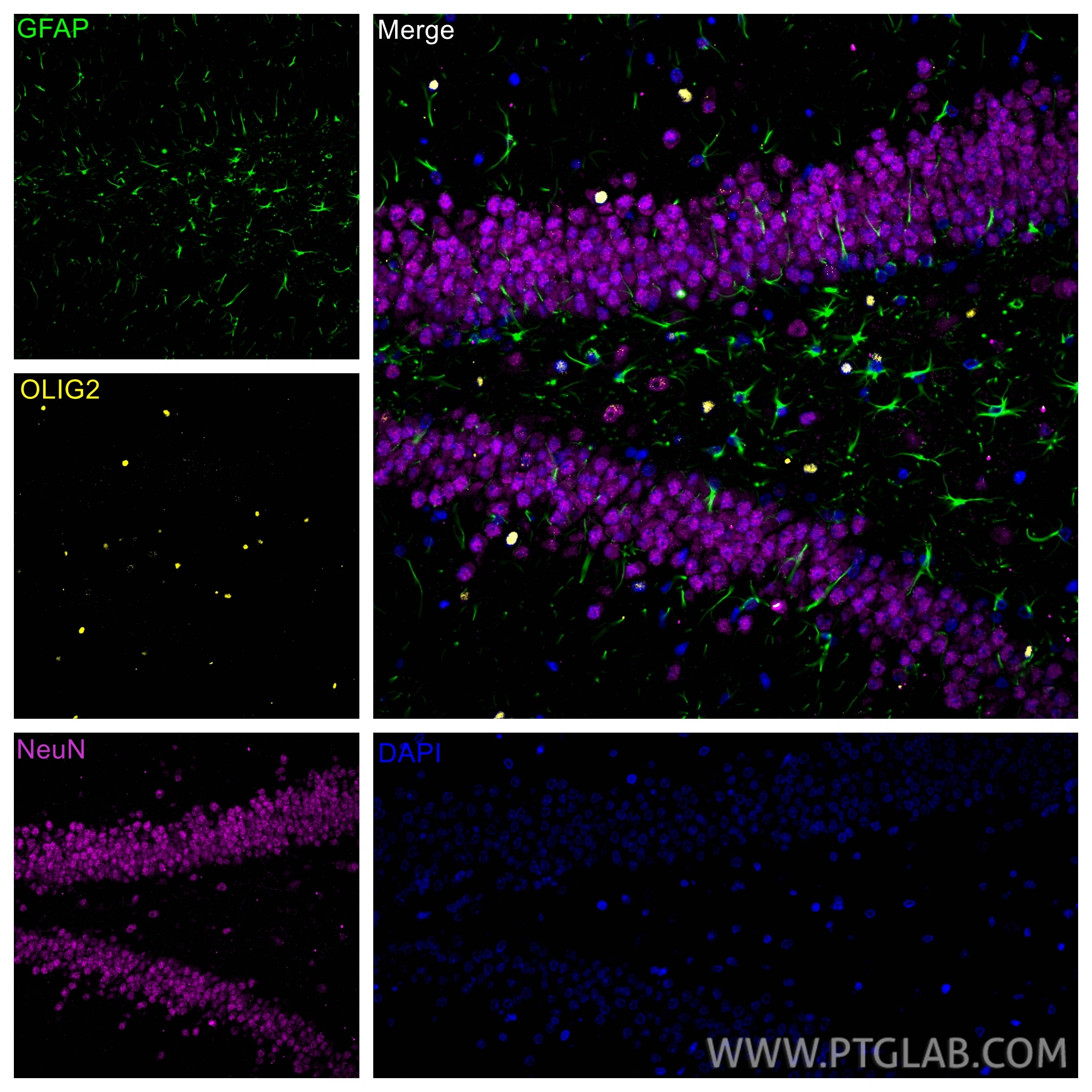
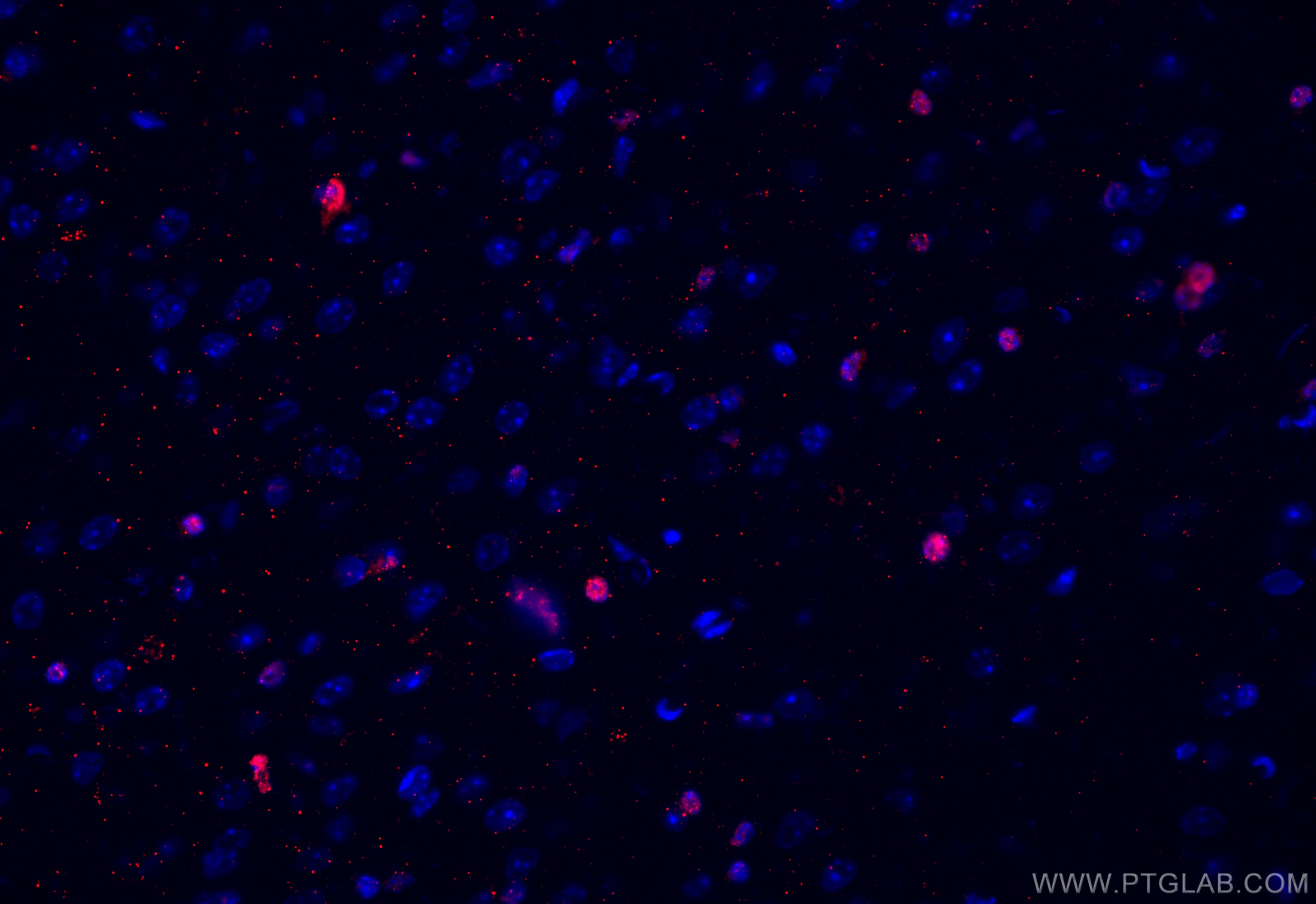
| Positive IF-P detected in | rat brain tissue |
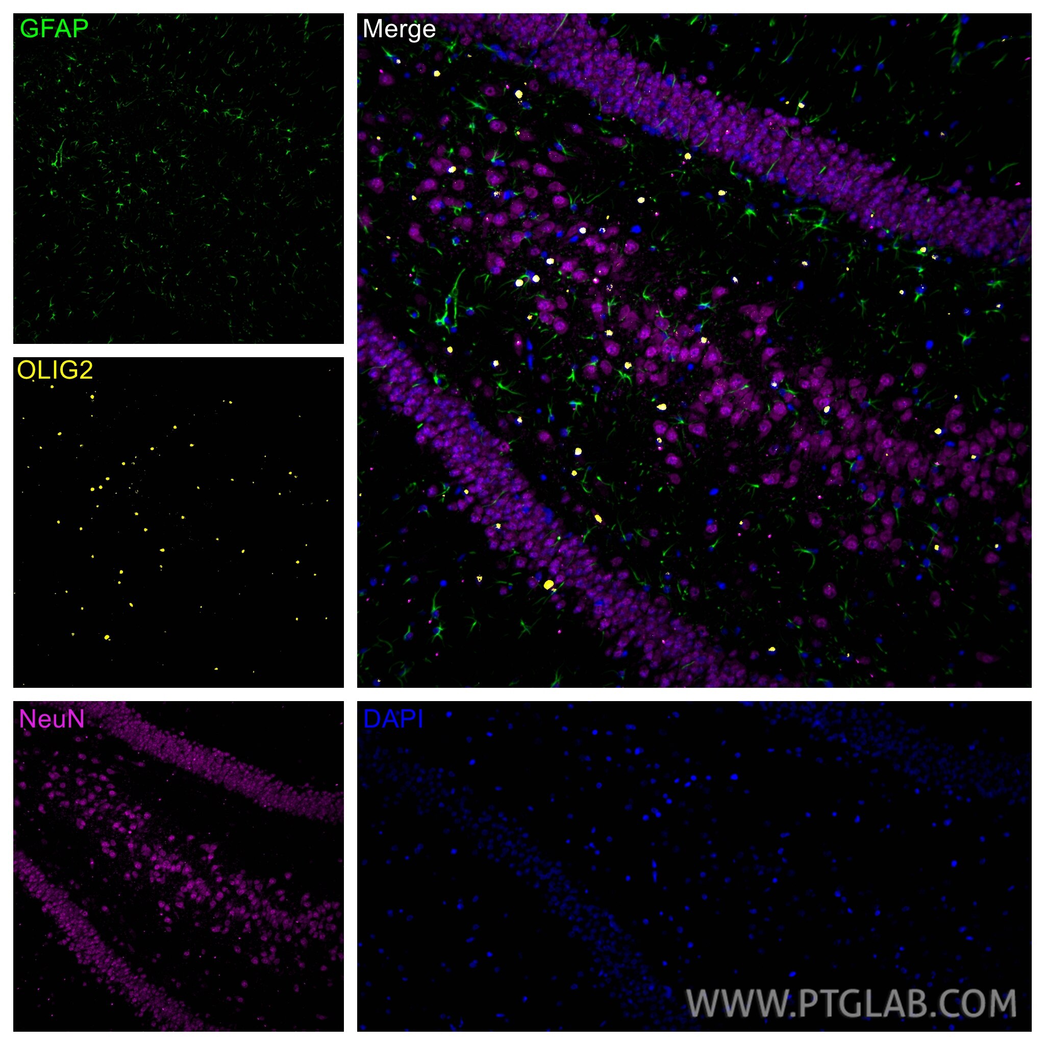
| Positive IF-Fro detected in | mouse brain tissue |
Recommended dilution
| Application | Dilution |
|---|---|
| Western Blot (WB) | WB : 1:1000-1:8000 |
| Immunoprecipitation (IP) | IP : 0.5-4.0 ug for 1.0-3.0 mg of total protein lysate |
| Immunohistochemistry (IHC) | IHC : 1:500-1:2000 |
| Immunofluorescence (IF)-P | IF-P : 1:200-1:800 |
| Immunofluorescence (IF)-FRO | IF-FRO : 1:200-1:800 |
| It is recommended that this reagent should be titrated in each testing system to obtain optimal results. | |
| Sample-dependent, Check data in validation data gallery. | |
Product Information
13999-1-AP targets OLIG2 in WB, IHC, IF-P, IF-Fro, IP, ELISA applications and shows reactivity with human, mouse, rat samples.
| Tested Reactivity | human, mouse, rat |
| Cited Reactivity | human, mouse, rat, bovine, sheep |
| Host / Isotype | Rabbit / IgG |
| Class | Polyclonal |
| Type | Antibody |
| Immunogen |
CatNo: Ag5089 Product name: Recombinant human OLIG2 protein Source: e coli.-derived, PGEX-4T Tag: GST Domain: 1-323 aa of BC047511 Sequence: MDSDASLVSSRPSSPEPDDLFLPARSKGSSGSAFTGGTVSSSTPSDCPPELSAELRGAMGSAGAHPGDKLGGSGFKSSSSSTSSSTSSAAASSTKKDKKQMTEPELQQLRLKINSRERKRMHDLNIAMDGLREVMPYAHGPSVRKLSKITTLLLARNYILMLTNSLEEMKRLVSEIYGGHHAGFHPSACGGLAHSAPLPAATAHPAAAAHAAHHPAVHHPILPPAAAAAAAAAAAAAVSSASLPGSGLPSVGSIRPPHGLLKSPSAAAAAPLGGGGGGSGASGGFQHWGGMPCPCSMCQVPPPHHHVSAMGAGSLPRLTSDAK Predict reactive species |
| Full Name | oligodendrocyte lineage transcription factor 2 |
| Calculated Molecular Weight | 32 kDa |
| Observed Molecular Weight | 32-36 kDa |
| GenBank Accession Number | BC047511 |
| Gene Symbol | OLIG2 |
| Gene ID (NCBI) | 10215 |
| RRID | AB_2157541 |
| Conjugate | Unconjugated |
| Form | Liquid |
| Purification Method | Antigen affinity purification |
| UNIPROT ID | Q13516 |
| Storage Buffer | PBS with 0.02% sodium azide and 50% glycerol, pH 7.3. |
| Storage Conditions | Store at -20°C. Stable for one year after shipment. Aliquoting is unnecessary for -20oC storage. 20ul sizes contain 0.1% BSA. |
Background Information
What is the specificity of Olig2?
Oligodendrocyte transcription factor 2 (OLIG2) is expressed by cells found in the central nervous system (CNS) called oligodendrocyte precursor cells (OPCs), which form the myelin sheaths wrapping the axons of neurons in the brain and spinal cord. OPCs differentiate into oligodendrocytes that form the myelin, providing metabolic support and saltatory conduction. OLIG2 is expressed broadly throughout their development to OPCs. This protein can be used to identify many cells of the oligodendrocyte lineage. It is found mostly in the nucleoplasm but also in the cytoplasm.
What is the function of OLIG2?
The basic helix-loop-helix structure of OLIG2 allows it to function as a transcription factor, determining cell fate in the development of neural tissue, where it is located at the pMN domain in the embryonic spinal cord. Expression of OLIG2 causes neural precursors to develop into oligodendrocytes or into motor neurons and expression is then maintained postnatally. OLIG2 co-operates with other factors to cause this differentiation from precursors, although overexpression alone can cause differentiation to the oligodendrocyte lineage.1 The continued expression of OLIG2 in OPCs indicates an ongoing role in the maintenance of their stemness.
What is the involvement of OLIG2 in disease?
The expression of OLIG2 in glioblastoma, the most common type of malignant brain tumor in adults, is well characterized, where the pathological function is an extension of the normal function. Stem-like cells that propagate the tumor growth have been shown to be OLIG2-expressing, and are one of the key transcription factors involved in the re-programming of differentiated cells of the tumor to stem-like cells.2 OLIG2 has also been associated with demyelinating diseases like multiple sclerosis (MS), as OPCs have been shown to be involved in the process of remyelination.3
1. Liu, Z. et al. Induction of oligodendrocyte differentiation by Olig2 and Sox10: Evidence for reciprocal interactions and dosage-dependent mechanisms. Dev. Biol. 302, 683-693 (2007).
2. Wegener, A. et al. Gain of Olig2 function in oligodendrocyte progenitors promotes remyelination. Brain 138, 120-35 (2015).
3. Ettle, B., Schlachetzki, J. C. M. & Winkler, J. Oligodendroglia and Myelin in Neurodegenerative Diseases: More Than Just Bystanders? Mol. Neurobiol. 53, 3046-3062 (2016).
Protocols
| Product Specific Protocols | |
|---|---|
| IF protocol for OLIG2 antibody 13999-1-AP | Download protocol |
| IHC protocol for OLIG2 antibody 13999-1-AP | Download protocol |
| IP protocol for OLIG2 antibody 13999-1-AP | Download protocol |
| WB protocol for OLIG2 antibody 13999-1-AP | Download protocol |
| Standard Protocols | |
|---|---|
| Click here to view our Standard Protocols |
Publications
What published studies show
OLIG2 Polyclonal antibody (Cat# 13999-1-AP) has 128 citations spanning journals such as Nature, Science, Cell, Neuron, Nat Commun, Nat Neurosci, Cell Metab, Immunity, Dev Cell, and J Neuroinflammation. Research fields include neuroscience (oligodendrocyte biology, myelination, white matter injury), oncology (glioma, glioblastoma), neurodegeneration (Alzheimer's, ALS, Krabbe disease), immunology (microglial activation), and stem cell biology. Cited applications include IF, IHC, and WB across human, mouse, and rat models.
| Species | Application | Title |
|---|---|---|
Cancer Cell Macrophage-mediated brain-bone marrow crosstalk promotes chronic stress-induced glioma growth. | ||
Science Mapping transcription factor functions in astrocytes using in vivo gain-of-function Perturb-seq. | ||
Reviews
What customers say
All 8 reviews are 5-star. Users report excellent performance across multiple applications: Western blot (1:2000–1:4000), flow cytometry (1:200), immunohistochemistry (1:200), and immunofluorescence (1:100–1:500). Common praise includes clear nuclear staining, reliable signal in brain tissue, organoids, and human nuclei. One user noted it did not work on zebrafish retinal cryosections due to low sequence homology, which was expected. Overall, the antibody is described as working well with strong, specific results in its intended species and applications.
The reviews below have been submitted by verified Proteintech customers who received an incentive for providing their feedback.
Burban (Verified Customer) (04-10-2026) | Works well for WB
|
Su (Verified Customer) (01-26-2026) | The Ab was conjugated with PE/Cy7 and stained oversnight in the dilution of 1:200 and 1:100 in human brain nuclei and analysed using spectral flow cytometer. the antibody works in both dilution.
|
Georgia (Verified Customer) (01-20-2026) | Lysates subject to SDS-PAGE followed by western blot with 13999-1-AP used at a 1:4000 dilution
|
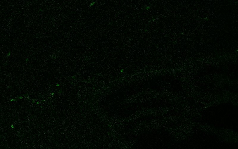
Luqing (Verified Customer) (11-21-2024) | looks great using free-floating brain slices
 |
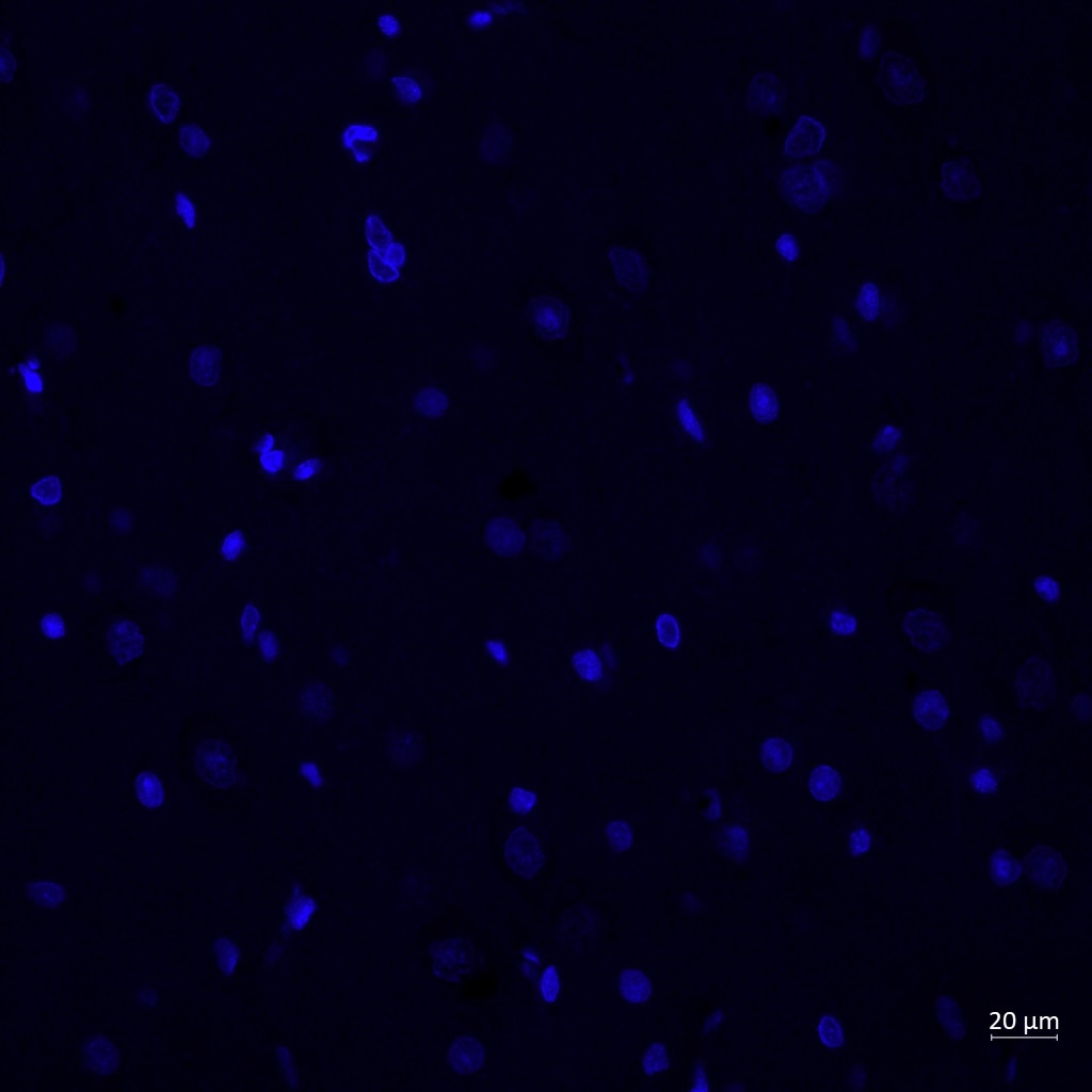
Reyes (Verified Customer) (03-01-2024) | Olig2 (in red) marked the nuclei of tiny cells (oligodendrocytes) around the neurons (in green)
 |
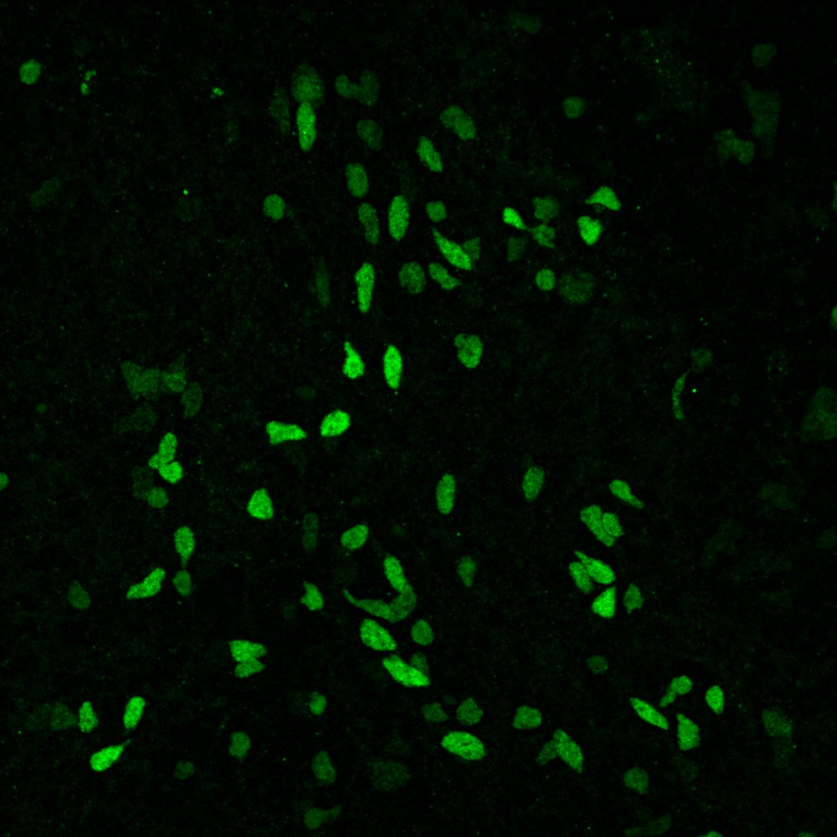
Clarisse (Verified Customer) (08-29-2022) | This antibody works very well (clear nuclear signal).
 |
An (Verified Customer) (09-17-2020) | Tried a free aliquot of this antibody to perform IHC on retinal cryosections of zebrafish. Did not work, although it was to be expected as the sequence homology was not great.
|
Alexander (Verified Customer) (08-05-2019) | This is a great product that has a clear nuclear stain at a 1:200 dilution and 1 hour primary incubation at room temp and 1 hour secondary incubation at room temp.
|











