
Recombinant Human RON/MST1R protein (rFc Tag)
Species
Human
Purity
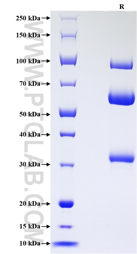
>90 %, SDS-PAGE
Tag
rFc Tag
Activity
not tested
Cat no : Eg3493
Validation Data Gallery
Product Information
| Purity | >90 %, SDS-PAGE |
| Endotoxin | <0.1 EU/μg protein, LAL method |
| Activity |
Not tested |
| Expression | CHO-derived Human RON protein Glu25-Leu571 (Accession# Q04912-1) with a rabbit IgG Fc tag at the C-terminus. |
| GeneID | 4486 |
| Accession | Q04912-1 |
| PredictedSize | 84.9 kDa |
| SDS-PAGE | 30-33 kDa, 55-65 kDa and 90-100 kDa, reducing (R) conditions |
| Formulation | Lyophilized from 0.22 μm filtered solution in PBS, pH 7.4. Normally 5% trehalose and 5% mannitol are added as protectants before lyophilization. |
| Reconstitution | Briefly centrifuge the tube before opening. Reconstitute at 0.1-0.5 mg/mL in sterile water. |
| Storage Conditions |
It is recommended that the protein be aliquoted for optimal storage. Avoid repeated freeze-thaw cycles.
|
| Shipping | The product is shipped at ambient temperature. Upon receipt, store it immediately at the recommended temperature. |
Background
RON, also known as CD136 or Macrophage-Stimulating Protein Receptor (MST1R), is a type I transmembrane protein belonging to the group B of the scavenger receptor cysteine-rich (SRCR) superfamily. It is primarily expressed on cells of epithelial origin, as well as on specific immune cells such as macrophages. Its activation occurs upon binding to its ligand, Macrophage-Stimulating Protein (MSP), which triggers intracellular signaling cascades. These pathways are critically involved in regulating key cellular processes including proliferation, migration, survival, and the inhibition of apoptosis. Under normal physiological conditions, CD136 signaling contributes to tissue repair, inflammation resolution, and maintaining epithelial homeostasis. However, dysregulated CD136 expression and signaling are strongly implicated in oncogenesis, where it promotes tumor growth, invasive metastasis, and confers resistance to conventional therapies in various carcinomas.
References:
1. Kowal, Krzysztof et al. Folia histochemica et cytobiologica vol. 49,3 (2011): 365-74.
2. Kang, Chang Moo et al. Pancreas vol. 43,2 (2014): 183-9.
3. Huang, Lingtong et al. Frontiers in immunology vol. 11(2020): 569082.