Tested Applications
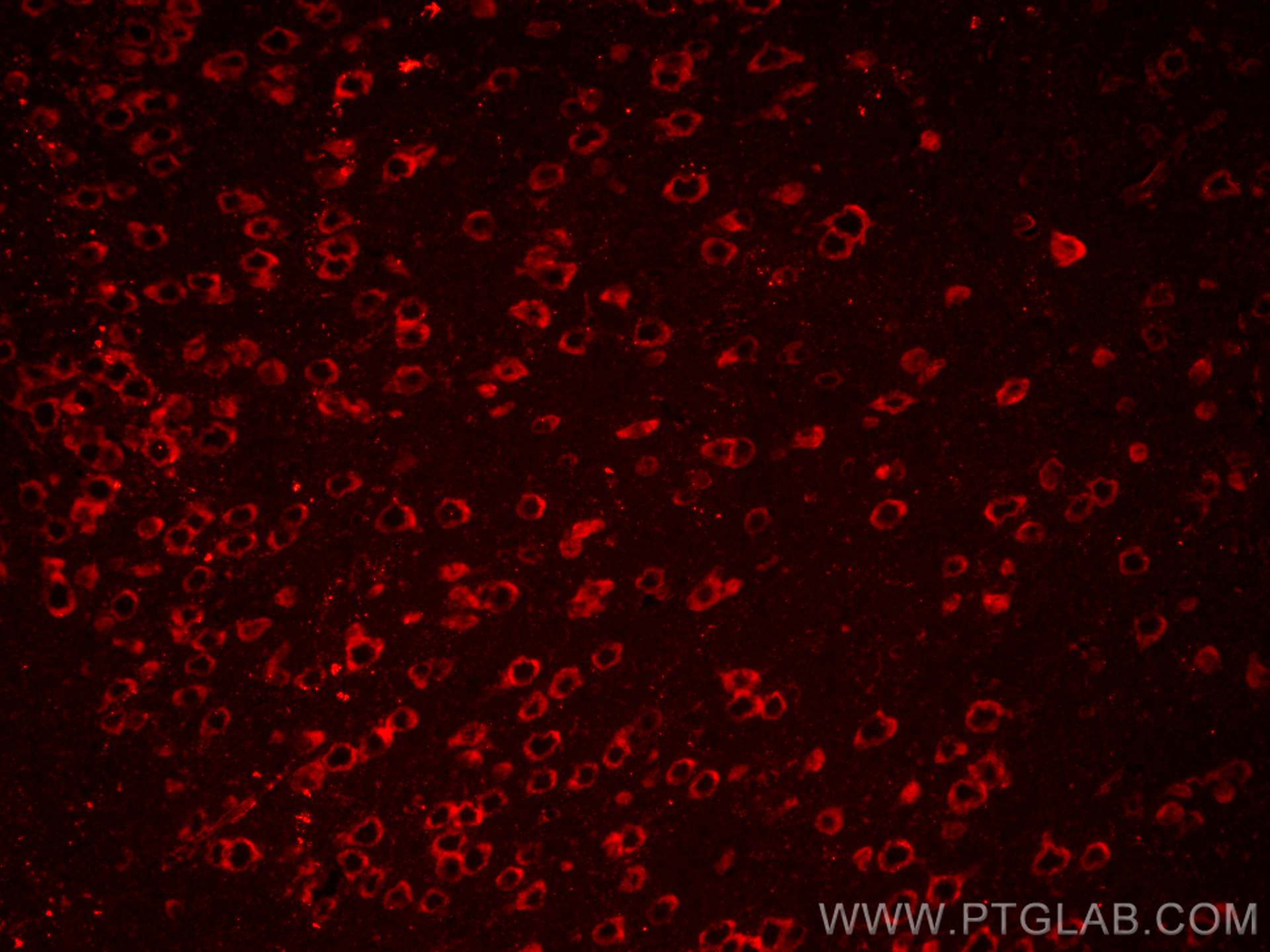
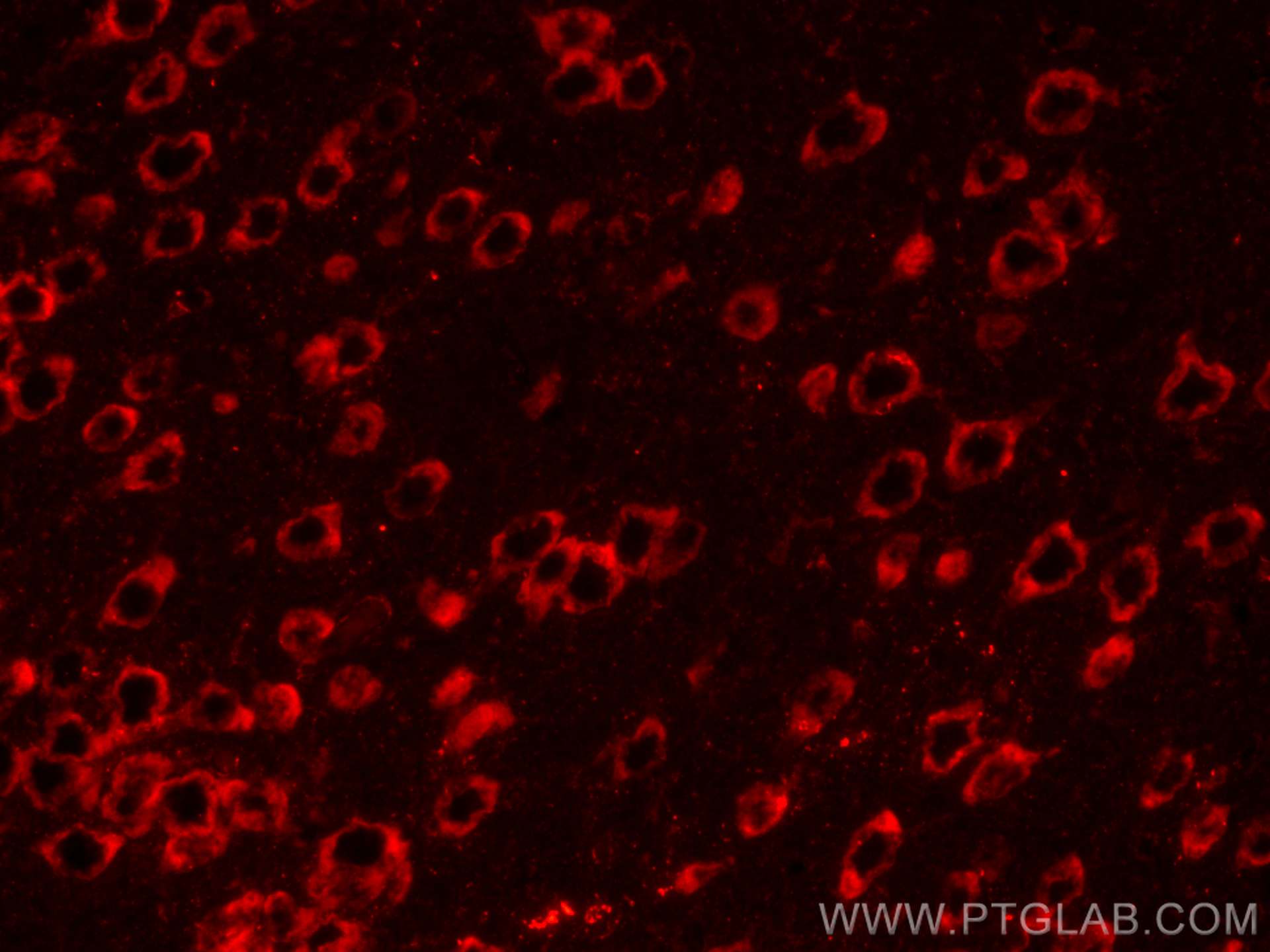
| Positive IF-P detected in | mouse brain tissue |
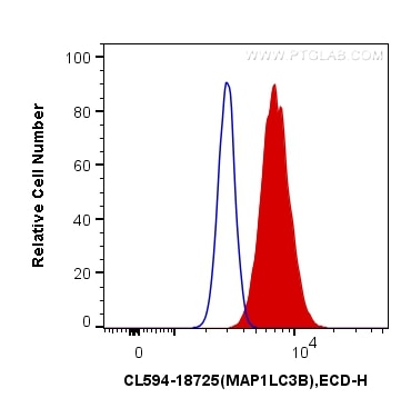
| Positive FC (Intra) detected in | HeLa cells |
Recommended dilution
| Application | Dilution |
|---|---|
| Immunofluorescence (IF)-P | IF-P : 1:50-1:500 |
| Flow Cytometry (FC) (INTRA) | FC (INTRA) : 0.20 ug per 10^6 cells in a 100 µl suspension |
| It is recommended that this reagent should be titrated in each testing system to obtain optimal results. | |
| Sample-dependent, Check data in validation data gallery. | |
Product Information
CL594-18725 targets LC3B-Specific in IF-P, FC (Intra) applications and shows reactivity with human, mouse, rat samples.
| Tested Reactivity | human, mouse, rat |
| Host / Isotype | Rabbit / IgG |
| Class | Polyclonal |
| Type | Antibody |
| Immunogen |
Peptide Predict reactive species |
| Full Name | microtubule-associated protein 1 light chain 3 beta |
| Calculated Molecular Weight | 15 kDa |
| GenBank Accession Number | NM_022818 |
| Gene Symbol | LC3B |
| Gene ID (NCBI) | 81631 |
| ENSEMBL Gene ID | ENSG00000140941 |
| RRID | AB_2919848 |
| Conjugate | CoraLite®594 Fluorescent Dye |
| Excitation/Emission Maxima Wavelengths | 588 nm / 604 nm |
| Excitation Laser | Yellow-Green Laser (561 nm) |
| Form | Liquid |
| Purification Method | Antigen affinity purification |
| UNIPROT ID | Q9GZQ8 |
| Storage Buffer | PBS with 50% glycerol, 0.05% Proclin300, 0.5% BSA, pH 7.3. |
| Storage Conditions | Store at -20°C. Avoid exposure to light. Stable for one year after shipment. Aliquoting is unnecessary for -20oC storage. |
Background Information
LC3B, also named as MAP1LC3B, MAP1A/1BLC3, belongs to the MAP1 LC3 family. It is a subunit of neuronal microtubule-associated MAP1A and MAP1B proteins, which are involved in microtubule assembly and important for neurogenesis. In cell biology, autophagy, or autophagocytosis, is a catabolic process involving the degradation of a cell's own components through the lysosomalmachinery. It is a major mechanism by which a starving cell reallocates nutrients from unnecessary processes to more-essential processes. Two forms of LC3, called LC3-I (17-19kd) and -II(14-16kd), were produced post-translationally in various cells. LC3-I is cytosolic, whereas LC3-II is membrane bound. The precursor molecule is cleaved by APG4B/ATG4B to form the cytosolic form, LC3-I. This is activated by APG7L/ATG7, transferred to ATG3 and conjugated to phospholipid to form the membrane-bound form, LC3-II. The amount of LC3-II is correlated with the extent of autophagosome formation. LC3-II is the first mammalian protein identified that specifically associates with autophagosome membranes. MAP1LC3 has 3 isoforms MAP1LC3A, MAP1LC3B and MAP1LC3C. MAP1LC3A and MAP1LC3C are produced by the proteolytic cleavage after the conserved C-terminal Gly residue, like their rat counterpart, MAP1LC3B does not undergo C-terminal cleavage and exists in a single modified form. This antibody is specific to LC3B.
Protocols
| Product Specific Protocols | |
|---|---|
| FC protocol for CL594 LC3B-Specific antibody CL594-18725 | Download protocol |
| IF protocol for CL594 LC3B-Specific antibody CL594-18725 | Download protocol |
| Standard Protocols | |
|---|---|
| Click here to view our Standard Protocols |