
Tested Applications
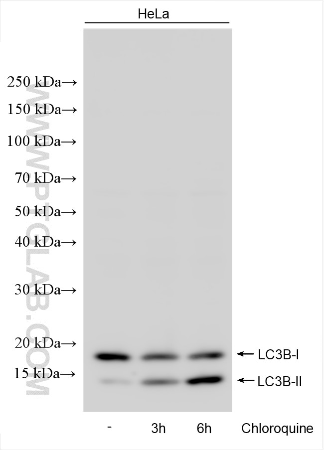
| Positive WB detected in | HeLa cells, Chloroquine treated HeLa cells |
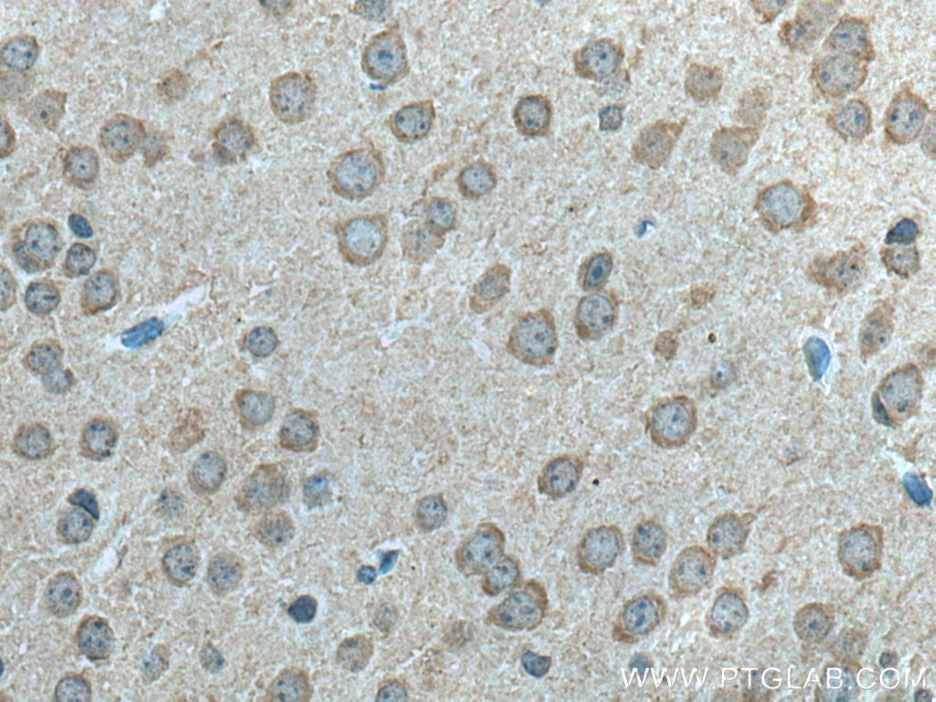
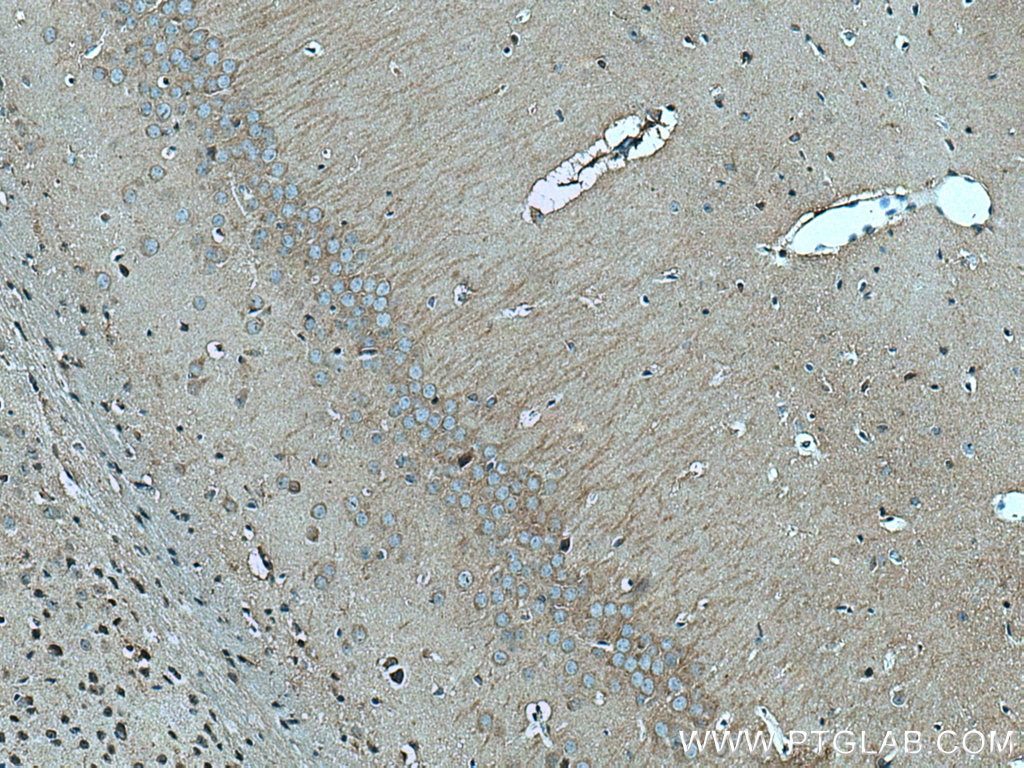
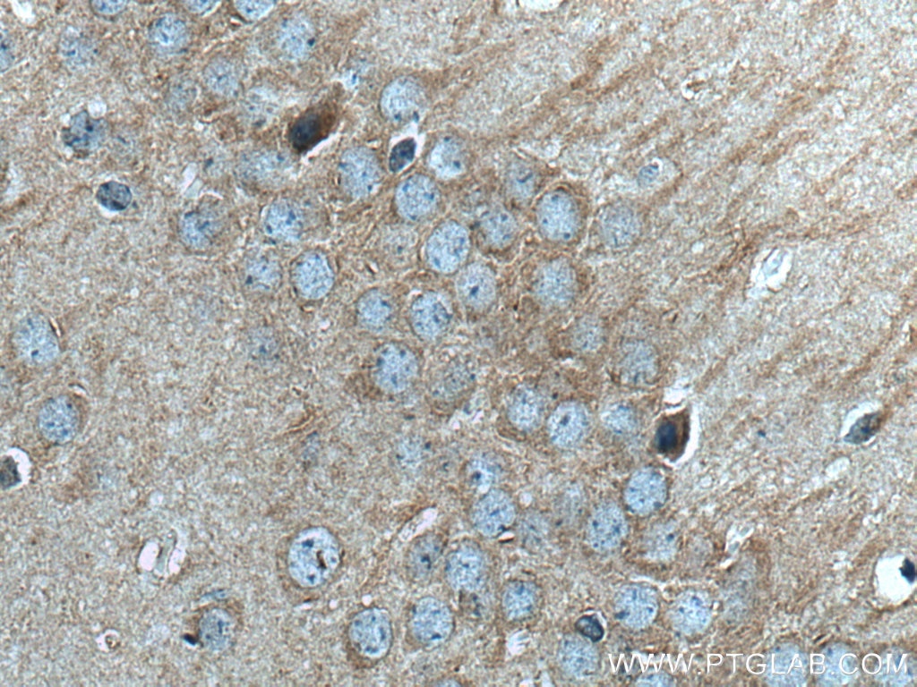
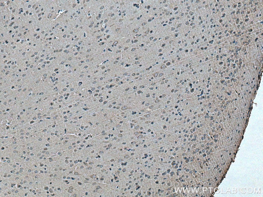
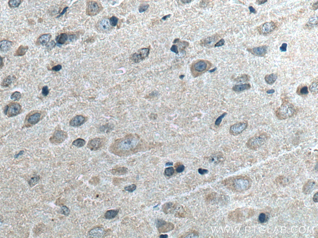
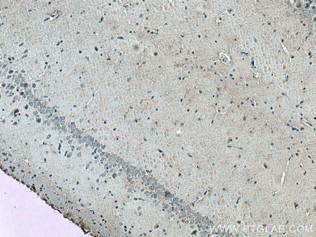
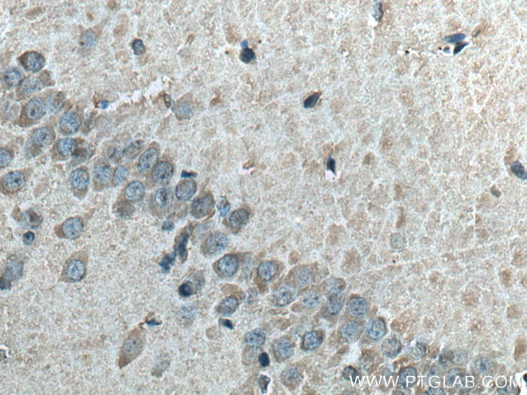
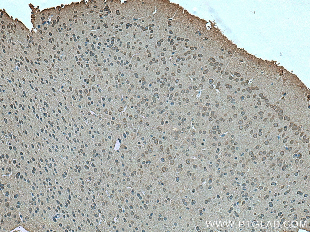
| Positive IHC detected in | mouse brain tissue, rat brain tissue Note: suggested antigen retrieval with TE buffer pH 9.0; (*) Alternatively, antigen retrieval may be performed with citrate buffer pH 6.0 |
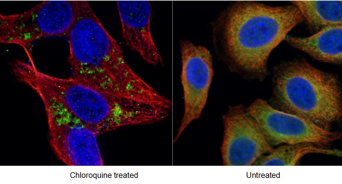
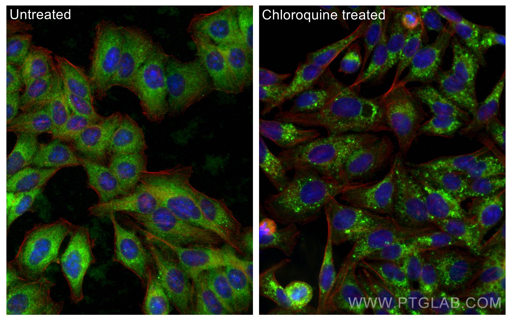
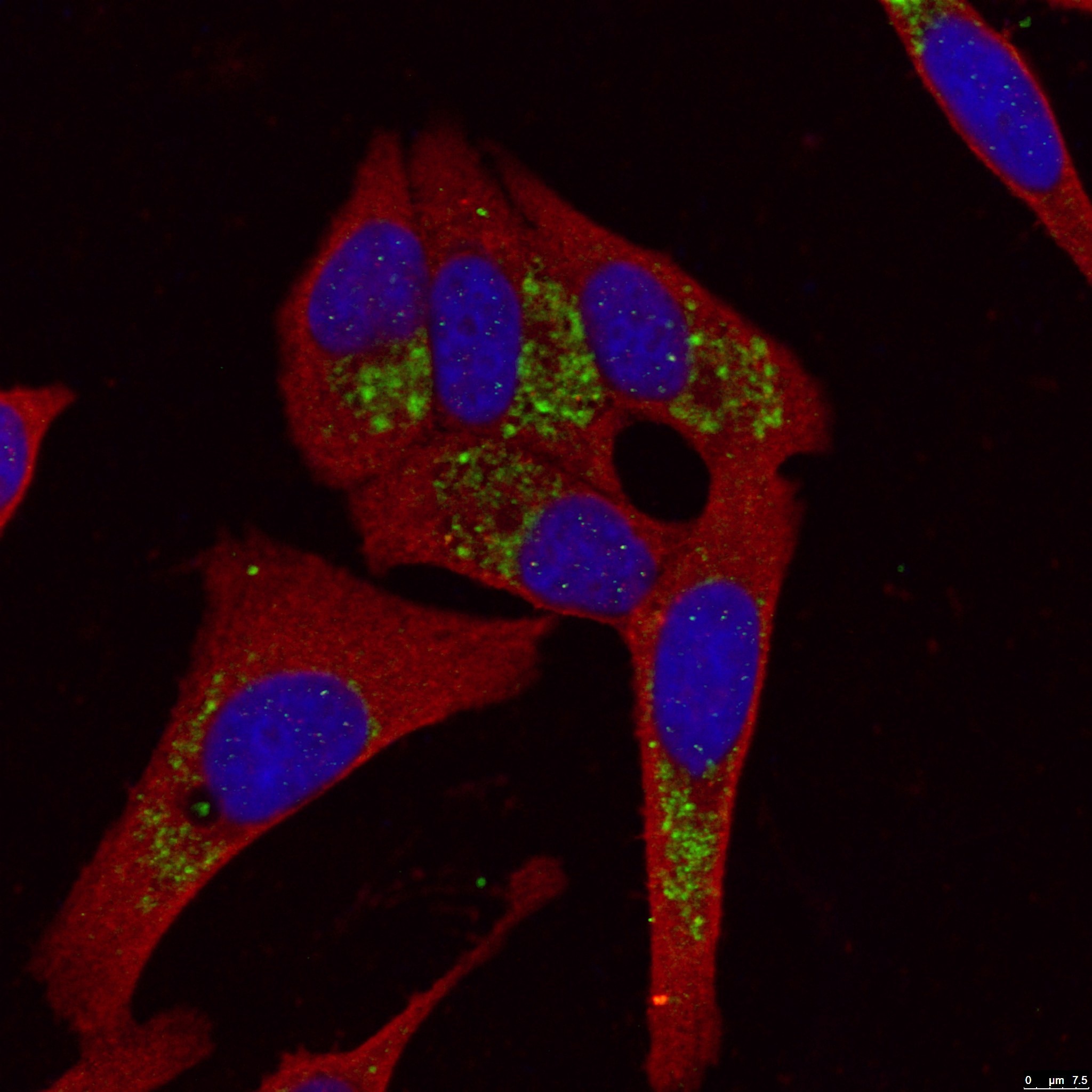
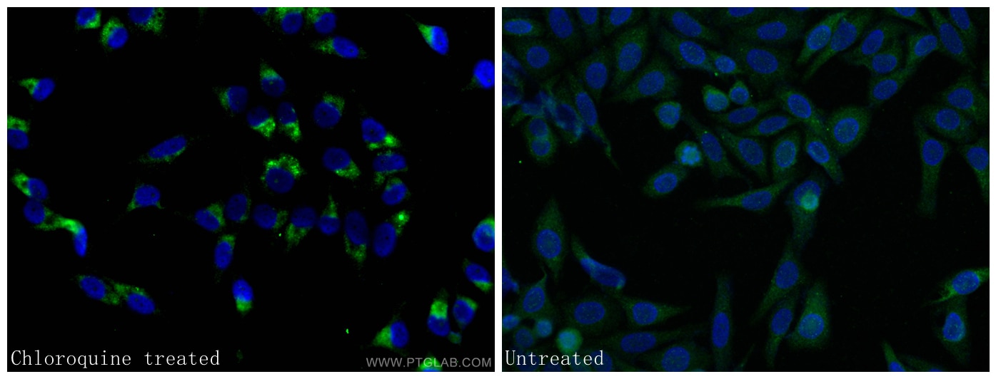
| Positive IF/ICC detected in | Chloroquine treated HepG2 cells, HepG2 cells |
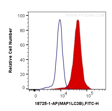
| Positive FC (Intra) detected in | HeLa cells |
Recommended dilution
| Application | Dilution |
|---|---|
| Western Blot (WB) | WB : 1:500-1:3000 |
| Immunohistochemistry (IHC) | IHC : 1:50-1:500 |
| Immunofluorescence (IF)/ICC | IF/ICC : 1:50-1:500 |
| Flow Cytometry (FC) (INTRA) | FC (INTRA) : 0.40 ug per 10^6 cells in a 100 µl suspension |
| It is recommended that this reagent should be titrated in each testing system to obtain optimal results. | |
| Sample-dependent, Check data in validation data gallery. | |
Product Information
18725-1-AP targets LC3B-Specific in WB, IHC, IF/ICC, FC (Intra), IP, CoIP, ELISA applications and shows reactivity with human, mouse, rat samples.
| Tested Reactivity | human, mouse, rat |
| Cited Reactivity | human, mouse, rat, pig, chicken, zebrafish, bovine |
| Host / Isotype | Rabbit / IgG |
| Class | Polyclonal |
| Type | Antibody |
| Immunogen |
Peptide Predict reactive species |
| Full Name | microtubule-associated protein 1 light chain 3 beta |
| Calculated Molecular Weight | 15 kDa |
| Observed Molecular Weight | 15 kDa, 18 kDa |
| GenBank Accession Number | NM_022818 |
| Gene Symbol | LC3B |
| Gene ID (NCBI) | 81631 |
| ENSEMBL Gene ID | ENSG00000140941 |
| RRID | AB_2137745 |
| Conjugate | Unconjugated |
| Form | Liquid |
| Purification Method | Antigen affinity purification |
| UNIPROT ID | Q9GZQ8 |
| Storage Buffer | PBS with 0.02% sodium azide and 50% glycerol, pH 7.3. |
| Storage Conditions | Store at -20°C. Stable for one year after shipment. Aliquoting is unnecessary for -20oC storage. 20ul sizes contain 0.1% BSA. |
Background Information
LC3B, also named as MAP1LC3B, MAP1A/1BLC3, belongs to the MAP1 LC3 family. It is a subunit of neuronal microtubule-associated MAP1A and MAP1B proteins, which are involved in microtubule assembly and important for neurogenesis. In cell biology, autophagy, or autophagocytosis, is a catabolic process involving the degradation of a cell's own components through the lysosomalmachinery. It is a major mechanism by which a starving cell reallocates nutrients from unnecessary processes to more-essential processes. Two forms of LC3, called LC3-I (17-19kd) and -II(14-16kd), were produced post-translationally in various cells. LC3-I is cytosolic, whereas LC3-II is membrane bound. The precursor molecule is cleaved by APG4B/ATG4B to form the cytosolic form, LC3-I. This is activated by APG7L/ATG7, transferred to ATG3 and conjugated to phospholipid to form the membrane-bound form, LC3-II. The amount of LC3-II is correlated with the extent of autophagosome formation. LC3-II is the first mammalian protein identified that specifically associates with autophagosome membranes. MAP1LC3 has 3 isoforms MAP1LC3A, MAP1LC3B and MAP1LC3C. MAP1LC3A and MAP1LC3C are produced by the proteolytic cleavage after the conserved C-terminal Gly residue, like their rat counterpart, MAP1LC3B does not undergo C-terminal cleavage and exists in a single modified form. This antibody is specific to LC3B.
Protocols
| Product Specific Protocols | |
|---|---|
| FC protocol for LC3B-Specific antibody 18725-1-AP | Download protocol |
| IF protocol for LC3B-Specific antibody 18725-1-AP | Download protocol |
| IHC protocol for LC3B-Specific antibody 18725-1-AP | Download protocol |
| WB protocol for LC3B-Specific antibody 18725-1-AP | Download protocol |
| Standard Protocols | |
|---|---|
| Click here to view our Standard Protocols |
Publications
What published studies show
| Species | Application | Title |
|---|---|---|
Cell Res Suppression of MAPK11 or HIPK3 reduces mutant Huntingtin levels in Huntington's disease models. | ||
Am J Respir Crit Care Med Acid Sphingomyelinase Inhibition Attenuates Cell Death in Mechanically-Ventilated Newborn Rat Lung. | ||
Nat Microbiol Chloroquine ameliorates carbon tetrachloride-induced acute liver injury in mice via the concomitant inhibition of inflammation and induction of apoptosis | ||
ACS Nano Nanoenabled Disruption of Multiple Barriers in Antigen Cross-Presentation of Dendritic Cells via Calcium Interference for Enhanced Chemo-Immunotherapy. | ||
Autophagy TRIM59 regulates autophagy through modulating both the transcription and the ubiquitination of BECN1. | ||
Autophagy STYK1 promotes autophagy through enhancing the assembly of autophagy-specific class III phosphatidylinositol 3-kinase complex I. |
Reviews
What customers say
The reviews below have been submitted by verified Proteintech customers who received an incentive for providing their feedback.
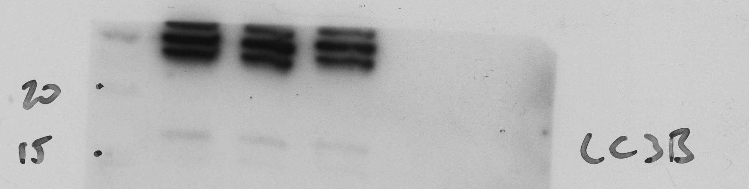
Jacob (Verified Customer) (07-23-2026) | Has a very strong non-specific banding pattern above 20kDa, so the LC3B was faint. 2*
 |
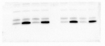
Alicia (Verified Customer) (09-30-2025) | Control microglia derived from iPSC with and without bafilomycin. Drug treated microglia derived from iPSC with and without bafilomycin.
 |
Allegra (Verified Customer) (09-30-2025) | Work for cell lysates only in concentration 1:500 and this is only slightly detectable. In extracellular vesicles nearly no signal.
|
Azita (Verified Customer) (06-02-2021) | Western blot analysis using LC3B-Specific Polyclonal antibody in NSC34 cell line at dilution of 1:300. Immunohistochemistry labelling of primary rat cortical neurons using LC3B-Specific Polyclonal antibody at dilution of 1:500 showed strong labelling.
|
Tanusree (Verified Customer) (12-03-2019) | The antibody works great in Western blotting analysis.
|















