Tested Applications
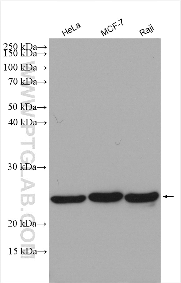
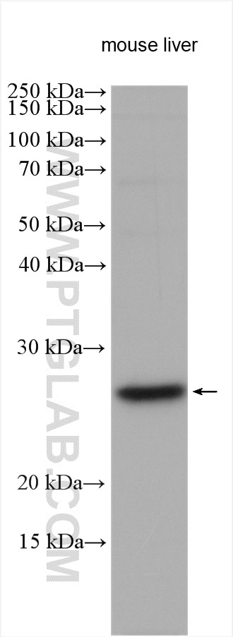
| Positive WB detected in | HeLa cells, mouse liver tissue, MCF-7 cells, Raji cells |
| Positive IP detected in | mouse brain tissue |
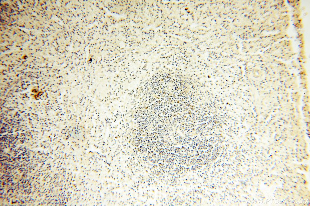
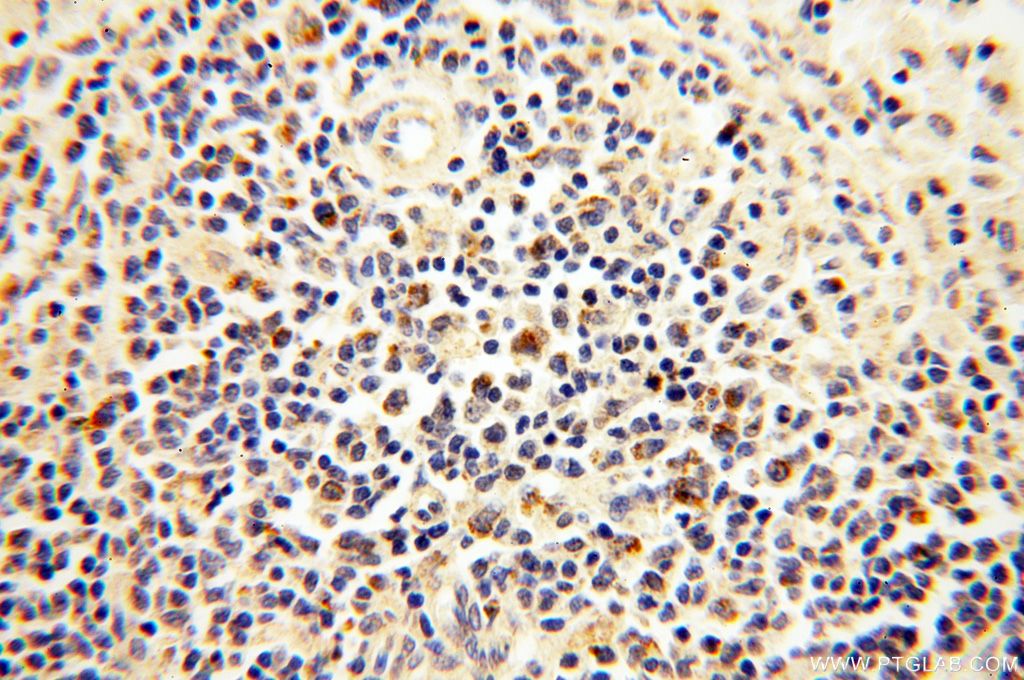
| Positive IHC detected in | human spleen tissue Note: suggested antigen retrieval with TE buffer pH 9.0; (*) Alternatively, antigen retrieval may be performed with citrate buffer pH 6.0 |
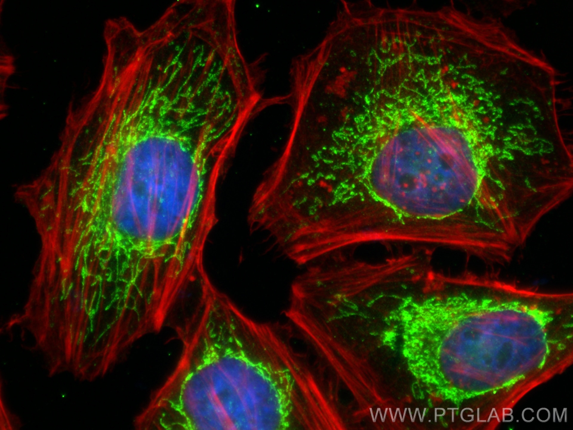
| Positive IF/ICC detected in | HeLa cells |
Recommended dilution
| Application | Dilution |
|---|---|
| Western Blot (WB) | WB : 1:500-1:2000 |
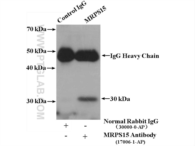
| Immunoprecipitation (IP) | IP : 0.5-4.0 ug for 1.0-3.0 mg of total protein lysate |
| Immunohistochemistry (IHC) | IHC : 1:20-1:200 |
| Immunofluorescence (IF)/ICC | IF/ICC : 1:200-1:800 |
| It is recommended that this reagent should be titrated in each testing system to obtain optimal results. | |
| Sample-dependent, Check data in validation data gallery. | |
Published Applications
| WB | See 13 publications below |
Product Information
17006-1-AP targets MRPS15 in WB, IHC, IF/ICC, IP, ELISA applications and shows reactivity with human, mouse, rat samples.
| Tested Reactivity | human, mouse, rat |
| Cited Reactivity | human, mouse |
| Host / Isotype | Rabbit / IgG |
| Class | Polyclonal |
| Type | Antibody |
| Immunogen |
CatNo: Ag10702 Product name: Recombinant human MRPS15 protein Source: e coli.-derived, PGEX-4T Tag: GST Domain: 1-257 aa of BC031336 Sequence: MLRVAWRTLSLIRTRAVTQVLVPGLPGGGSAKFPFNQWGLQPRSLLLQAARGYVVRKPAQSRLDDDPPPSTLLKDYQNVPGIEKVDDVVKRLLSLEMANKKEMLKIKQEQFMKKIVANPEDTRSLEARIIALSVKIRSYEEHLEKHRKDKAHKRYLLMSIDQRKKMLKNLRNTNYDVFEKICWGLGIEYTFPPLYYRRAHRRFVTKKALCIRVFQETQKLKKRRRALKAAAAAQKQAKRRNPDSPAKAIPKTLKDSQ Predict reactive species |
| Full Name | mitochondrial ribosomal protein S15 |
| Calculated Molecular Weight | 257 aa, 30 kDa |
| Observed Molecular Weight | 25-30 kDa |
| GenBank Accession Number | BC031336 |
| Gene Symbol | MRPS15 |
| Gene ID (NCBI) | 64960 |
| RRID | AB_2301068 |
| Conjugate | Unconjugated |
| Form | Liquid |
| Purification Method | Antigen affinity purification |
| UNIPROT ID | P82914 |
| Storage Buffer | PBS with 0.02% sodium azide and 50% glycerol, pH 7.3. |
| Storage Conditions | Store at -20°C. Stable for one year after shipment. Aliquoting is unnecessary for -20oC storage. 20ul sizes contain 0.1% BSA. |
Background Information
MRPS15, also named as RPMS15 or DC37, is a 257 amino acid protein, which belongs to the ribosomal protein S15P family. MRPS15 localizes in the Mitochondrion and is a component of the mitochondrial ribosome small subunit (28S) which comprises a 12S rRNA and about 30 distinct proteins. MRPS15 is involved into the mitochondrial proteins biosynthesis and ribosomal biogenesis.
Protocols
| Product Specific Protocols | |
|---|---|
| IF protocol for MRPS15 antibody 17006-1-AP | Download protocol |
| IHC protocol for MRPS15 antibody 17006-1-AP | Download protocol |
| IP protocol for MRPS15 antibody 17006-1-AP | Download protocol |
| WB protocol for MRPS15 antibody 17006-1-AP | Download protocol |
| Standard Protocols | |
|---|---|
| Click here to view our Standard Protocols |
Publications
| Species | Application | Title |
|---|---|---|
Mol Cell Identification of TMEM126A as OXA1L-interacting protein reveals cotranslational quality control in mitochondria | ||
Cell Metab Initial steps in RNA processing and ribosome assembly occur at mitochondrial DNA nucleoids. | ||
Hum Mol Genet Mutations in the MRPS28 gene encoding the small mitoribosomal subunit protein bS1m in a patient with intrauterine growth retardation, craniofacial dysmorphism and multisystemic involvement. |