Tested Applications
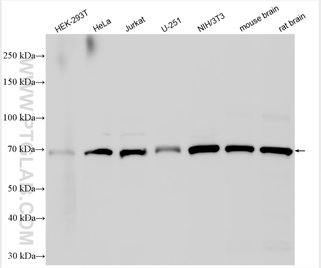
| Positive WB detected in | HEK-293T cells, HeLa cells, Jurkat cells, U-251 cells, NIH/3T3 cells, mouse brain tissue, rat brain tissue |
| Positive IP detected in | Jurkat cells |
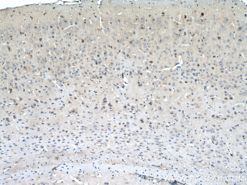
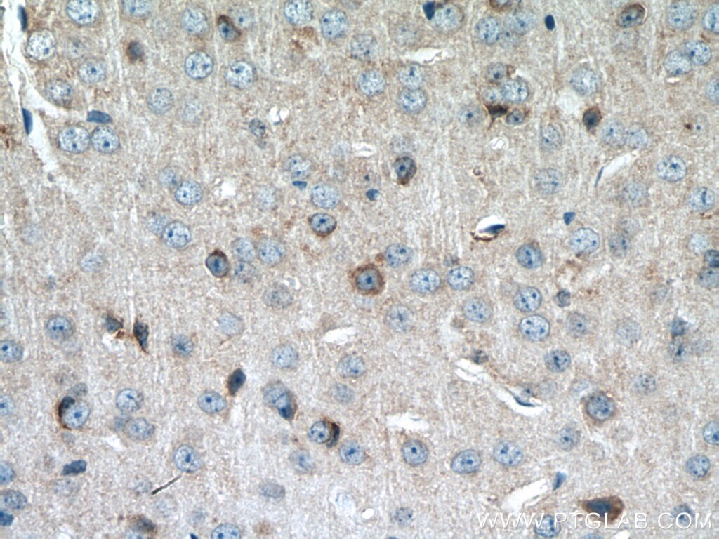
| Positive IHC detected in | mouse brain tissue Note: suggested antigen retrieval with TE buffer pH 9.0; (*) Alternatively, antigen retrieval may be performed with citrate buffer pH 6.0 |
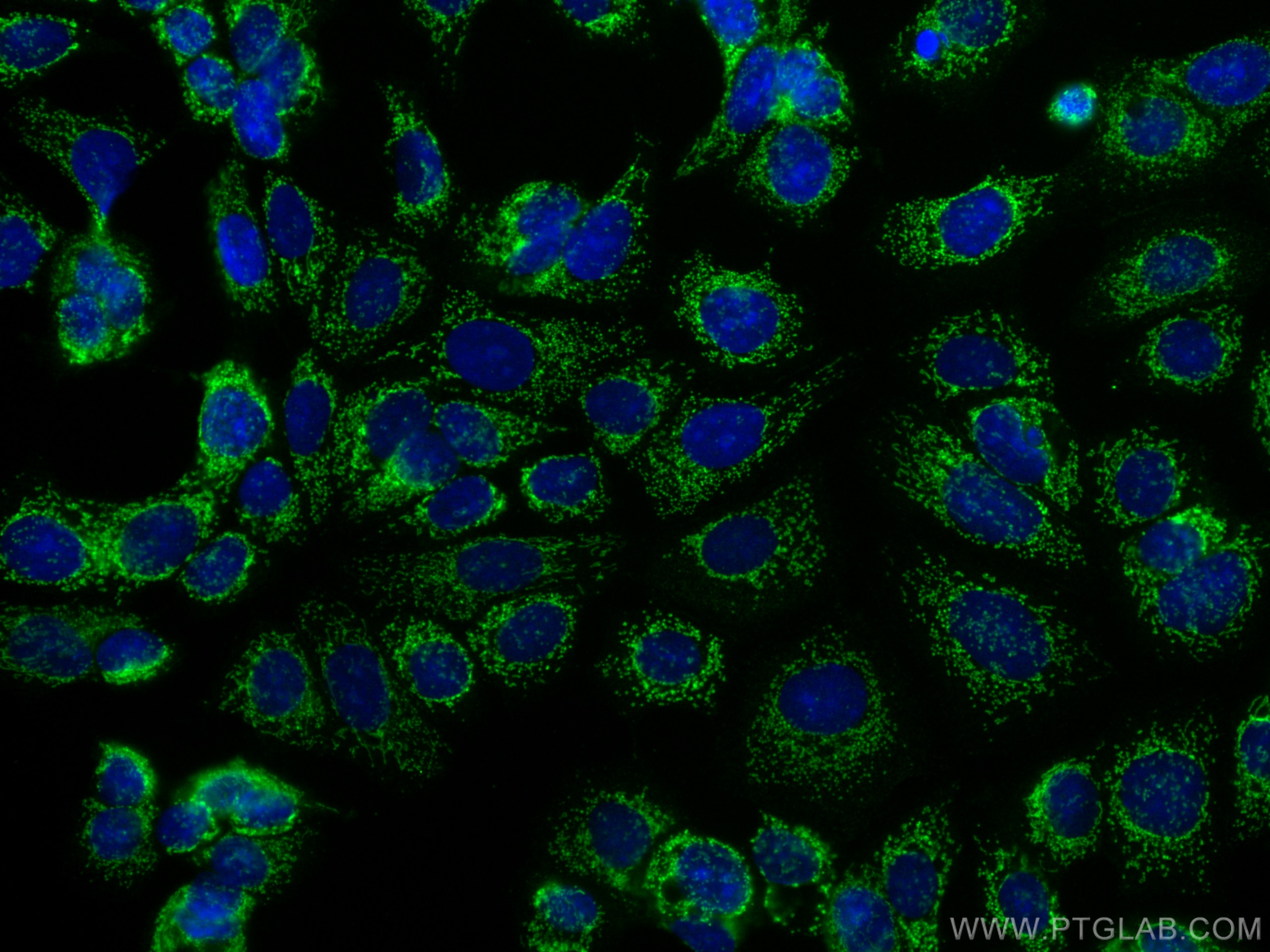
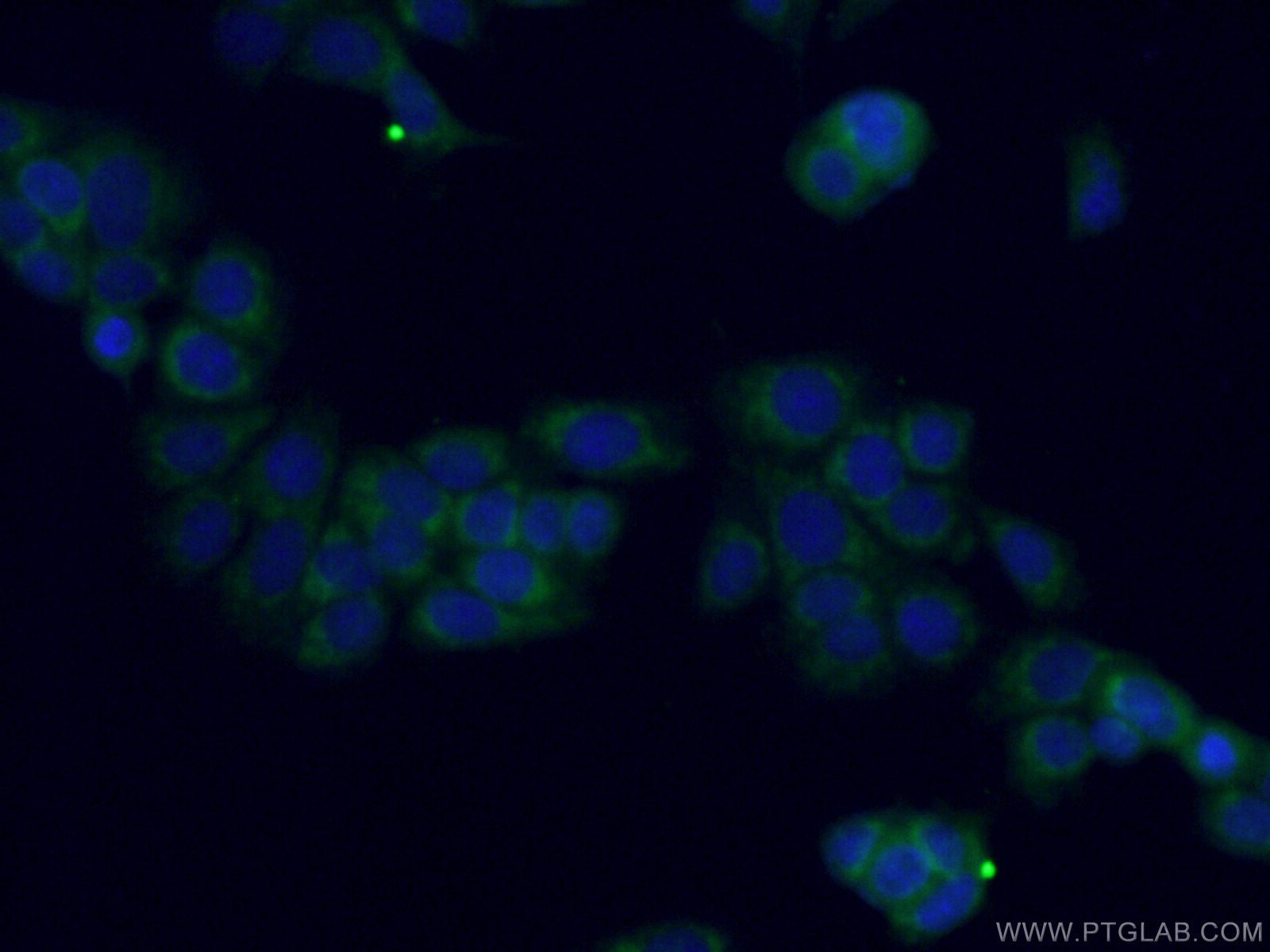
| Positive IF/ICC detected in | MCF-7 cells |
Recommended dilution
| Application | Dilution |
|---|---|
| Western Blot (WB) | WB : 1:2000-1:10000 |
| Immunoprecipitation (IP) | IP : 0.5-4.0 ug for 1.0-3.0 mg of total protein lysate |
| Immunohistochemistry (IHC) | IHC : 1:50-1:500 |
| Immunofluorescence (IF)/ICC | IF/ICC : 1:50-1:500 |
| It is recommended that this reagent should be titrated in each testing system to obtain optimal results. | |
| Sample-dependent, Check data in validation data gallery. | |
Published Applications
| WB | See 2 publications below |
Product Information
27832-1-AP targets MTSS1L in WB, IHC, IF/ICC, IP, ELISA applications and shows reactivity with human, mouse, rat samples.
| Tested Reactivity | human, mouse, rat |
| Cited Reactivity | human |
| Host / Isotype | Rabbit / IgG |
| Class | Polyclonal |
| Type | Antibody |
| Immunogen |
CatNo: Ag27216 Product name: Recombinant human MTSS1L protein Source: e coli.-derived, PGEX-4T Tag: GST Domain: 196-246 aa of BC002770 Sequence: TFITFLQPVVNGELTMLGEITHLQGIIDDLVVLTAEPHKLPPASEQVIKDL Predict reactive species |
| Full Name | metastasis suppressor 1-like |
| Observed Molecular Weight | 68-70 kDa |
| GenBank Accession Number | BC002770 |
| Gene Symbol | MTSS1L |
| Gene ID (NCBI) | 92154 |
| RRID | AB_2880986 |
| Conjugate | Unconjugated |
| Form | Liquid |
| Purification Method | Antigen affinity purification |
| UNIPROT ID | Q765P7 |
| Storage Buffer | PBS with 0.02% sodium azide and 50% glycerol, pH 7.3. |
| Storage Conditions | Store at -20°C. Stable for one year after shipment. Aliquoting is unnecessary for -20oC storage. 20ul sizes contain 0.1% BSA. |
Background Information
MTSS2, also known as MTSS1L, contains a conserved N-terminal domain, called an IRSP53 /MIM homology domain (IMD) or inverse BAR domain, that is involved in plasma membrane dynamics (PMID: 20875796). MTSS2 potentiated PDGF-mediated formation of membrane ruffles and lamellipodia in fibroblasts, acting via RAC1 activation (PMID:14752106). MTSS2 is implicated in intellectual developmental disorders with ocular anomalies and distinctive facial features(PMID: 36067766).
Protocols
| Product Specific Protocols | |
|---|---|
| IF protocol for MTSS1L antibody 27832-1-AP | Download protocol |
| IHC protocol for MTSS1L antibody 27832-1-AP | Download protocol |
| IP protocol for MTSS1L antibody 27832-1-AP | Download protocol |
| WB protocol for MTSS1L antibody 27832-1-AP | Download protocol |
| Standard Protocols | |
|---|---|
| Click here to view our Standard Protocols |
Publications
| Species | Application | Title |
|---|---|---|
Cell Discov m6Am methyltransferase PCIF1 is essential for aggressiveness of gastric cancer cells by inhibiting TM9SF1 mRNA translation. | ||
Acta Biochim Biophys Sin (Shanghai) MicroRNA-23a promotes colorectal cancer cell migration and proliferation by targeting at MARK1. |