Recombinant Mouse RAGE protein (His Tag)
Species
Mouse
Purity
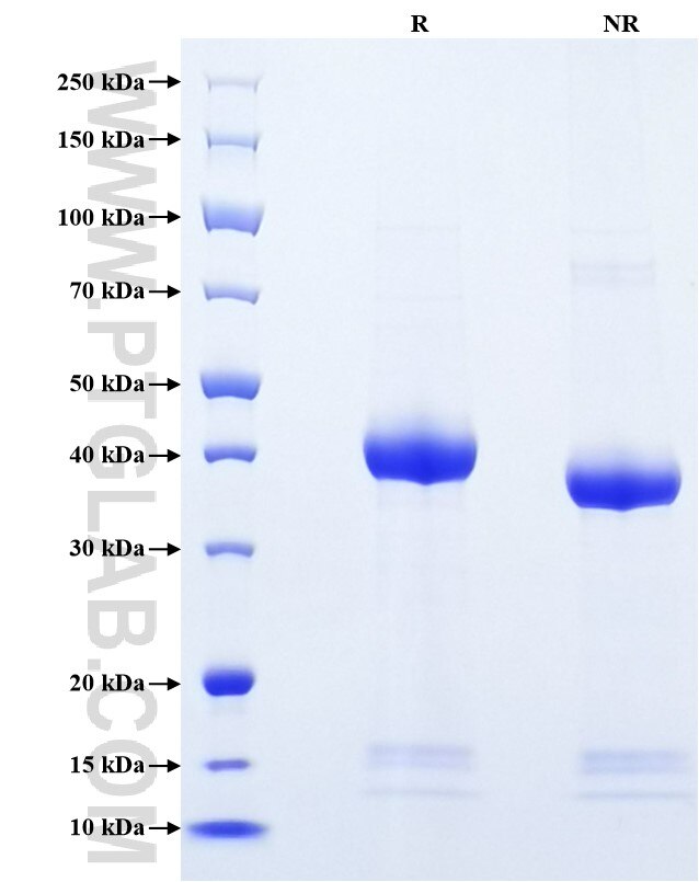
>90 %, SDS-PAGE
Tag
His Tag
Activity
not tested
Cat no : Eg0873
Validation Data Gallery
Product Information
| Purity | >90 %, SDS-PAGE |
| Endotoxin | <0.1 EU/μg protein, LAL method |
| Activity |
Not tested |
| Expression | HEK293-derived Mouse RAGE protein Gly23-Ala340 (Accession# Q62151-1) with a His tag at the C-terminus. |
| GeneID | 11596 |
| Accession | Q62151-1 |
| PredictedSize | 34.8 kDa |
| SDS-PAGE | 37-45 kDa, reducing (R) conditions |
| Formulation | Lyophilized from 0.22 μm filtered solution in PBS, pH 7.4. Normally 5% trehalose and 5% mannitol are added as protectants before lyophilization. |
| Reconstitution | Briefly centrifuge the tube before opening. Reconstitute at 0.1-0.5 mg/mL in sterile water. |
| Storage Conditions |
It is recommended that the protein be aliquoted for optimal storage. Avoid repeated freeze-thaw cycles.
|
| Shipping | The product is shipped at ambient temperature. Upon receipt, store it immediately at the recommended temperature. |
Background
RAGE (receptor for advanced glycation endproducts), also called AGER, is a transmembrane receptor of the immunoglobulin super family. It mediates interactions of advanced glycosylation end products (AGE) which accumulate in vascular tissue in aging in diabetes. Acts as a mediator of vascular inflammation such as atherosclerosis and a complication of diabetes. AGE/RAGE signal regulates production or expression of TNF-alpha, oxidative stress, and endothelial dysfunction in type 2 diabetes. Interaction with S100A12 on endothelium, mononuclear phagocytes, and lymphocytes triggers cellular activation. Interaction with S100B after myocardial infarction regulates myocyte apoptosis by activating ERK1/2 and p53/TP53 signaling. ABPP-initiated RAGE signaling, especially stimulation of P38 mitogen-activated protein kinase (MAPK), delivers ABPP as a complex with RAGE to the intraneuronal. RAGE has higher expression in lung tissues, in particular in alveolar type I cells, and is lost in idiopathic pulmonary fibrosis (IPF).
References:
1. Neeper M. et al. (1992) J Biol Chem. 267(21):14998-5004.
2. Hofmann MA. et al. (1999) Cell. 97(7):889-901.
3. Markus A. et al. (2008) Am J Respir Cell Mol Bio. 39(3):337-45.
4. Xue G. et al. (2008) Am J Physiol Heart Circ Physiol. 295(2):H491-8.
5. Takuma K. et al. (2009) Proc Natl Acad Sci U S A. 106(47):20021-6.
6. Fang F. et al. (2010) FASEB J. 24(4):1043-55.
