Tested Applications
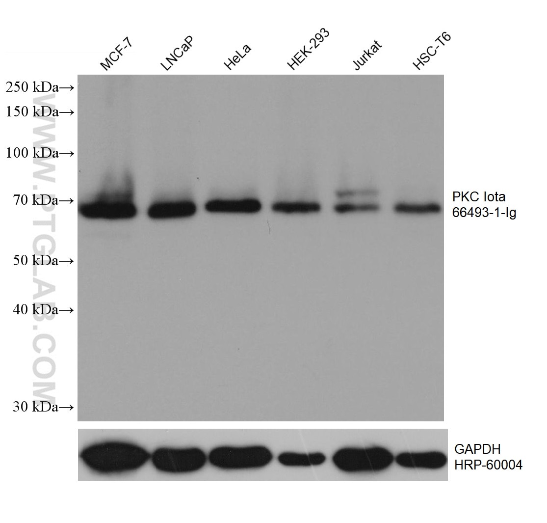
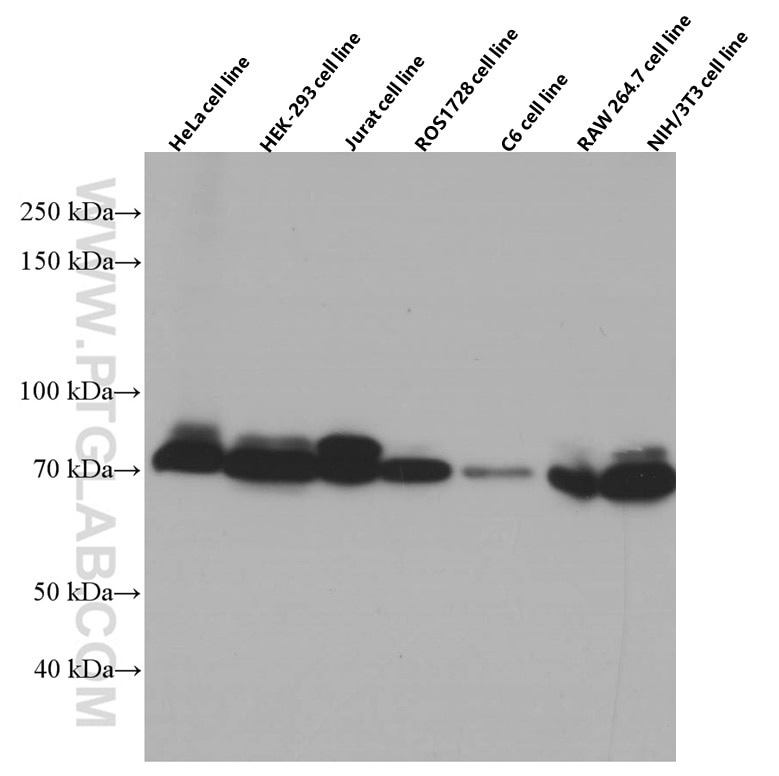
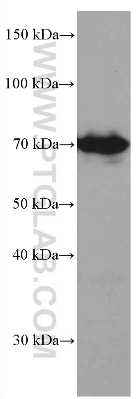
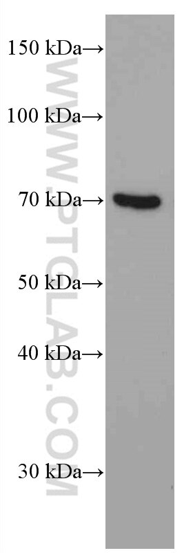
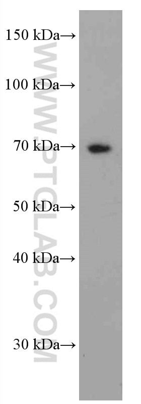
| Positive WB detected in | MCF-7 cells, HeLa cells, HEK-293 cells, ROS1728 cells, C6 cells, LNCaP cells, Jurkat cells, HSC-T6 cells, RAW 264.7 cells, NIH/3T3 cells |
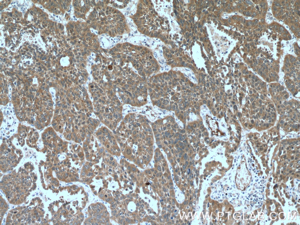
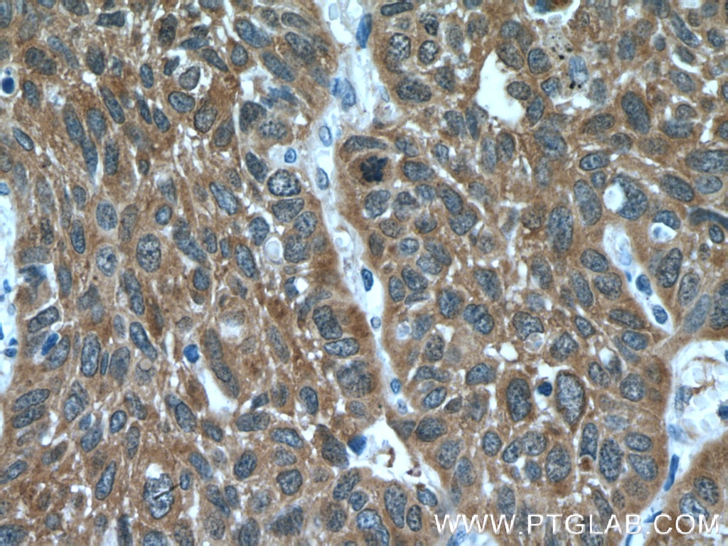
| Positive IHC detected in | human lung cancer tissue Note: suggested antigen retrieval with TE buffer pH 9.0; (*) Alternatively, antigen retrieval may be performed with citrate buffer pH 6.0 |
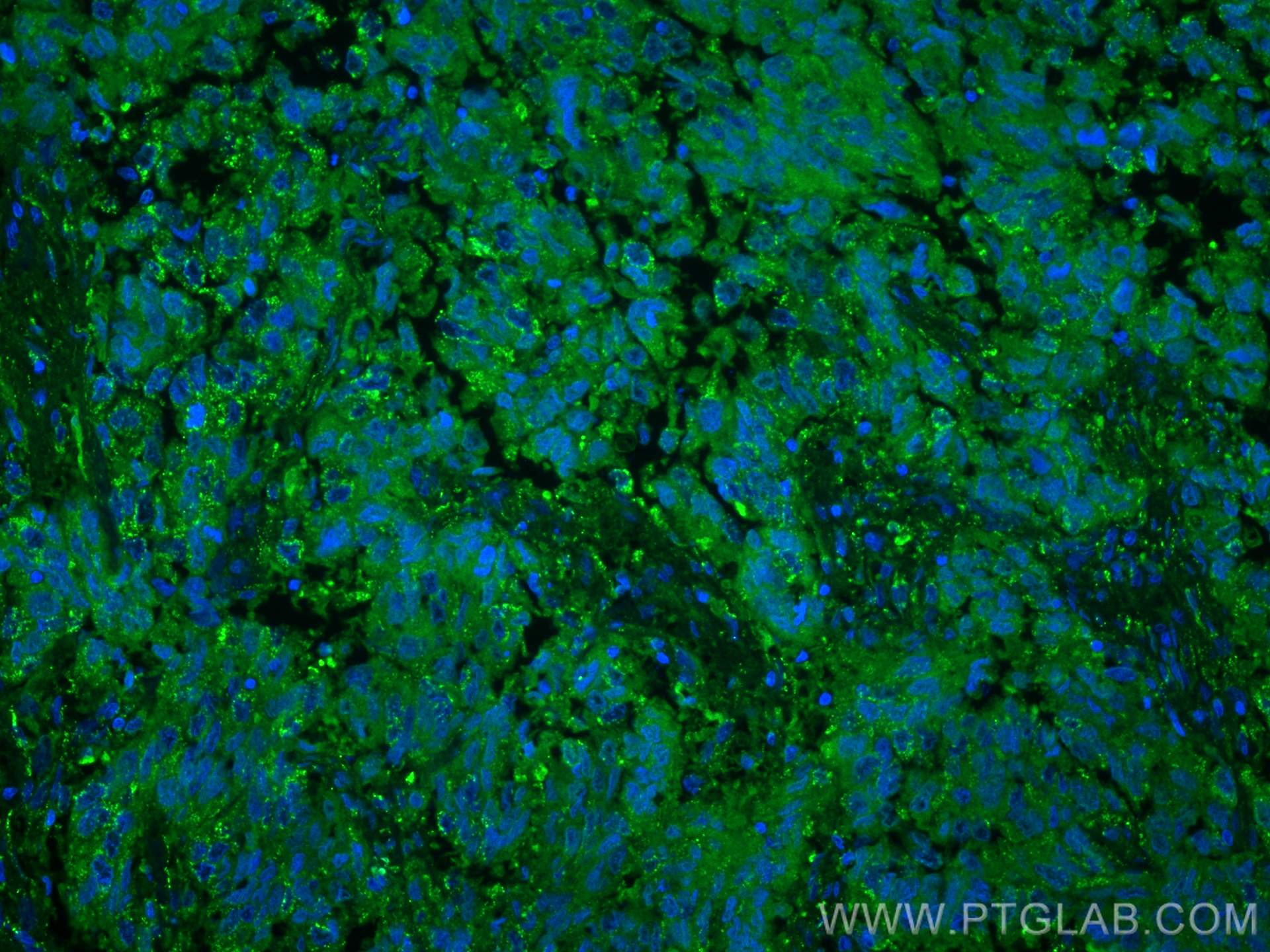
| Positive IF-P detected in | human lung cancer tissue |
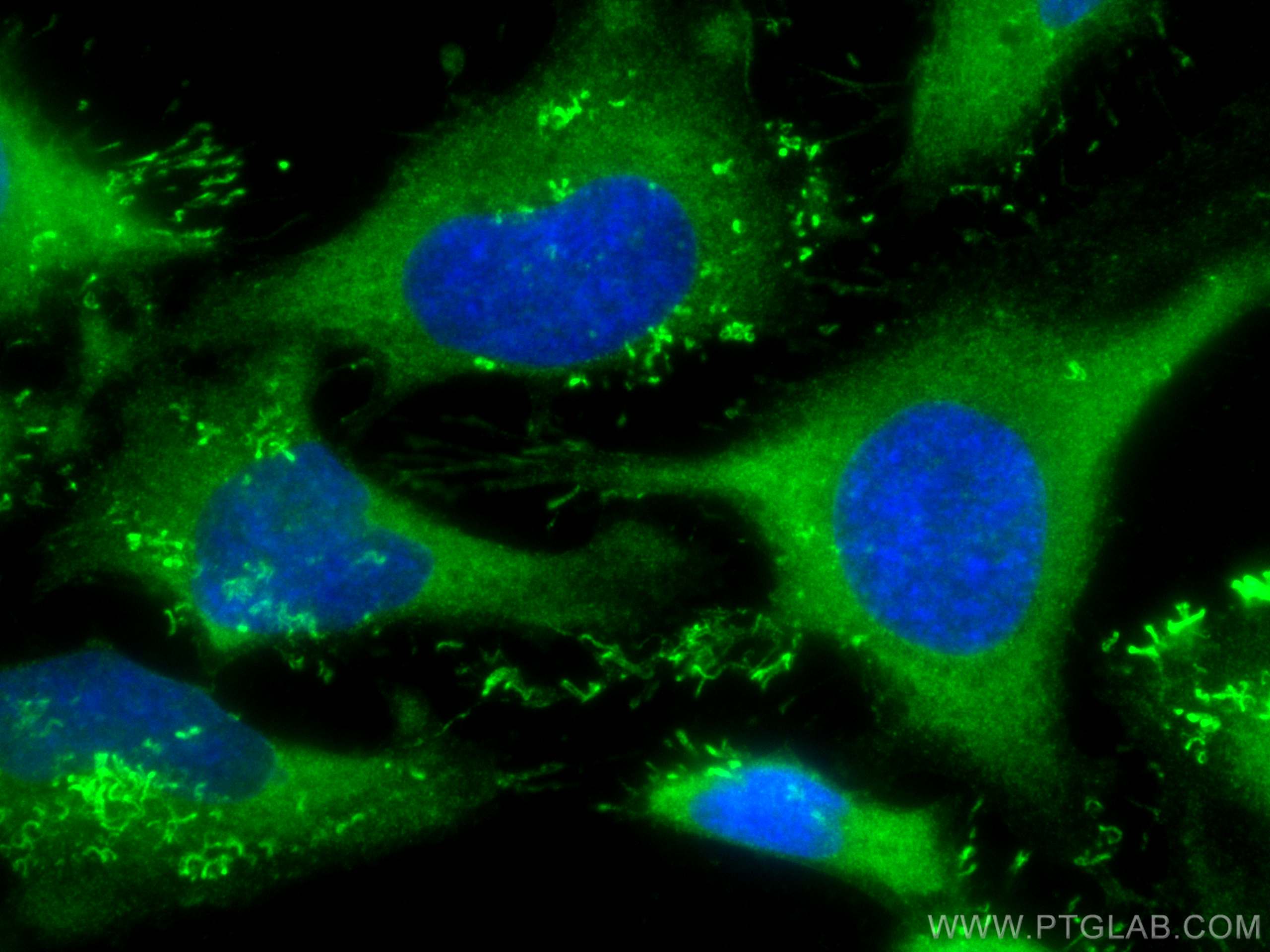
| Positive IF/ICC detected in | HeLa cells |
Recommended dilution
| Application | Dilution |
|---|---|
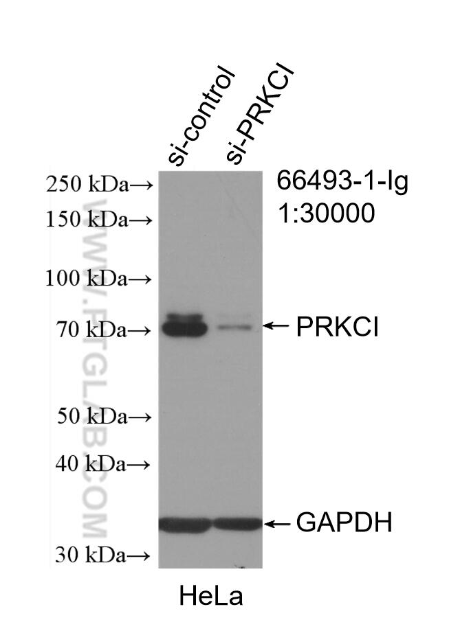
| Western Blot (WB) | WB : 1:5000-1:50000 |
| Immunohistochemistry (IHC) | IHC : 1:150-1:600 |
| Immunofluorescence (IF)-P | IF-P : 1:200-1:800 |
| Immunofluorescence (IF)/ICC | IF/ICC : 1:400-1:1600 |
| It is recommended that this reagent should be titrated in each testing system to obtain optimal results. | |
| Sample-dependent, Check data in validation data gallery. | |
Published Applications
| KD/KO | See 2 publications below |
| WB | See 3 publications below |
| IHC | See 1 publications below |
Product Information
66493-1-Ig targets PKC Iota in WB, IHC, IF/ICC, IF-P, ELISA applications and shows reactivity with human, mouse, rat samples.
| Tested Reactivity | human, mouse, rat |
| Cited Reactivity | human |
| Host / Isotype | Mouse / IgG2a |
| Class | Monoclonal |
| Type | Antibody |
| Immunogen |
CatNo: Ag4990 Product name: Recombinant human PRKCI protein Source: e coli.-derived, PGEX-4T Tag: GST Domain: 238-587 aa of BC022016 Sequence: SSLGLQDFDLLRVIGRGSYAKVLLVRLKKTDRIYAMKVVKKELVNDDEDIDWVQTEKHVFEQASNHPFLVGLHSCFQTESRLFFVIEYVNGGDLMFHMQRQRKLPEEHARFYSAEISLALNYLHERGIIYRDLKLDNVLLDSEGHIKLTDYGMCKEGLRPGDTTSTFCGTPNYIAPEILRGEDYGFSVDWWALGVLMFEMMAGRSPFDIVGSSDNPDQNTEDYLFQVILEKQIRIPRSMSVKAASVLKSFLNKDPKERLGCLPQTGFADIQGHPFFRNVDWDMMEQKQVVPPFKPNISGEFGLDNFDSQFTNERVQLTPDDDDIVRKIDQSEFEGFEYINPLLMSAEECV Predict reactive species |
| Full Name | protein kinase C, iota |
| Calculated Molecular Weight | 68 kDa |
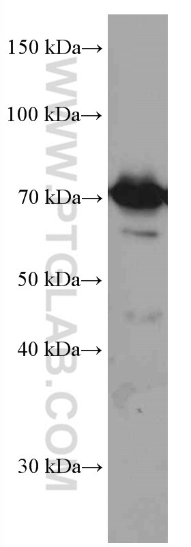
| Observed Molecular Weight | 70 kDa |
| GenBank Accession Number | BC022016 |
| Gene Symbol | PKC Iota |
| Gene ID (NCBI) | 5584 |
| RRID | AB_2881858 |
| Conjugate | Unconjugated |
| Form | Liquid |
| Purification Method | Protein A purification |
| UNIPROT ID | P41743 |
| Storage Buffer | PBS with 0.02% sodium azide and 50% glycerol, pH 7.3. |
| Storage Conditions | Store at -20°C. Stable for one year after shipment. Aliquoting is unnecessary for -20oC storage. 20ul sizes contain 0.1% BSA. |
Background Information
The atypical protein kinase C isoform PRKC iota (PRKCI) is a member of the protein kinase C (PKC) family of serine/threonine protein kinases. PKC family comprises at least eight members, which are differentially expressed and are involved in a wide variety of cellular processes. PRKC iota is calcium-independent and phospholipid-dependent. It is not activated by phorbolesters or diacylglycerol. This kinase can be recruited to vesicle tubular clusters (VTCs) by direct interaction with the small GTPase RAB2, where this kinase phosphorylates glyceraldehyde-3-phosphate dehydrogenase (GAPD/GAPDH) and plays a role in microtubule dynamics in the early secretory pathway. This kinase is found to be necessary for BCL-ABL-mediated resistance to drug-induced apoptosis and therefore protects leukemia cells against drug-induced apoptosis. PRKC iota plays a key role in cell proliferation, differentiation, and carcinogenesis, and it has been shown to be a human oncogene.
Protocols
| Product Specific Protocols | |
|---|---|
| IF protocol for PKC Iota antibody 66493-1-Ig | Download protocol |
| IHC protocol for PKC Iota antibody 66493-1-Ig | Download protocol |
| WB protocol for PKC Iota antibody 66493-1-Ig | Download protocol |
| Standard Protocols | |
|---|---|
| Click here to view our Standard Protocols |
Publications
| Species | Application | Title |
|---|---|---|
Int J Mol Sci Regulation of a PRMT5/NF-κB Axis by Phosphorylation of PRMT5 at Serine 15 in Colorectal Cancer.
| ||
Cell Commun Signal Prkci promotes colorectal cancer metastasis by phosphorylating and stabilizing Tgfbr1 to activate TGF-β signaling | ||
Neoplasia Prkci activates Jak2/Stat3 signaling to promote tumor angiogenesis: Short Name: Prkci in tumor angiogenesis |