")
")
")
")
")
")


Tested Applications
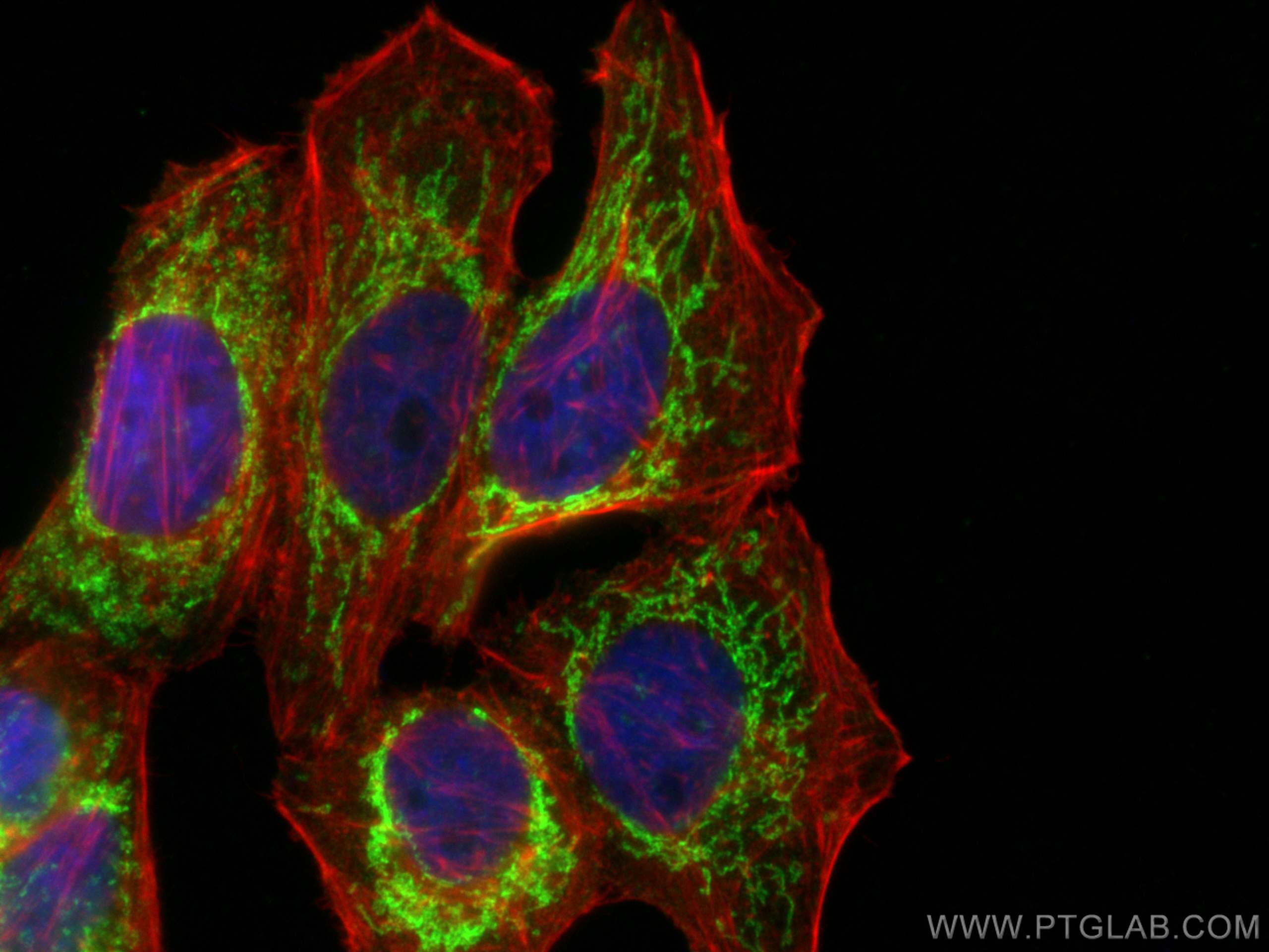
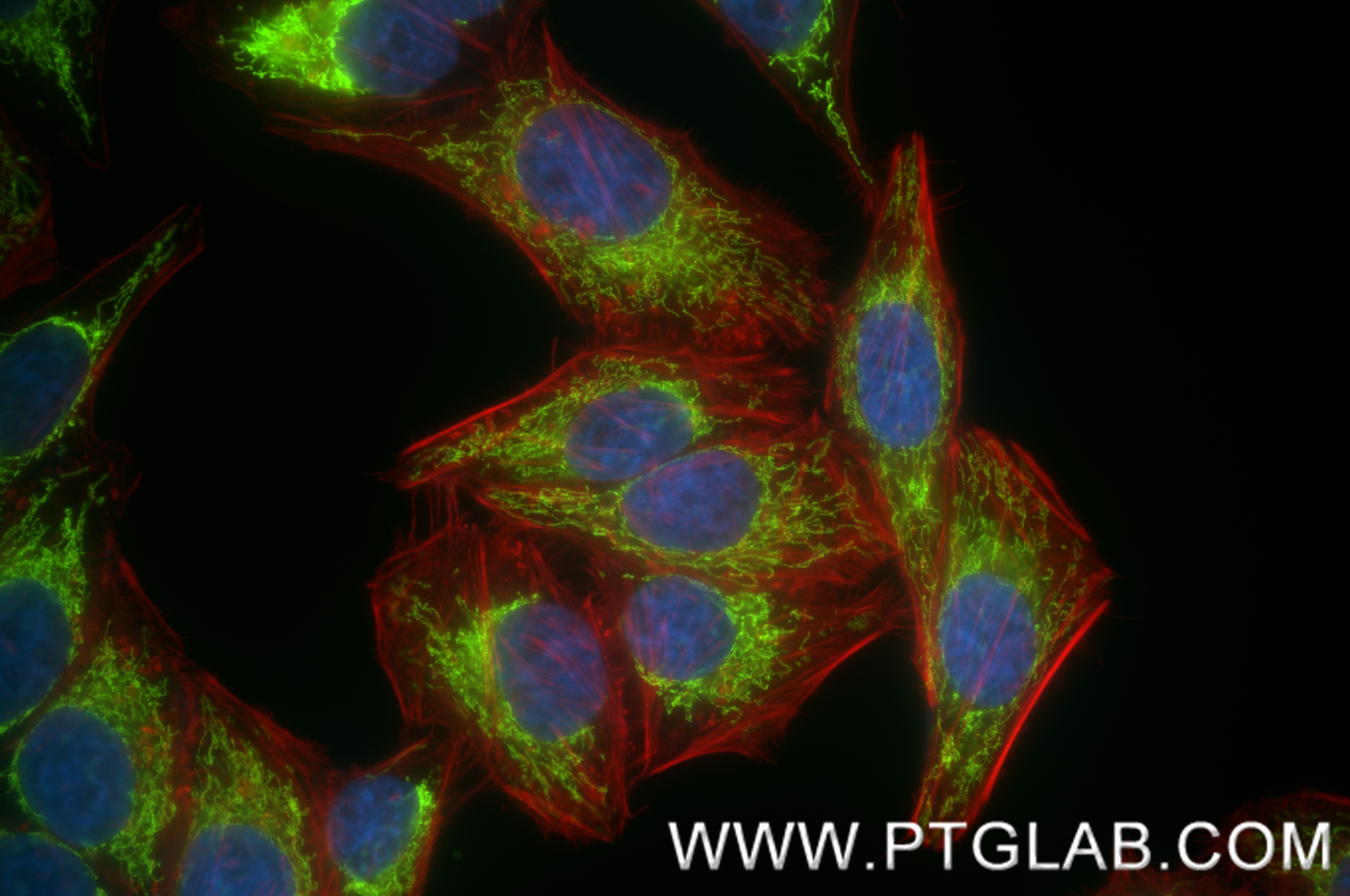
| Positive IF/ICC detected in | HepG2 cells |
Recommended dilution
| Application | Dilution |
|---|---|
| Immunofluorescence (IF)/ICC | IF/ICC : 1:200-1:800 |
| It is recommended that this reagent should be titrated in each testing system to obtain optimal results. | |
Product Information
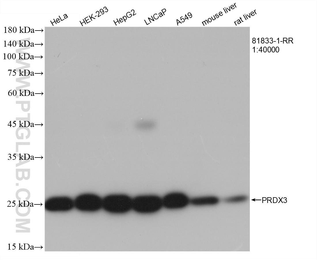
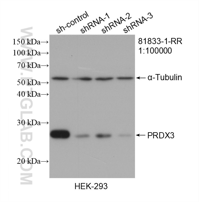
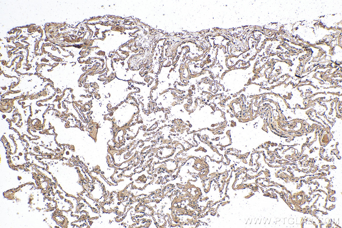
81833-1-PBS targets PRDX3 in WB, IHC, IF/ICC, Indirect ELISA applications and shows reactivity with human, mouse, rat samples.
| Tested Reactivity | human, mouse, rat |
| Host / Isotype | Rabbit / IgG |
| Class | Recombinant |
| Type | Antibody |
| Immunogen |
CatNo: Ag1062 Product name: Recombinant human PRX3 protein Source: e coli.-derived, PGEX-4T Tag: GST Domain: 1-256 aa of BC007062 Sequence: MAAAVGRLLRASVARHVSAIPWGISATAALRPAACGRTSLTNLLCSGSSQAKLFSTSSSCHAPAVTQHAPYFKGTAVVNGEFKDLSLDDFKGKYLVLFFYPLDFTFVCPTEIVAFSDKANEFHDVNCEVVAVSVDSHFSHLAWINTPRKNGGLGHMNIALLSDLTKQISRDYGVLLEGSGLALRGLFIIDPNGVIKHLSVNDLPVGRSVEETLRLVKAFQYVETHGEVCPANWTPDSPTIKPSPAASKEYFQKVNQ Predict reactive species |
| Full Name | peroxiredoxin 3 |
| Calculated Molecular Weight | 27 kDa |
| Observed Molecular Weight | 28 kDa |
| GenBank Accession Number | BC007062 |
| Gene Symbol | PRDX3 |
| Gene ID (NCBI) | 10935 |
| RRID | AB_2935583 |
| Conjugate | Unconjugated |
| Form | Liquid |
| Purification Method | Protein A purification |
| UNIPROT ID | P30048 |
| Storage Buffer | PBS only, pH 7.3. |
| Storage Conditions | Store at -80°C. |
Background Information
PRDX3(Peroxiredoxin 3), also named as AOP1, HBC189 and MER5, belongs to the ahpC/TSA family. It is involved in redox regulation of the cell and protects radical-sensitive enzymes from oxidative damage by a radical-generating system. PRDX3 is required for MYC-mediated proliferation, transformation, and apoptosis after glucose withdrawal and essential for maintaining mitochondrial mass and membrane potential in transformed rat and human cells (PMID:12011429). PRDX3 acts synergistically with MAP3K13 to regulate the activation of NF-kappa-B in the cytosol.
Protocols
| Product Specific Protocols | |
|---|---|
| IF protocol for PRDX3 antibody 81833-1-PBS | Download protocol |
| Standard Protocols | |
|---|---|
| Click here to view our Standard Protocols |