Tested Applications
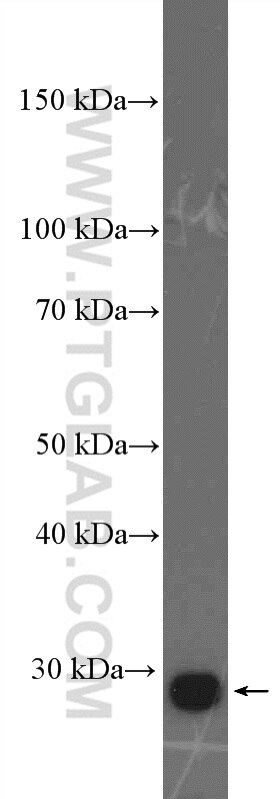
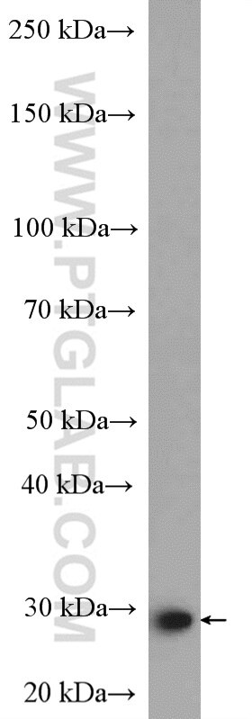
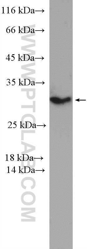
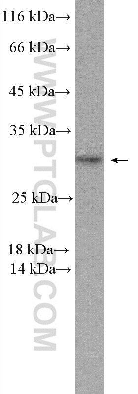
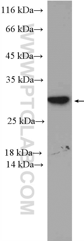
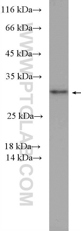
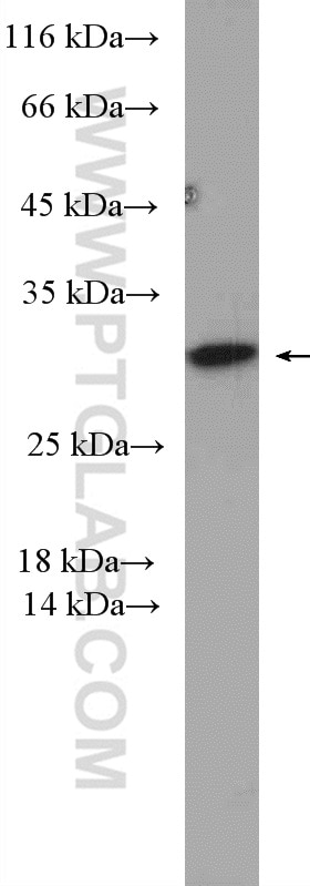
| Positive WB detected in | A549 cells, MCF-7 cells, MDA-MB-453s cells, HepG2 cells, Jurkat cells, mouse spleen tissue |
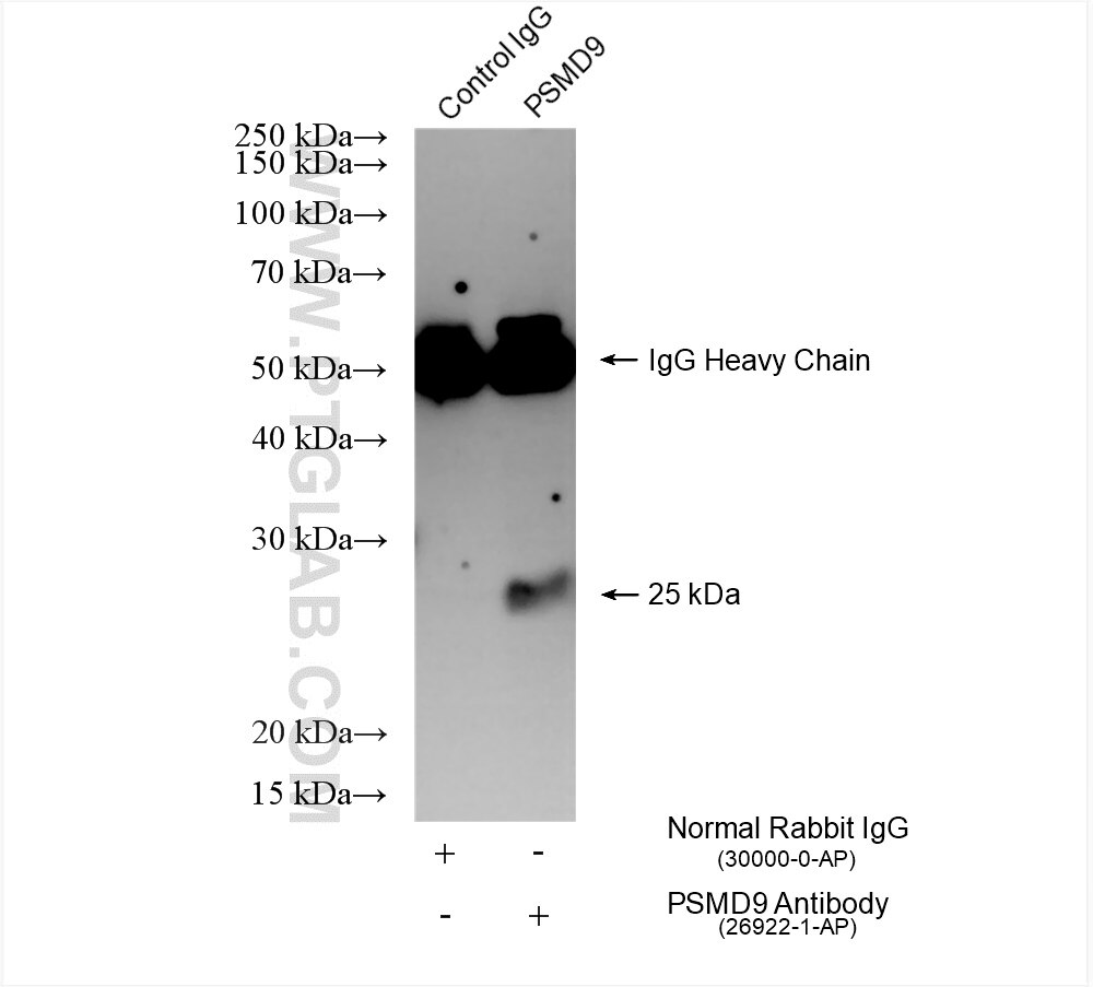
| Positive IP detected in | A549 cells |
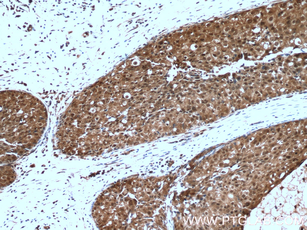
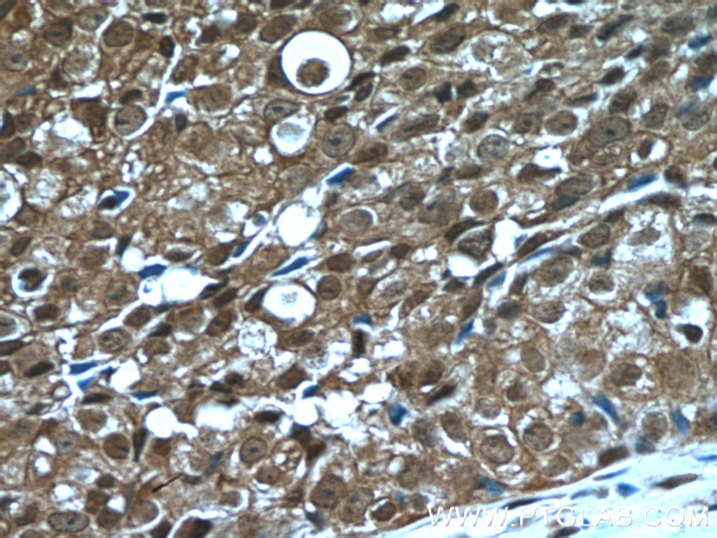
| Positive IHC detected in | human breast cancer tissue Note: suggested antigen retrieval with TE buffer pH 9.0; (*) Alternatively, antigen retrieval may be performed with citrate buffer pH 6.0 |
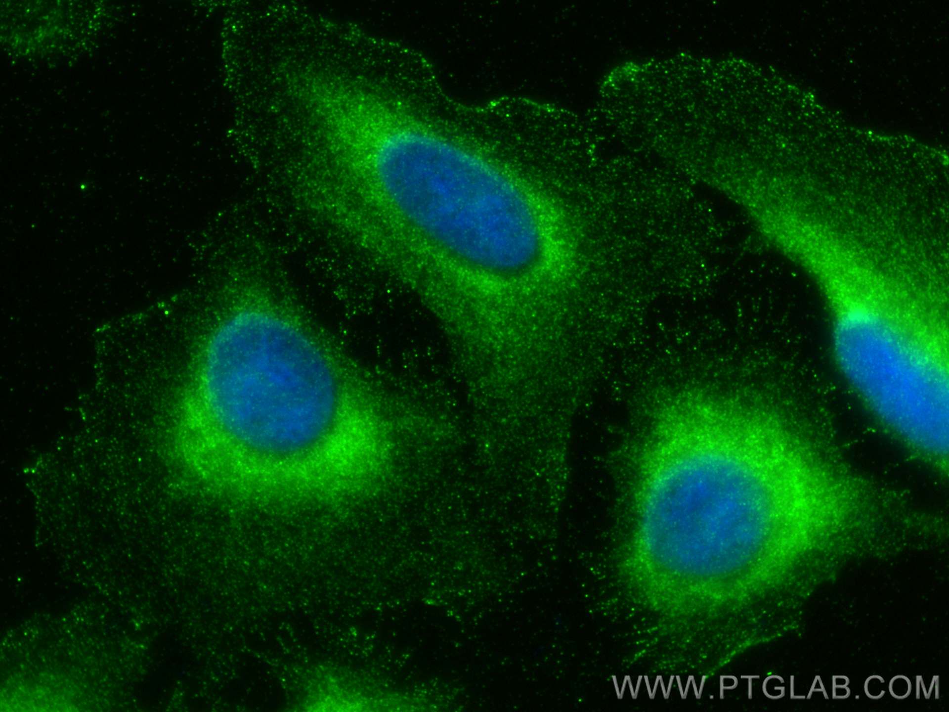
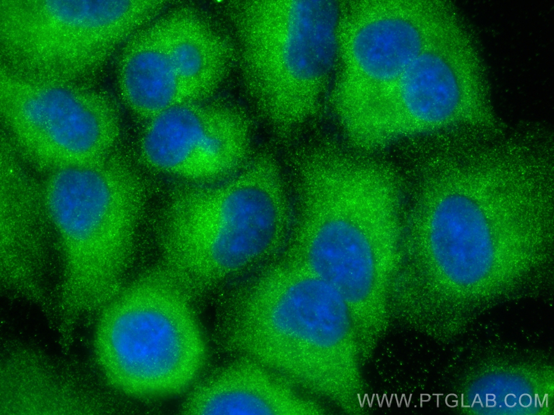
| Positive IF/ICC detected in | U2OS cells, A549 cells |
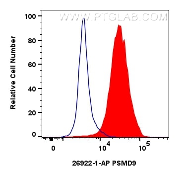
| Positive FC (Intra) detected in | HeLa cells |
Recommended dilution
| Application | Dilution |
|---|---|
| Western Blot (WB) | WB : 1:1000-1:6000 |
| Immunoprecipitation (IP) | IP : 0.5-4.0 ug for 1.0-3.0 mg of total protein lysate |
| Immunohistochemistry (IHC) | IHC : 1:50-1:500 |
| Immunofluorescence (IF)/ICC | IF/ICC : 1:50-1:500 |
| Flow Cytometry (FC) (INTRA) | FC (INTRA) : 0.80 ug per 10^6 cells in a 100 µl suspension |
| It is recommended that this reagent should be titrated in each testing system to obtain optimal results. | |
| Sample-dependent, Check data in validation data gallery. | |
Product Information
26922-1-AP targets PSMD9 in WB, IHC, IF/ICC, FC (Intra), IP, ELISA applications and shows reactivity with human, mouse samples.
| Tested Reactivity | human, mouse |
| Host / Isotype | Rabbit / IgG |
| Class | Polyclonal |
| Type | Antibody |
| Immunogen |
CatNo: Ag25638 Product name: Recombinant human PSMD9 protein Source: e coli.-derived, PGEX-4T Tag: GST Domain: 1-102 aa of BC004213 Sequence: MSDEEARQSGGSSQAGVVTVSDVQELMRRKEEIEAQIKANYDVLESQKGIGMNEPLVDCEGYPRSDVDLYQVRTARHNIICLQNDHKAVMKQVEEALHQLHA Predict reactive species |
| Full Name | proteasome (prosome, macropain) 26S subunit, non-ATPase, 9 |
| Calculated Molecular Weight | 27 kDa |
| Observed Molecular Weight | 25-30 kDa |
| GenBank Accession Number | BC004213 |
| Gene Symbol | PSMD9 |
| Gene ID (NCBI) | 5715 |
| RRID | AB_2880687 |
| Conjugate | Unconjugated |
| Form | Liquid |
| Purification Method | Antigen affinity purification |
| UNIPROT ID | O00233 |
| Storage Buffer | PBS with 0.02% sodium azide and 50% glycerol, pH 7.3. |
| Storage Conditions | Store at -20°C. Stable for one year after shipment. Aliquoting is unnecessary for -20oC storage. 20ul sizes contain 0.1% BSA. |
Background Information
PSMD9 is a ubiquitous protein of eukaryotic cells and is a chaperon of the 26S proteasome complex, which degrades ubiquitinated proteins in eukaryotic cells and contributes to the degradation of intracellular proteins into antigenic peptides for antigen presentation by MHC class I cells. The 26S mammalian base sub-complex involves three distinct modules which have ATPase subunits distinctly associated to three chaperones, one of which is PSMD9 regulating the modules assembly. The PSMD9 ubiquitous regulatory role within the proteasome implies its potential pleiotropic effects within different physio-pathological systems. PSMD9 is known to form a stable subcomplex with PSMC3 and PSMC6, two of the AAA-ATPases, assisting in the assembly of the 20S and 19S particles to form the holo complex.
Protocols
| Product Specific Protocols | |
|---|---|
| FC protocol for PSMD9 antibody 26922-1-AP | Download protocol |
| IF protocol for PSMD9 antibody 26922-1-AP | Download protocol |
| IHC protocol for PSMD9 antibody 26922-1-AP | Download protocol |
| IP protocol for PSMD9 antibody 26922-1-AP | Download protocol |
| WB protocol for PSMD9 antibody 26922-1-AP | Download protocol |
| Standard Protocols | |
|---|---|
| Click here to view our Standard Protocols |
Reviews
The reviews below have been submitted by verified Proteintech customers who received an incentive for providing their feedback.
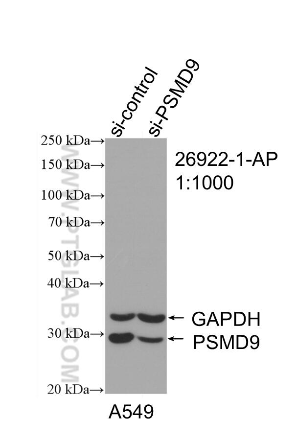
FH Tanel (Verified Customer) (01-02-2026) | Both antibodies work great in WB and IF. Single band in A549 and H1299 cells, for WB one could dilute even more up to 1:2000. In IF, there is nuclear and cytoplasmic distribution.
|