The Proteintech guarantee covers Proteintech antibodies in any species and any application, including those not listed on the datasheet. If the antibody doesn’t perform, you can receive a hassle-free refund or credit note.
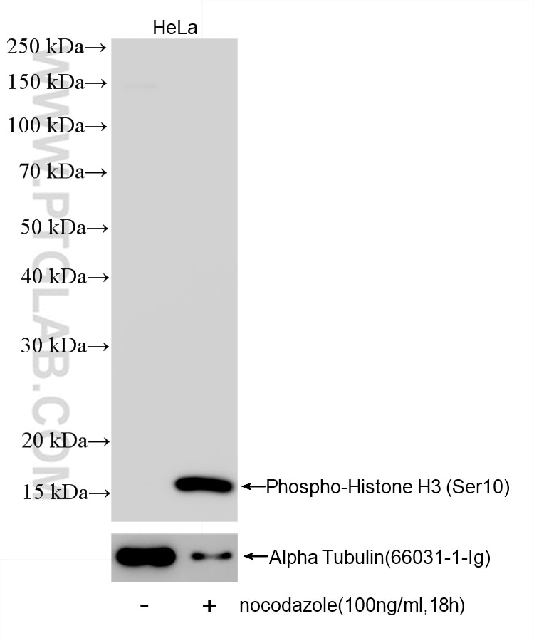
WB analysis of HeLa using 82828-10-RR (same clone as 82828-10-PBS)
Nocodazole treated HeLa cells were subjected to SDS PAGE followed by western blot with 82828-10-RR (Phospho-Histone H3 (Ser10) antibody) at dilution of 1:2000 incubated at room temperature for 1.5 hours. The membrane was stripped and reblotted with Alpha Tubulin Monoclonal antibody (66031-1-Ig) as loading control. This data was developed using the same antibody clone with 82828-10-PBS in a different storage buffer formulation.
This validation data has been generated in-house and reviewed by Proteintech scientists. Find out more on our antibody validation policy
Nocodazole treated HeLa cells were subjected to SDS PAGE followed by western blot with 82828-10-RR (Phospho-Histone H3 (Ser10) antibody) at dilution of 1:2000 incubated at room temperature for 1.5 hours. The membrane was stripped and reblotted with Alpha Tubulin Monoclonal antibody (66031-1-Ig) as loading control. This data was developed using the same antibody clone with 82828-10-PBS in a different storage buffer formulation.
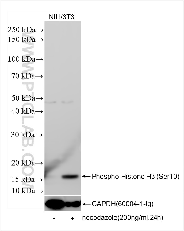
WB analysis of NIH/3T3 using 82828-10-RR (same clone as 82828-10-PBS)
Nocodazole treated and untreated NIH/3T3 cells were subjected to SDS PAGE followed by western blot with 82828-10-RR (Phospho-Histone H3 (Ser10) antibody) at dilution of 1:2000 incubated at room temperature for 1.5 hours. The membrane was stripped and reblotted with GAPDH Monoclonal antibody (60004-1-Ig) as loading control. This data was developed using the same antibody clone with 82828-10-PBS in a different storage buffer formulation.
This validation data has been generated in-house and reviewed by Proteintech scientists. Find out more on our antibody validation policy
Nocodazole treated and untreated NIH/3T3 cells were subjected to SDS PAGE followed by western blot with 82828-10-RR (Phospho-Histone H3 (Ser10) antibody) at dilution of 1:2000 incubated at room temperature for 1.5 hours. The membrane was stripped and reblotted with GAPDH Monoclonal antibody (60004-1-Ig) as loading control. This data was developed using the same antibody clone with 82828-10-PBS in a different storage buffer formulation.
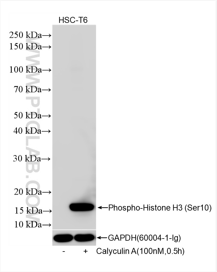
WB analysis of HSC-T6 using 82828-10-RR (same clone as 82828-10-PBS)
Calyculin A treated and untreated HSC-T6 cells were subjected to SDS PAGE followed by western blot with 82828-10-RR (Phospho-Histone H3 (Ser10) antibody) at dilution of 1:2000 incubated at room temperature for 1.5 hours. The membrane was stripped and reblotted with GAPDH Monoclonal antibody (60004-1-Ig) as loading control. This data was developed using the same antibody clone with 82828-10-PBS in a different storage buffer formulation.
This validation data has been generated in-house and reviewed by Proteintech scientists. Find out more on our antibody validation policy
Calyculin A treated and untreated HSC-T6 cells were subjected to SDS PAGE followed by western blot with 82828-10-RR (Phospho-Histone H3 (Ser10) antibody) at dilution of 1:2000 incubated at room temperature for 1.5 hours. The membrane was stripped and reblotted with GAPDH Monoclonal antibody (60004-1-Ig) as loading control. This data was developed using the same antibody clone with 82828-10-PBS in a different storage buffer formulation.
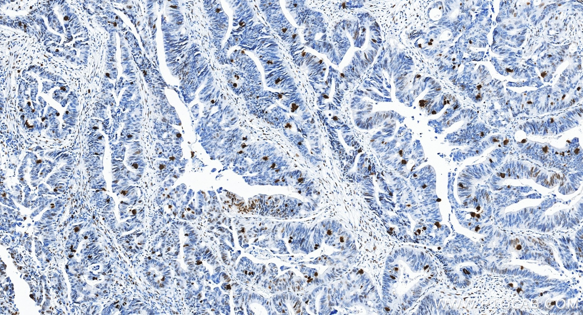
IHC staining of human colon cancer using 82828-10-RR (same clone as 82828-10-PBS)
Immunohistochemical analysis of paraffin-embedded human colon cancer tissue slide using 82828-10-RR (Phospho-Histone H3 (Ser10) antibody) at dilution of 1:20000 (under 20x lens). Heat mediated antigen retrieval with Tris-EDTA buffer (pH 9.0). This data was developed using the same antibody clone with 82828-10-PBS in a different storage buffer formulation.
This validation data has been generated in-house and reviewed by Proteintech scientists. Find out more on our antibody validation policy
Immunohistochemical analysis of paraffin-embedded human colon cancer tissue slide using 82828-10-RR (Phospho-Histone H3 (Ser10) antibody) at dilution of 1:20000 (under 20x lens). Heat mediated antigen retrieval with Tris-EDTA buffer (pH 9.0). This data was developed using the same antibody clone with 82828-10-PBS in a different storage buffer formulation.
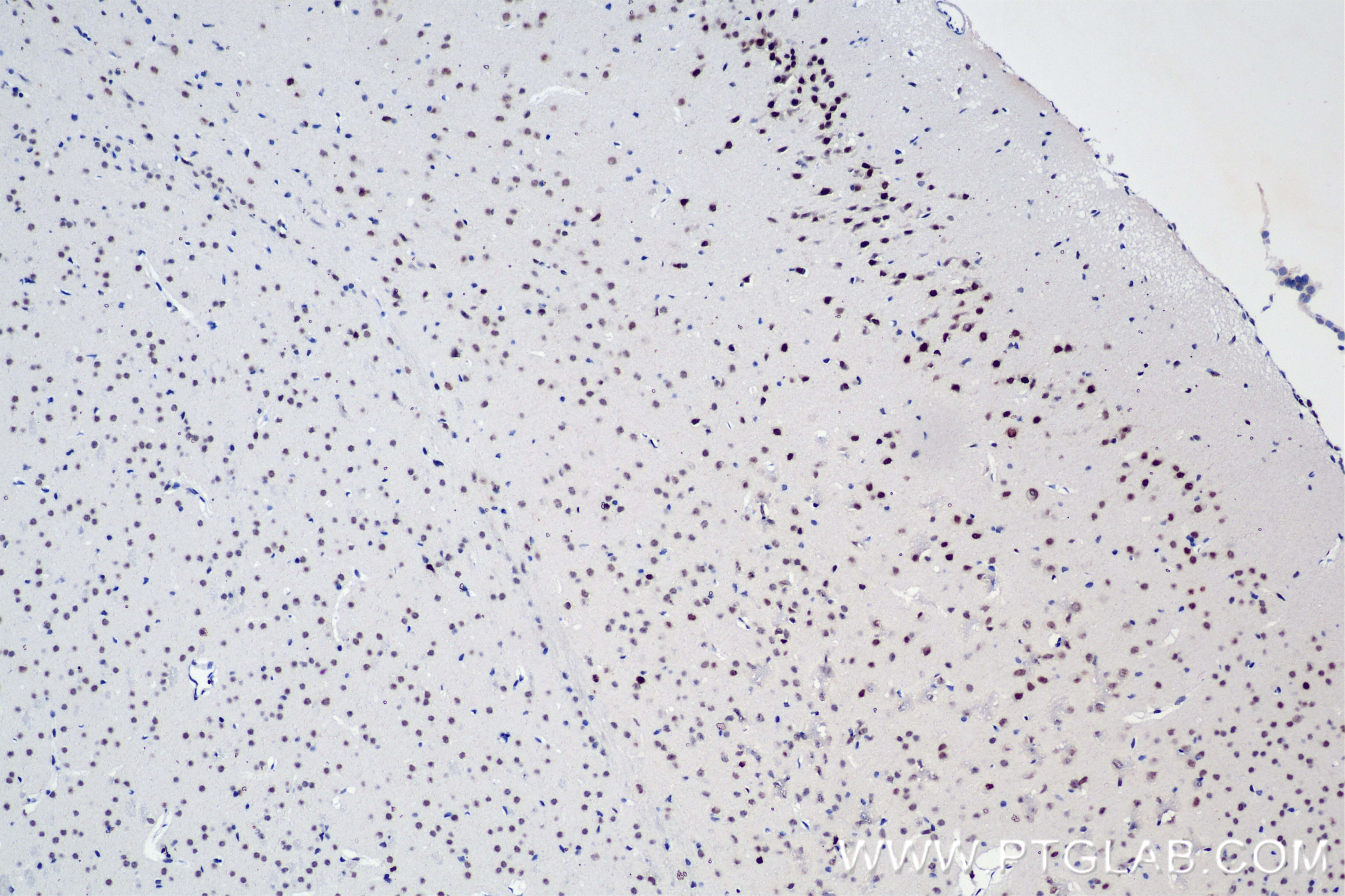
IHC staining of mouse brain using 82828-10-RR (same clone as 82828-10-PBS)
Immunohistochemical analysis of paraffin-embedded mouse brain tissue slide using 82828-10-RR (Phospho-Histone H3 (Ser10) antibody) at dilution of 1:1000 (under 10x lens). Heat mediated antigen retrieval with Tris-EDTA buffer (pH 9.0). This data was developed using the same antibody clone with 82828-10-PBS in a different storage buffer formulation.
This validation data has been generated in-house and reviewed by Proteintech scientists. Find out more on our antibody validation policy
Immunohistochemical analysis of paraffin-embedded mouse brain tissue slide using 82828-10-RR (Phospho-Histone H3 (Ser10) antibody) at dilution of 1:1000 (under 10x lens). Heat mediated antigen retrieval with Tris-EDTA buffer (pH 9.0). This data was developed using the same antibody clone with 82828-10-PBS in a different storage buffer formulation.
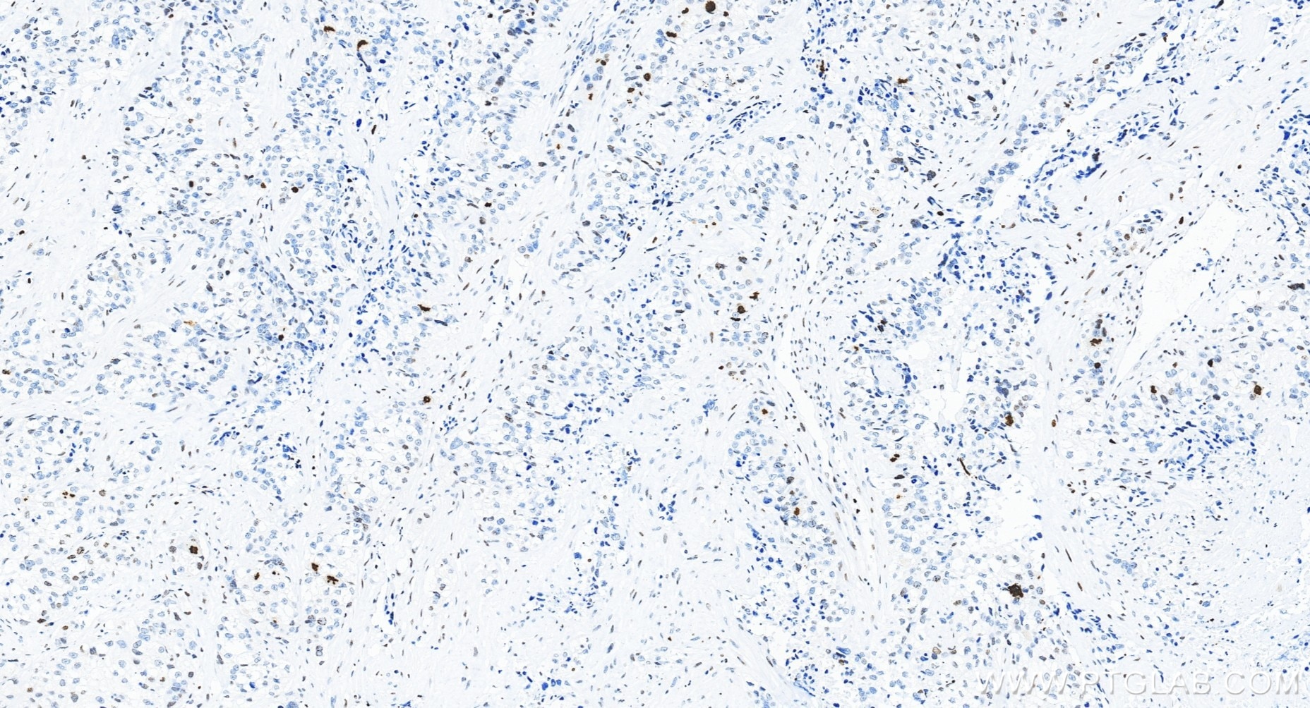
IHC staining of human ovary cancer using 82828-10-RR (same clone as 82828-10-PBS)
Immunohistochemical analysis of paraffin-embedded human ovary cancer tissue slide using 82828-10-RR (Phospho-Histone H3 (Ser10) antibody) at dilution of 1:20000 (under 20x lens). Heat mediated antigen retrieval with Tris-EDTA buffer (pH 9.0). This data was developed using the same antibody clone with 82828-10-PBS in a different storage buffer formulation.
This validation data has been generated in-house and reviewed by Proteintech scientists. Find out more on our antibody validation policy
Immunohistochemical analysis of paraffin-embedded human ovary cancer tissue slide using 82828-10-RR (Phospho-Histone H3 (Ser10) antibody) at dilution of 1:20000 (under 20x lens). Heat mediated antigen retrieval with Tris-EDTA buffer (pH 9.0). This data was developed using the same antibody clone with 82828-10-PBS in a different storage buffer formulation.
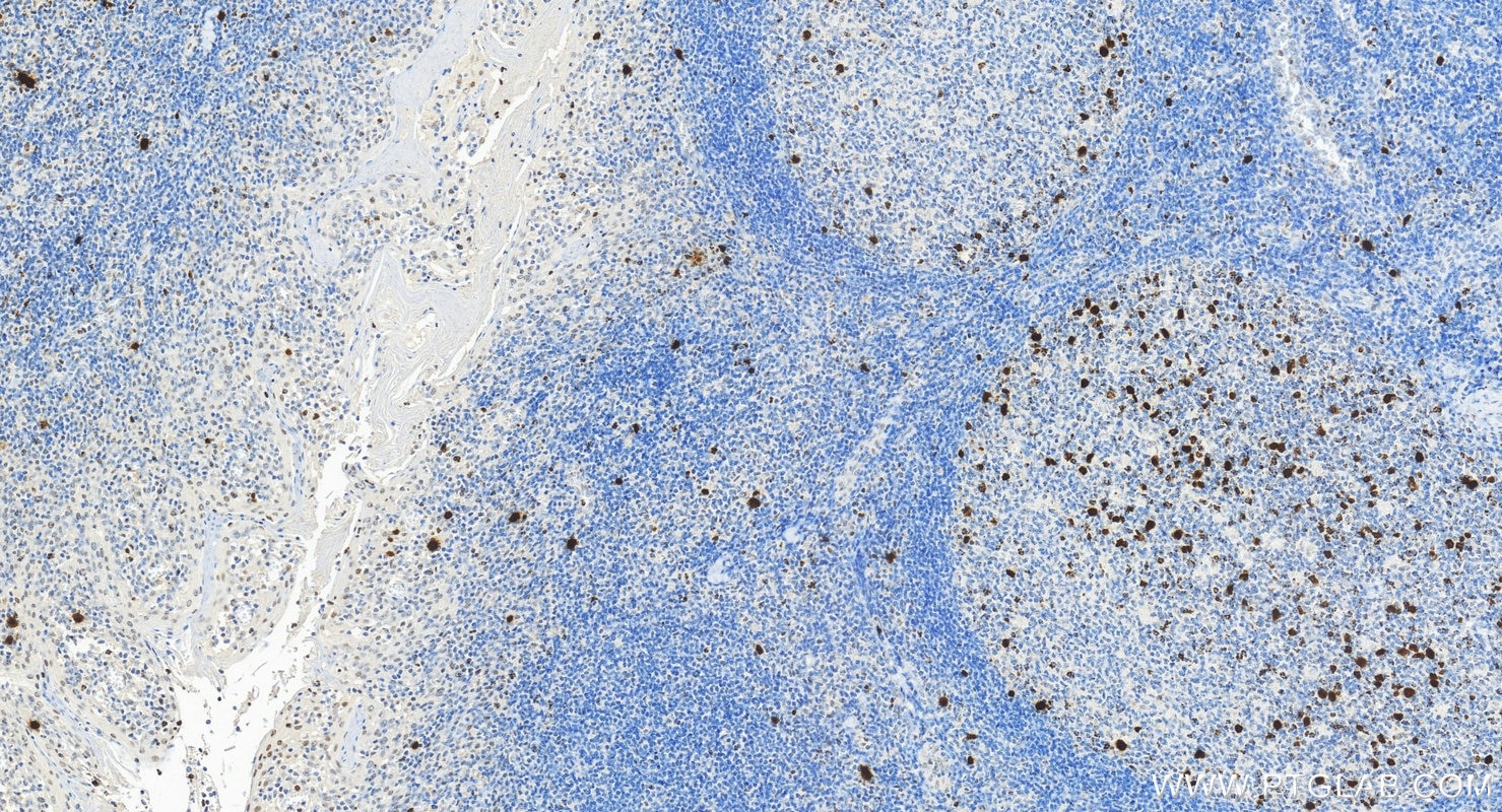
IHC staining of human tonsillitis using 82828-10-RR (same clone as 82828-10-PBS)
Immunohistochemical analysis of paraffin-embedded human tonsillitis tissue slide using 82828-10-RR (Phospho-Histone H3 (Ser10) antibody) at dilution of 1:4000 (under 20x lens). Heat mediated antigen retrieval with Tris-EDTA buffer (pH 9.0). This data was developed using the same antibody clone with 82828-10-PBS in a different storage buffer formulation.
This validation data has been generated in-house and reviewed by Proteintech scientists. Find out more on our antibody validation policy
Immunohistochemical analysis of paraffin-embedded human tonsillitis tissue slide using 82828-10-RR (Phospho-Histone H3 (Ser10) antibody) at dilution of 1:4000 (under 20x lens). Heat mediated antigen retrieval with Tris-EDTA buffer (pH 9.0). This data was developed using the same antibody clone with 82828-10-PBS in a different storage buffer formulation.
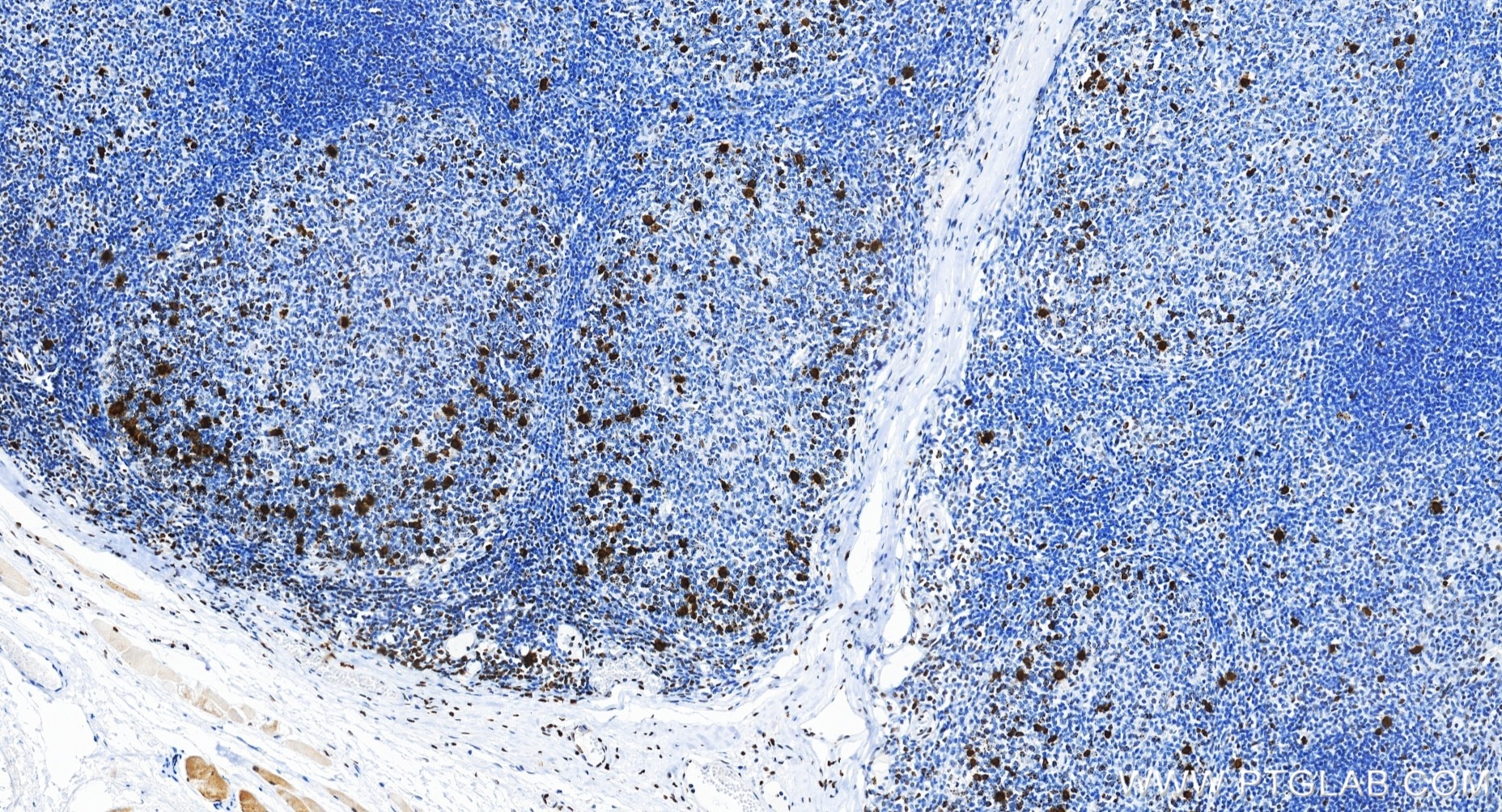
IHC staining of human tonsillitis using 82828-10-RR (same clone as 82828-10-PBS)
Immunohistochemical analysis of paraffin-embedded human tonsillitis tissue slide using 82828-10-RR (Phospho-Histone H3 (Ser10) antibody) at dilution of 1:20000 (under 20x lens). Heat mediated antigen retrieval with Tris-EDTA buffer (pH 9.0). This data was developed using the same antibody clone with 82828-10-PBS in a different storage buffer formulation.
This validation data has been generated in-house and reviewed by Proteintech scientists. Find out more on our antibody validation policy
Immunohistochemical analysis of paraffin-embedded human tonsillitis tissue slide using 82828-10-RR (Phospho-Histone H3 (Ser10) antibody) at dilution of 1:20000 (under 20x lens). Heat mediated antigen retrieval with Tris-EDTA buffer (pH 9.0). This data was developed using the same antibody clone with 82828-10-PBS in a different storage buffer formulation.
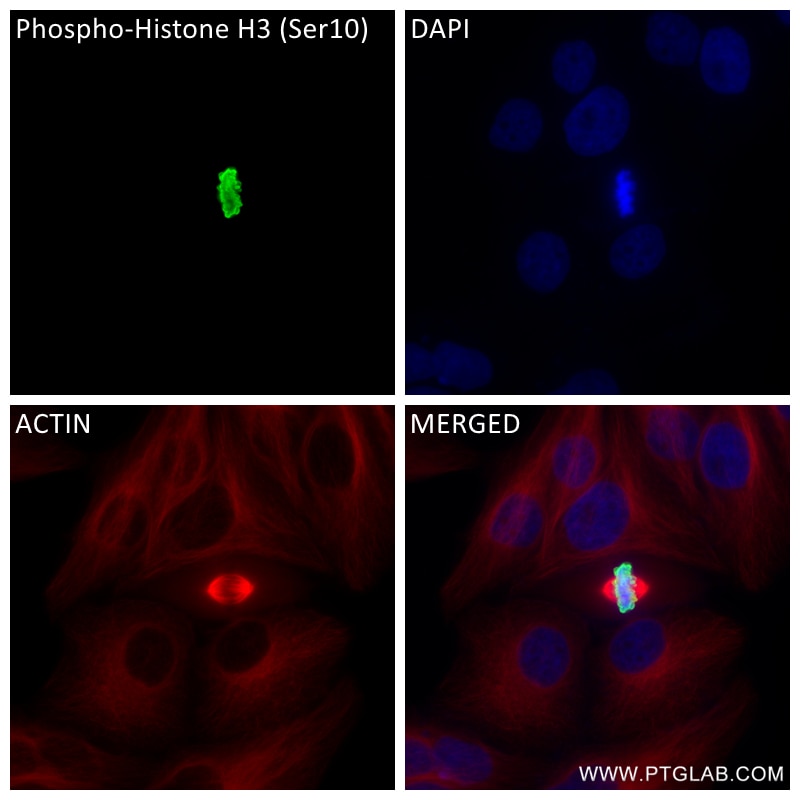
IF Staining of HepG2 using 82828-10-RR (same clone as 82828-10-PBS)
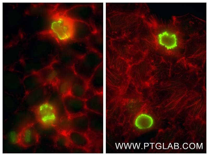
Immunofluorescent analysis of (4% PFA) fixed HepG2 cells using Phospho-Histone H3 (Ser10) antibody (82828-10-RR, Clone: 242921B1 ) at dilution of 1:800 and CoraLite®488-Conjugated Goat Anti-Rabbit IgG(H+L) (SA00013-2), Beta Tubulin antibody (66240-1-Ig, Clone: 1D4A4, red). This data was developed using the same antibody clone with 82828-10-PBS in a different storage buffer formulation.
This validation data has been generated in-house and reviewed by Proteintech scientists. Find out more on our antibody validation policy
Immunofluorescent analysis of (4% PFA) fixed HepG2 cells using Phospho-Histone H3 (Ser10) antibody (82828-10-RR, Clone: 242921B1 ) at dilution of 1:800 and CoraLite®488-Conjugated Goat Anti-Rabbit IgG(H+L) (SA00013-2), Beta Tubulin antibody (66240-1-Ig, Clone: 1D4A4, red). This data was developed using the same antibody clone with 82828-10-PBS in a different storage buffer formulation.
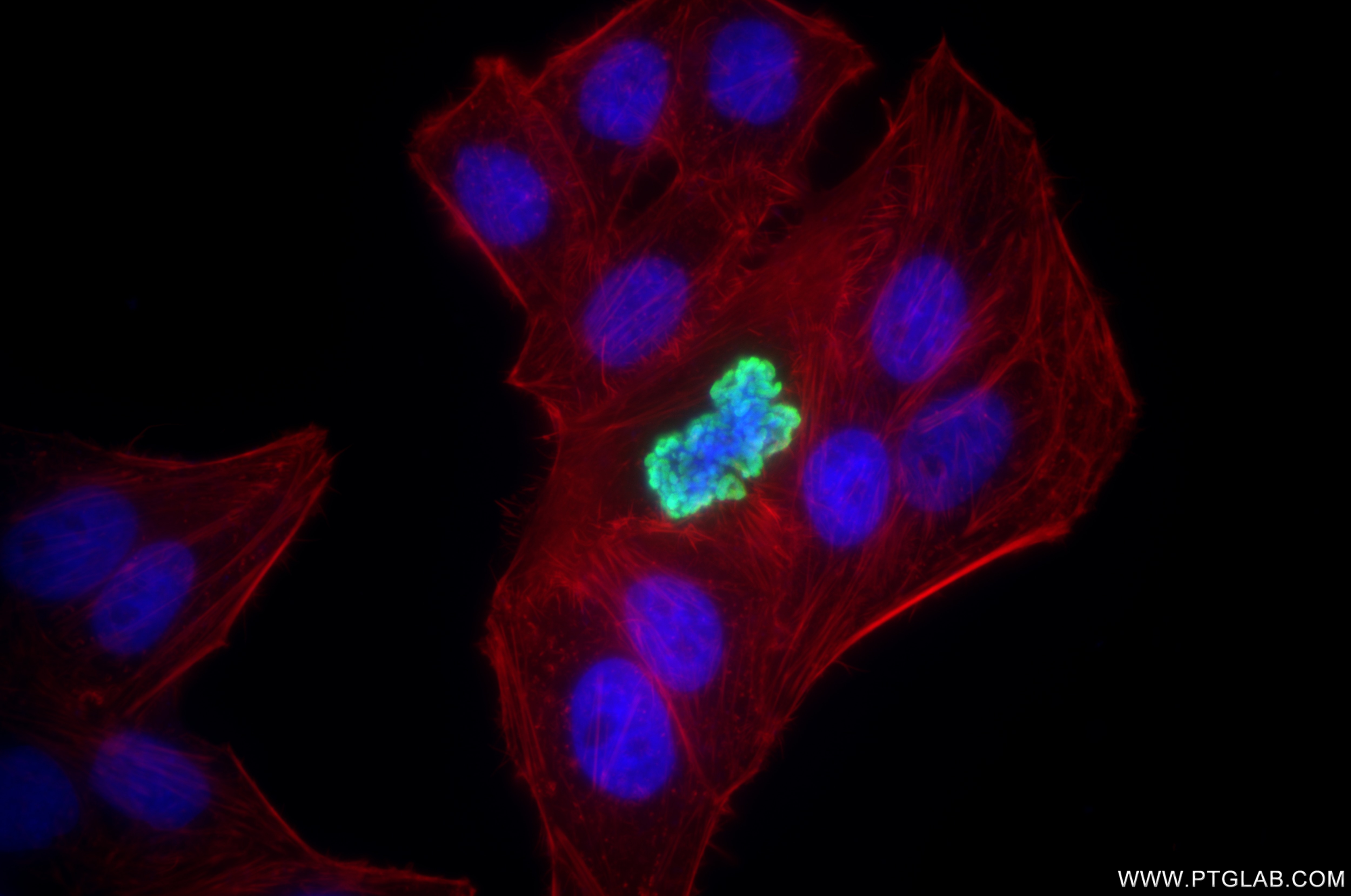
IF Staining of HepG2 using 82828-10-RR (same clone as 82828-10-PBS)
Immunofluorescent analysis of (4% PFA) fixed HepG2 cells using Phospho-Histone H3 (Ser10) antibody (82828-10-RR, Clone: 242921B1 ) at dilution of 1:800 and CoraLite®488-Conjugated Goat Anti-Rabbit IgG(H+L) (SA00013-2), CL594-Phalloidin (red). This data was developed using the same antibody clone with 82828-10-PBS in a different storage buffer formulation.
This validation data has been generated in-house and reviewed by Proteintech scientists. Find out more on our antibody validation policy
Immunofluorescent analysis of (4% PFA) fixed HepG2 cells using Phospho-Histone H3 (Ser10) antibody (82828-10-RR, Clone: 242921B1 ) at dilution of 1:800 and CoraLite®488-Conjugated Goat Anti-Rabbit IgG(H+L) (SA00013-2), CL594-Phalloidin (red). This data was developed using the same antibody clone with 82828-10-PBS in a different storage buffer formulation.
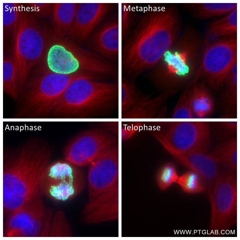
IF Staining of HepG2 using 82828-10-RR (same clone as 82828-10-PBS)
Immunofluorescent analysis of (4% PFA) fixed HepG2 cells using Phospho-Histone H3 (Ser10) antibody (82828-10-RR, Clone: 242921B1 ) at dilution of 1:800 and CoraLite®488-Conjugated Goat Anti-Rabbit IgG(H+L) (SA00013-2), Beta Tubulin antibody (66240-1-Ig, Clone: 1D4A4, red). This data was developed using the same antibody clone with 82828-10-PBS in a different storage buffer formulation.
This validation data has been generated in-house and reviewed by Proteintech scientists. Find out more on our antibody validation policy
Immunofluorescent analysis of (4% PFA) fixed HepG2 cells using Phospho-Histone H3 (Ser10) antibody (82828-10-RR, Clone: 242921B1 ) at dilution of 1:800 and CoraLite®488-Conjugated Goat Anti-Rabbit IgG(H+L) (SA00013-2), Beta Tubulin antibody (66240-1-Ig, Clone: 1D4A4, red). This data was developed using the same antibody clone with 82828-10-PBS in a different storage buffer formulation.
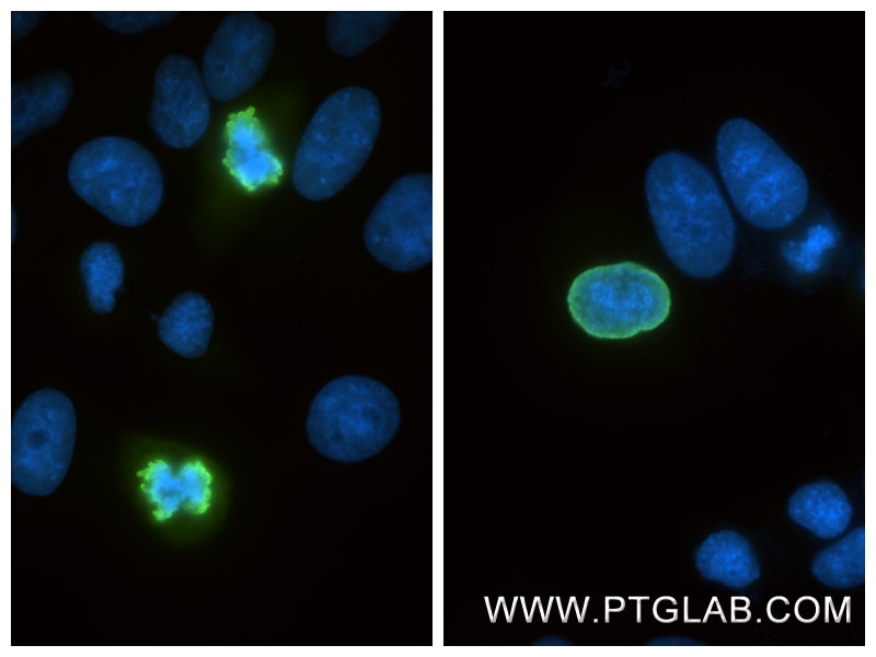
IF Staining of HepG2 using 82828-10-RR (same clone as 82828-10-PBS)
Immunofluorescent analysis of (4% PFA) fixed HepG2 cells using Phospho-Histone H3 (Ser10) antibody (82828-10-RR, Clone: 242921B1 ) at dilution of 1:3000 and CoraLite®488-Conjugated Goat Anti-Rabbit IgG(H+L) (SA00013-2). This data was developed using the same antibody clone with 82828-10-PBS in a different storage buffer formulation.
This validation data has been generated in-house and reviewed by Proteintech scientists. Find out more on our antibody validation policy
Immunofluorescent analysis of (4% PFA) fixed HepG2 cells using Phospho-Histone H3 (Ser10) antibody (82828-10-RR, Clone: 242921B1 ) at dilution of 1:3000 and CoraLite®488-Conjugated Goat Anti-Rabbit IgG(H+L) (SA00013-2). This data was developed using the same antibody clone with 82828-10-PBS in a different storage buffer formulation.
IF Staining of HepG2 using 82828-10-PBS
Immunofluorescent analysis of (4% PFA) fixed HepG2 cells using Phospho-Histone H3 (Ser10) antibody (82828-10-PBS, Clone: 242921B1 ) at dilution of 1:1600 and CoraLite®488-Conjugated Goat Anti-Rabbit IgG(H+L) (SA00013-2), CL594-Phalloidin (red).
This validation data has been generated in-house and reviewed by Proteintech scientists. Find out more on our antibody validation policy
Immunofluorescent analysis of (4% PFA) fixed HepG2 cells using Phospho-Histone H3 (Ser10) antibody (82828-10-PBS, Clone: 242921B1 ) at dilution of 1:1600 and CoraLite®488-Conjugated Goat Anti-Rabbit IgG(H+L) (SA00013-2), CL594-Phalloidin (red).
ChIP experiment of HeLa using 82828-10-RR (same clone as 82828-10-PBS)
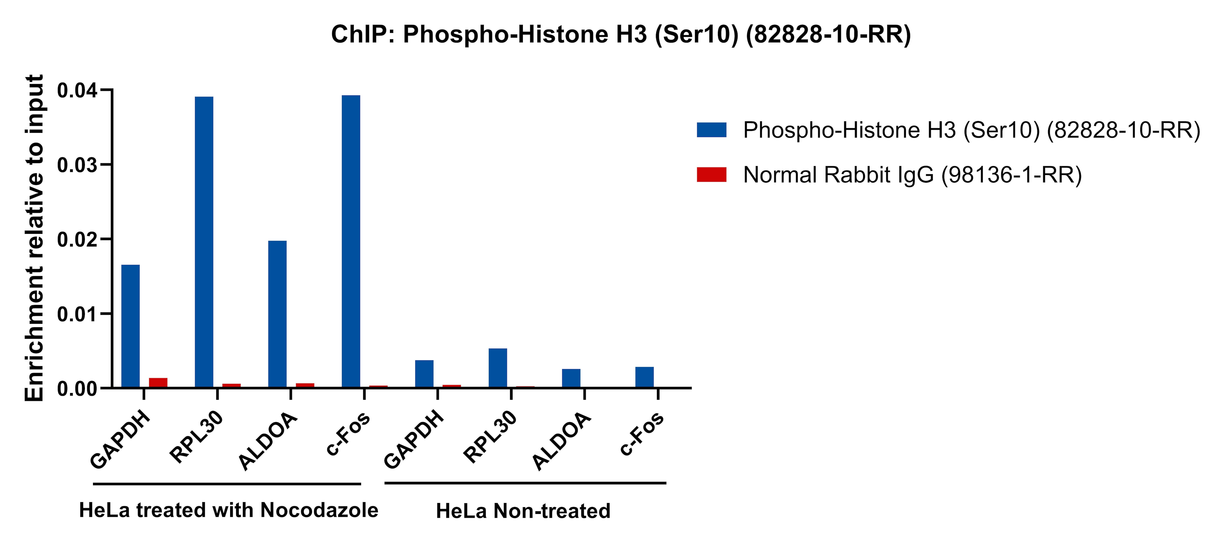
Chromatin was prepared from HeLa cells either non-treated or treated with Nocodazole (100 ng/ml, 16 h). Cells were fixed with formaldehyde for 10 minutes. The ChIP was performed with 20 µg of cross-linked chromatin, 5 µg of Phospho-Histone H3 (Ser10) (82828-10-RR) or 5 ug of Normal Rabbit IgG (98136-1-RR), and 20 µl of Protein A Magarose Beads. The immunoprecipitated DNA was quantified by real-time PCR. This data was developed using the same antibody clone with 82828-10-PBS in a different storage buffer formulation.
This validation data has been generated in-house and reviewed by Proteintech scientists. Find out more on our antibody validation policy
Chromatin was prepared from HeLa cells either non-treated or treated with Nocodazole (100 ng/ml, 16 h). Cells were fixed with formaldehyde for 10 minutes. The ChIP was performed with 20 µg of cross-linked chromatin, 5 µg of Phospho-Histone H3 (Ser10) (82828-10-RR) or 5 ug of Normal Rabbit IgG (98136-1-RR), and 20 µl of Protein A Magarose Beads. The immunoprecipitated DNA was quantified by real-time PCR. This data was developed using the same antibody clone with 82828-10-PBS in a different storage buffer formulation.
Phospho-histone-H3 (PHH3) is a core histone protein, which in its phosphorylated state forms the principal constituents of eukaryotic chromatin, with histone H3 being phosphorylated at serine (Ser) 10 or Ser28 as well as its phosphorylation of Ser10 being strongly correlated with the late G2 to M-phase transition in mammalian mitotic cells. On the basis of previous research, a few cell line- and animal model-based researches have displayed an increase in phosphorylation of histone H3 at Ser10 (H3S10ph), the only histone marker that is involved in carcinogenesis and cellular transformation. Histone H3 phosphorylation on serine-10 is specific to mitosis and phosphorylated histone H3 (PHH3) proliferation markers (as counts defined per area or as indices defined per cell numbers) are increasingly being used to evaluate proliferation in various tumors.
Protocols
Product Specific Protocols
IF protocol for Phospho-Histone H3 (Ser10) antibody 82828-10-PBS
This validation data has been generated in-house and reviewed by Proteintech scientists.
Find out more on our antibody validation policy
WB Figures
WB analysis of HeLa using 82828-10-RR (same clone as 82828-10-PBS)
Nocodazole treated HeLa cells were subjected to SDS PAGE followed by western blot with 82828-10-RR (Phospho-Histone H3 (Ser10) antibody) at dilution of 1:2000 incubated at room temperature for 1.5 hours. The membrane was stripped and reblotted with Alpha Tubulin Monoclonal antibody (66031-1-Ig) as loading control. This data was developed using the same antibody clone with 82828-10-PBS in a different storage buffer formulation.
WB analysis of NIH/3T3 using 82828-10-RR (same clone as 82828-10-PBS)
Nocodazole treated and untreated NIH/3T3 cells were subjected to SDS PAGE followed by western blot with 82828-10-RR (Phospho-Histone H3 (Ser10) antibody) at dilution of 1:2000 incubated at room temperature for 1.5 hours. The membrane was stripped and reblotted with GAPDH Monoclonal antibody (60004-1-Ig) as loading control. This data was developed using the same antibody clone with 82828-10-PBS in a different storage buffer formulation.
WB analysis of HSC-T6 using 82828-10-RR (same clone as 82828-10-PBS)
Calyculin A treated and untreated HSC-T6 cells were subjected to SDS PAGE followed by western blot with 82828-10-RR (Phospho-Histone H3 (Ser10) antibody) at dilution of 1:2000 incubated at room temperature for 1.5 hours. The membrane was stripped and reblotted with GAPDH Monoclonal antibody (60004-1-Ig) as loading control. This data was developed using the same antibody clone with 82828-10-PBS in a different storage buffer formulation.
IHC Figures
IHC staining of human colon cancer using 82828-10-RR (same clone as 82828-10-PBS)
Immunohistochemical analysis of paraffin-embedded human colon cancer tissue slide using 82828-10-RR (Phospho-Histone H3 (Ser10) antibody) at dilution of 1:20000 (under 20x lens). Heat mediated antigen retrieval with Tris-EDTA buffer (pH 9.0). This data was developed using the same antibody clone with 82828-10-PBS in a different storage buffer formulation.
IHC staining of mouse brain using 82828-10-RR (same clone as 82828-10-PBS)
Immunohistochemical analysis of paraffin-embedded mouse brain tissue slide using 82828-10-RR (Phospho-Histone H3 (Ser10) antibody) at dilution of 1:1000 (under 10x lens). Heat mediated antigen retrieval with Tris-EDTA buffer (pH 9.0). This data was developed using the same antibody clone with 82828-10-PBS in a different storage buffer formulation.
IHC staining of human ovary cancer using 82828-10-RR (same clone as 82828-10-PBS)
Immunohistochemical analysis of paraffin-embedded human ovary cancer tissue slide using 82828-10-RR (Phospho-Histone H3 (Ser10) antibody) at dilution of 1:20000 (under 20x lens). Heat mediated antigen retrieval with Tris-EDTA buffer (pH 9.0). This data was developed using the same antibody clone with 82828-10-PBS in a different storage buffer formulation.
IHC staining of human tonsillitis using 82828-10-RR (same clone as 82828-10-PBS)
Immunohistochemical analysis of paraffin-embedded human tonsillitis tissue slide using 82828-10-RR (Phospho-Histone H3 (Ser10) antibody) at dilution of 1:4000 (under 20x lens). Heat mediated antigen retrieval with Tris-EDTA buffer (pH 9.0). This data was developed using the same antibody clone with 82828-10-PBS in a different storage buffer formulation.
IHC staining of human tonsillitis using 82828-10-RR (same clone as 82828-10-PBS)
Immunohistochemical analysis of paraffin-embedded human tonsillitis tissue slide using 82828-10-RR (Phospho-Histone H3 (Ser10) antibody) at dilution of 1:20000 (under 20x lens). Heat mediated antigen retrieval with Tris-EDTA buffer (pH 9.0). This data was developed using the same antibody clone with 82828-10-PBS in a different storage buffer formulation.
IF/ICC Figures
IF Staining of HepG2 using 82828-10-RR (same clone as 82828-10-PBS)
Immunofluorescent analysis of (4% PFA) fixed HepG2 cells using Phospho-Histone H3 (Ser10) antibody (82828-10-RR, Clone: 242921B1 ) at dilution of 1:800 and CoraLite®488-Conjugated Goat Anti-Rabbit IgG(H+L) (SA00013-2), Beta Tubulin antibody (66240-1-Ig, Clone: 1D4A4, red). This data was developed using the same antibody clone with 82828-10-PBS in a different storage buffer formulation.
IF Staining of HepG2 using 82828-10-RR (same clone as 82828-10-PBS)
Immunofluorescent analysis of (4% PFA) fixed HepG2 cells using Phospho-Histone H3 (Ser10) antibody (82828-10-RR, Clone: 242921B1 ) at dilution of 1:800 and CoraLite®488-Conjugated Goat Anti-Rabbit IgG(H+L) (SA00013-2), CL594-Phalloidin (red). This data was developed using the same antibody clone with 82828-10-PBS in a different storage buffer formulation.
IF Staining of HepG2 using 82828-10-RR (same clone as 82828-10-PBS)
Immunofluorescent analysis of (4% PFA) fixed HepG2 cells using Phospho-Histone H3 (Ser10) antibody (82828-10-RR, Clone: 242921B1 ) at dilution of 1:800 and CoraLite®488-Conjugated Goat Anti-Rabbit IgG(H+L) (SA00013-2), Beta Tubulin antibody (66240-1-Ig, Clone: 1D4A4, red). This data was developed using the same antibody clone with 82828-10-PBS in a different storage buffer formulation.
IF Staining of HepG2 using 82828-10-RR (same clone as 82828-10-PBS)
Immunofluorescent analysis of (4% PFA) fixed HepG2 cells using Phospho-Histone H3 (Ser10) antibody (82828-10-RR, Clone: 242921B1 ) at dilution of 1:3000 and CoraLite®488-Conjugated Goat Anti-Rabbit IgG(H+L) (SA00013-2). This data was developed using the same antibody clone with 82828-10-PBS in a different storage buffer formulation.
IF Staining of HepG2 using 82828-10-PBS
Immunofluorescent analysis of (4% PFA) fixed HepG2 cells using Phospho-Histone H3 (Ser10) antibody (82828-10-PBS, Clone: 242921B1 ) at dilution of 1:1600 and CoraLite®488-Conjugated Goat Anti-Rabbit IgG(H+L) (SA00013-2), CL594-Phalloidin (red).
CHIP-QPCR Figures
ChIP experiment of HeLa using 82828-10-RR (same clone as 82828-10-PBS)
Chromatin was prepared from HeLa cells either non-treated or treated with Nocodazole (100 ng/ml, 16 h). Cells were fixed with formaldehyde for 10 minutes. The ChIP was performed with 20 µg of cross-linked chromatin, 5 µg of Phospho-Histone H3 (Ser10) (82828-10-RR) or 5 ug of Normal Rabbit IgG (98136-1-RR), and 20 µl of Protein A Magarose Beads. The immunoprecipitated DNA was quantified by real-time PCR. This data was developed using the same antibody clone with 82828-10-PBS in a different storage buffer formulation.
The species listed in Tested Reactivity are in-house verified and applicable species. For unlisted species, please refer to the homology analysis of the immunogen sequence and related species. For rabbit polyclonal antibodies, homology >70% is recommended. For mouse monoclonal antibodies and rabbit recombinant antibodies, homology >90% is recommended. Generally, the higher the homology, the greater the applicability. However, there will be certain differences in protein expression in different species, tissues or cells. Therefore, the homology analysis results are for reference only and do not serve as a guarantee.
At Proteintech, we pride ourselves on our antibody quality, customer service and transparency. As such, we are comparing our antibodies with other vendors, enabling easy identification and comparisons of key data to help you choose the suitable antibody for your needs.
We have selected the top cited antibodies from these vendors for you to compare.
Proteintech
Phospho-Histone H3 (Ser10) Recombinant antibody
Catalog Number
82828-10-PBS
Citations
-
Dilutions
IF/ICC : 1:800-1:3200
Applications
WB, IHC, IF/ICC, Indirect ELISA, ChIP-qPCR
Reactivity
human, mouse, rat
Product Guarantee
Covers any species including not listed on datasheet
Covers any applications including not listed on datasheet