

Tested Applications
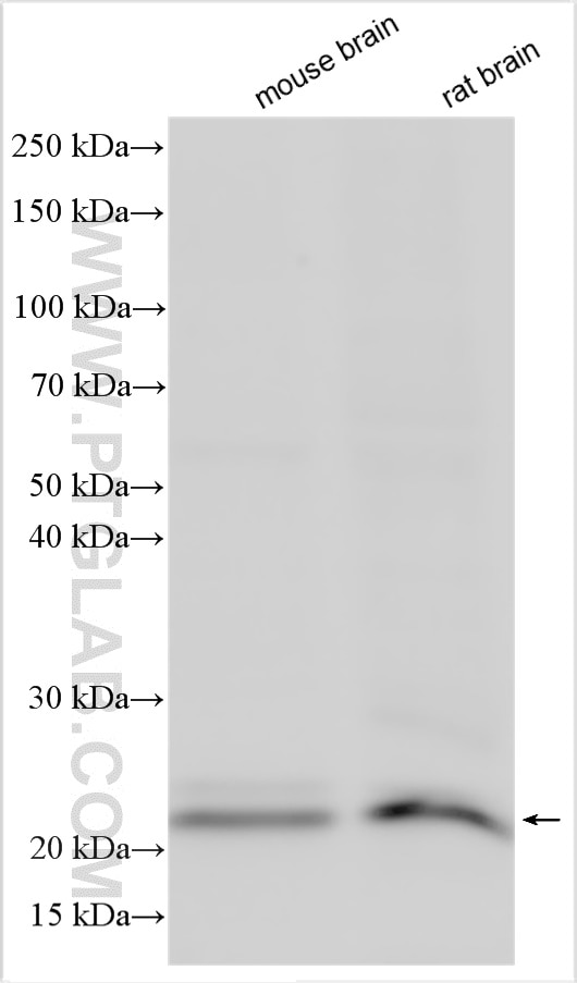
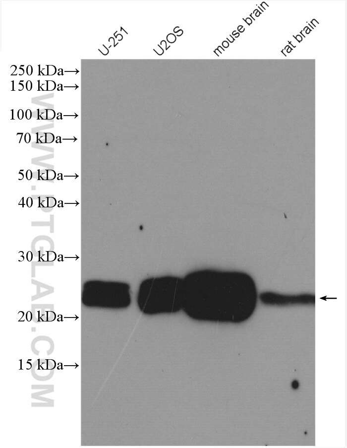
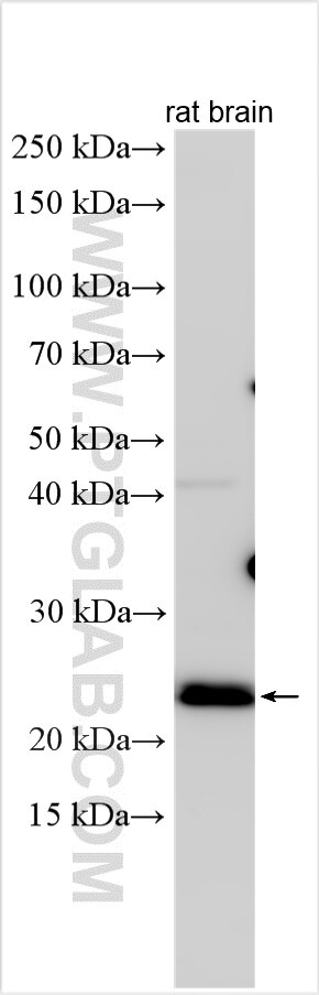
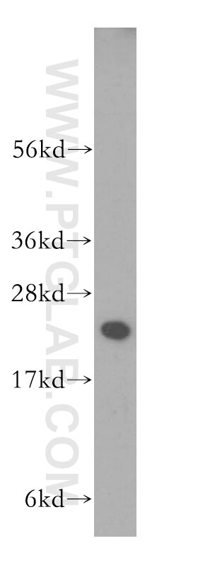
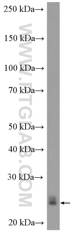
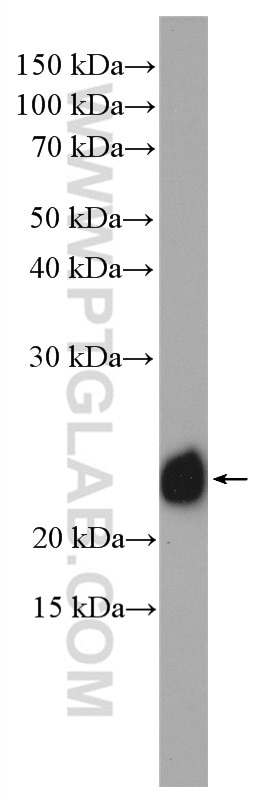
| Positive WB detected in | mouse brain tissue, human brain tissue, NIH/3T3 cells, U-251 cells, rat brain tissue, U2OS cells |
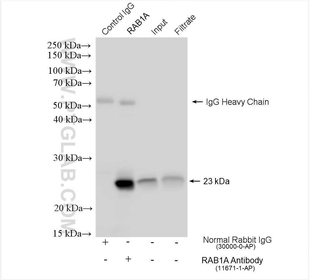
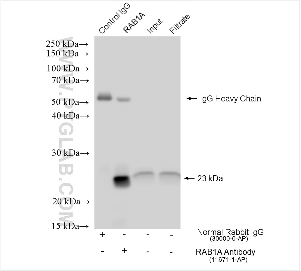
| Positive IP detected in | A549 cells, C2C12 cells |
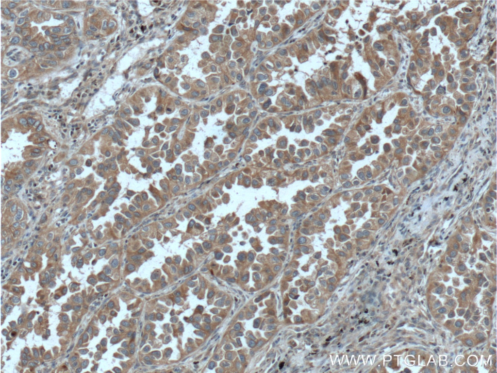
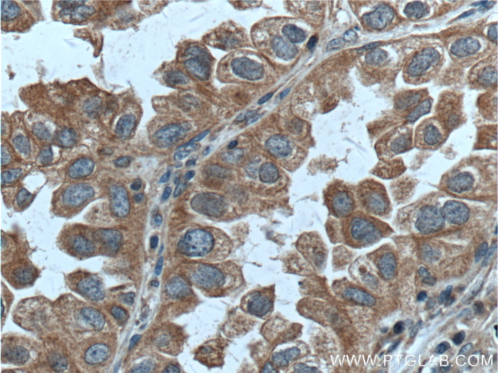
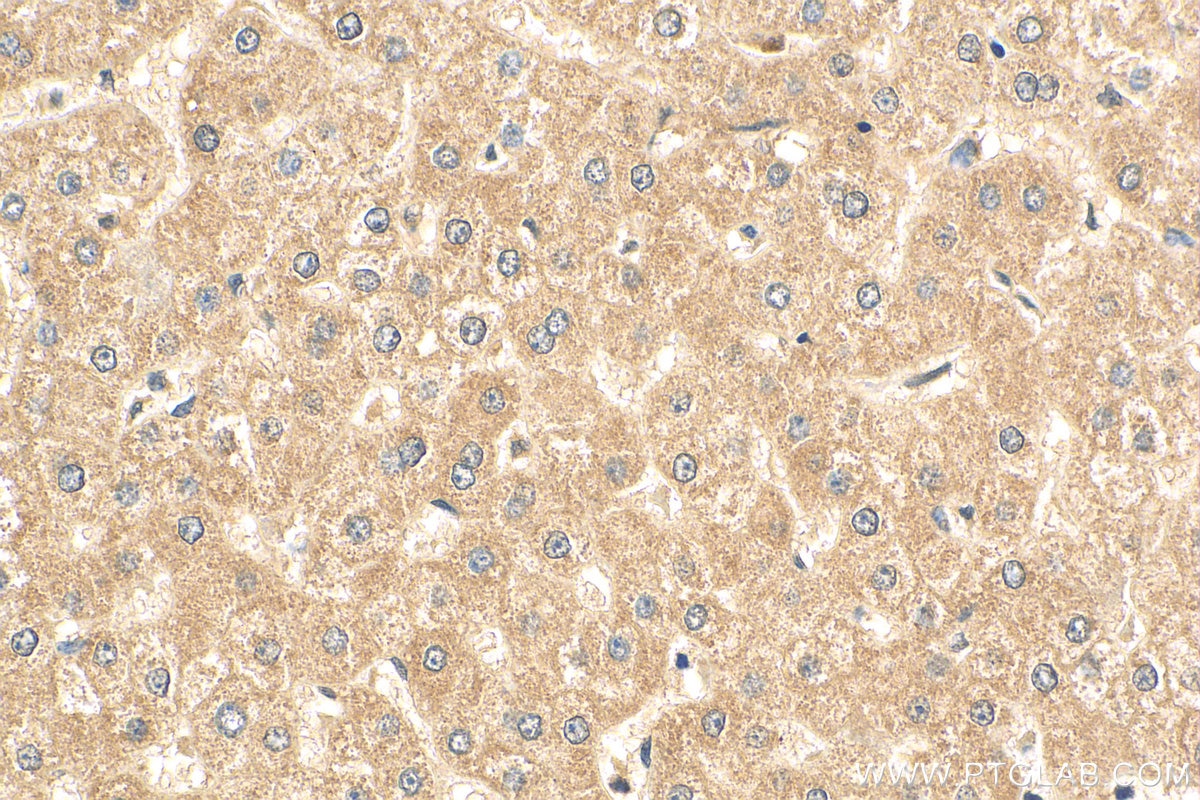
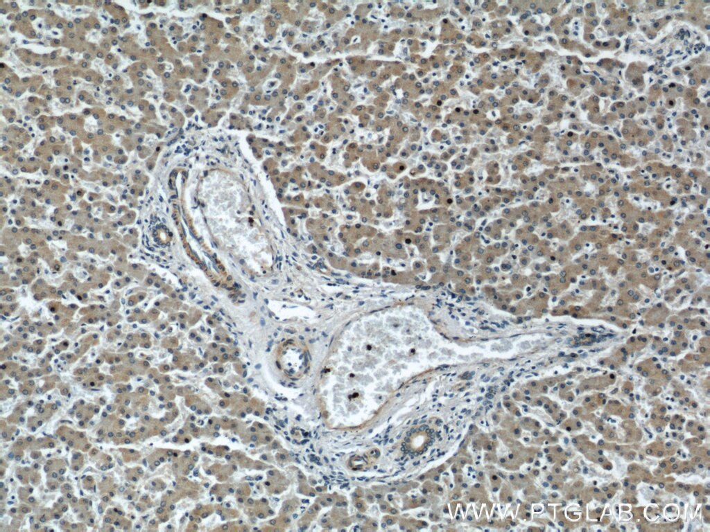
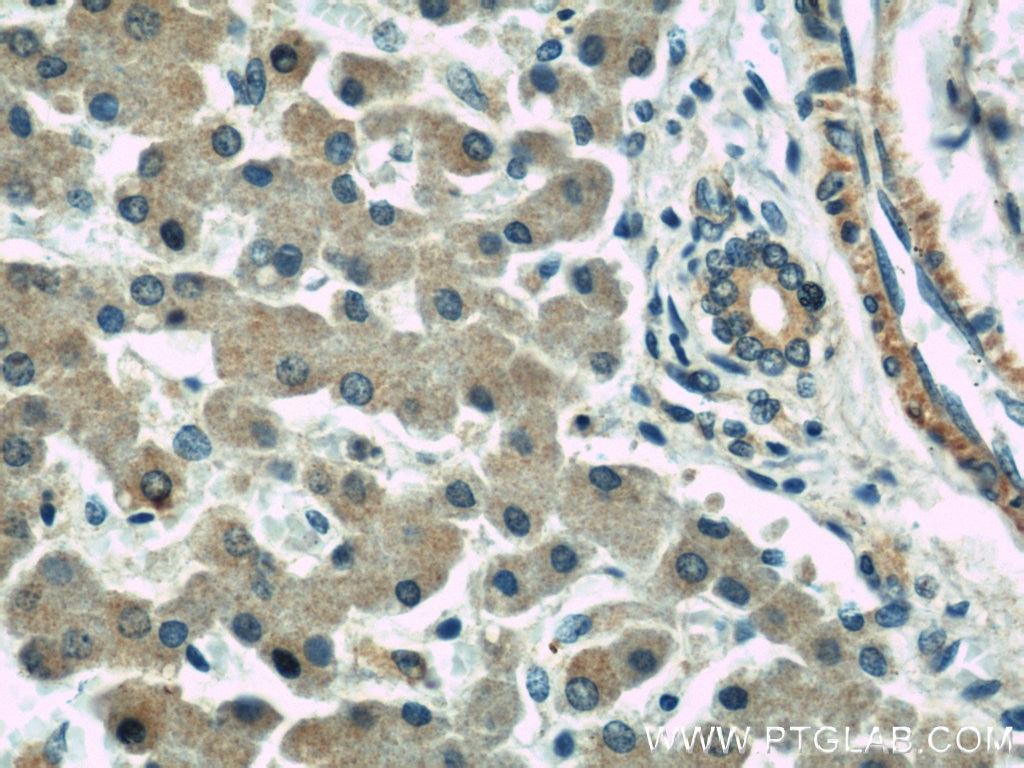
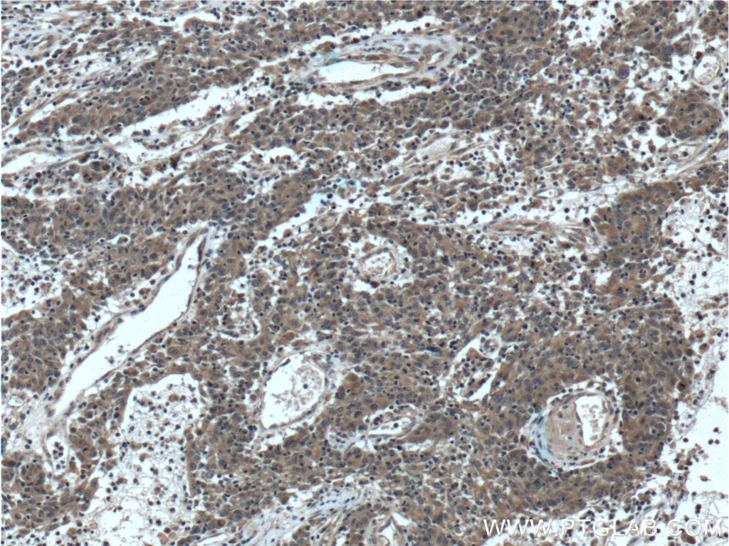
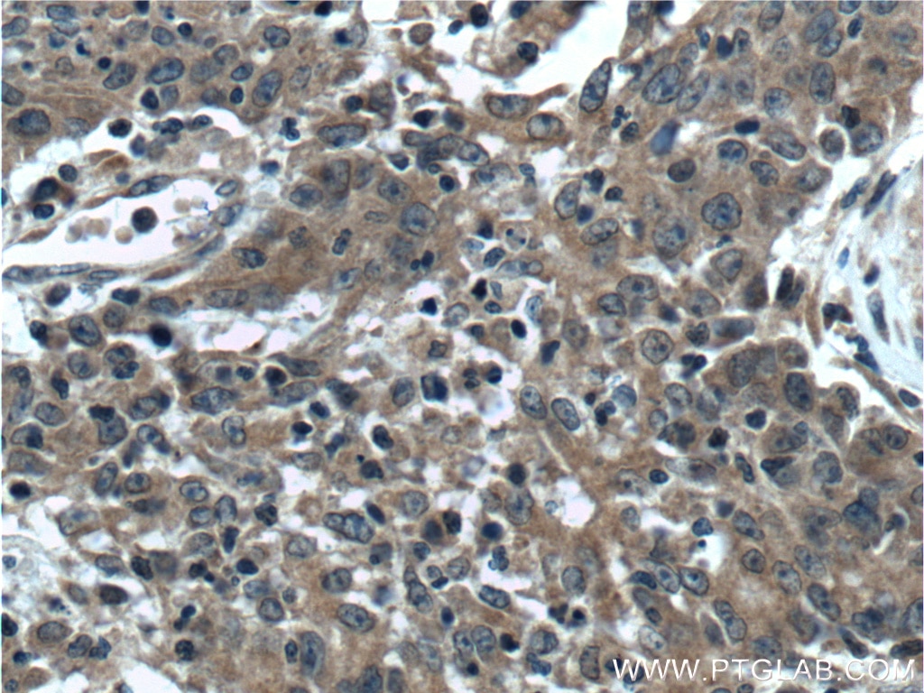
| Positive IHC detected in | human lung cancer tissue, human liver tissue, human colon cancer tissue Note: suggested antigen retrieval with TE buffer pH 9.0; (*) Alternatively, antigen retrieval may be performed with citrate buffer pH 6.0 |
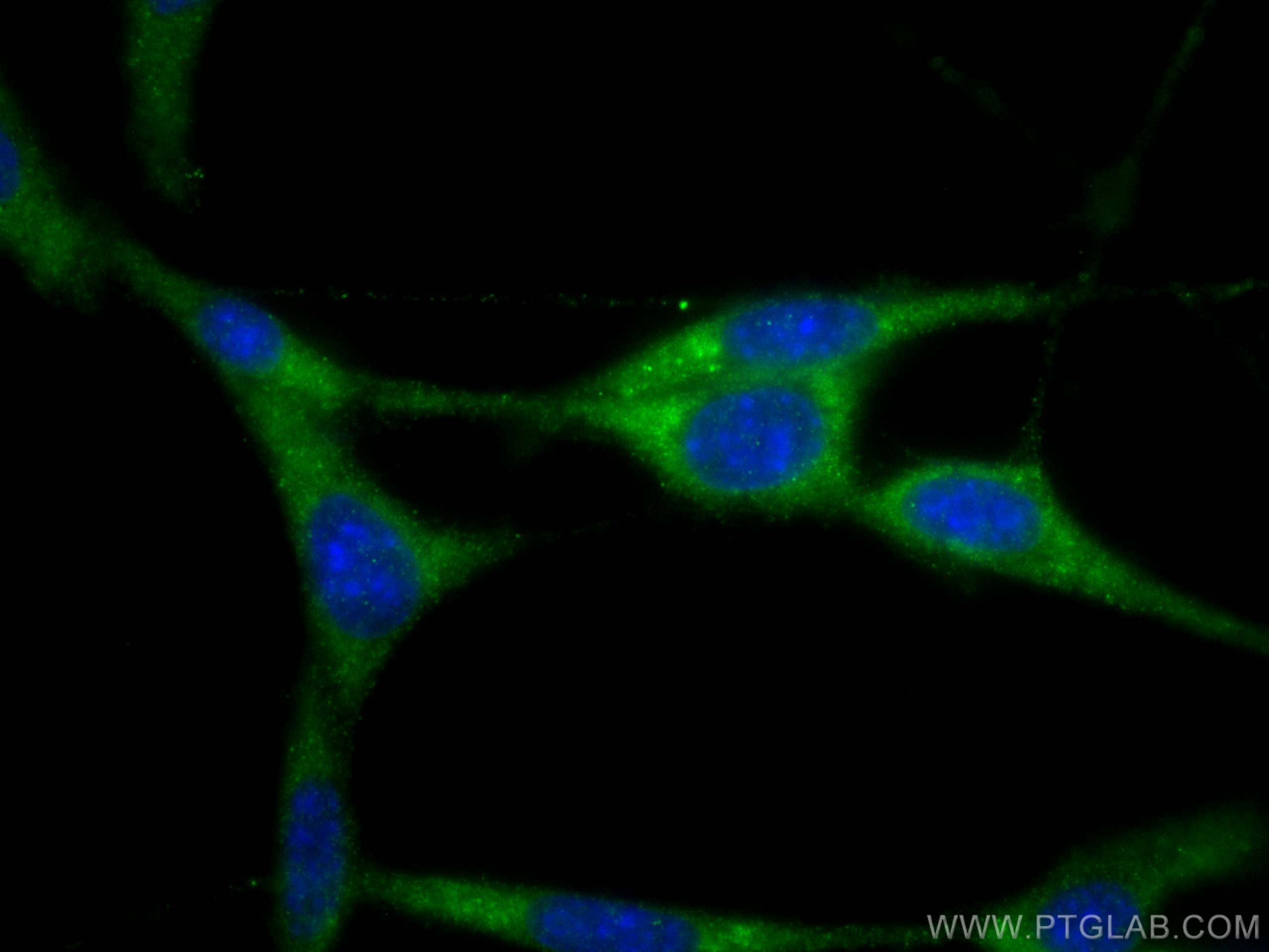
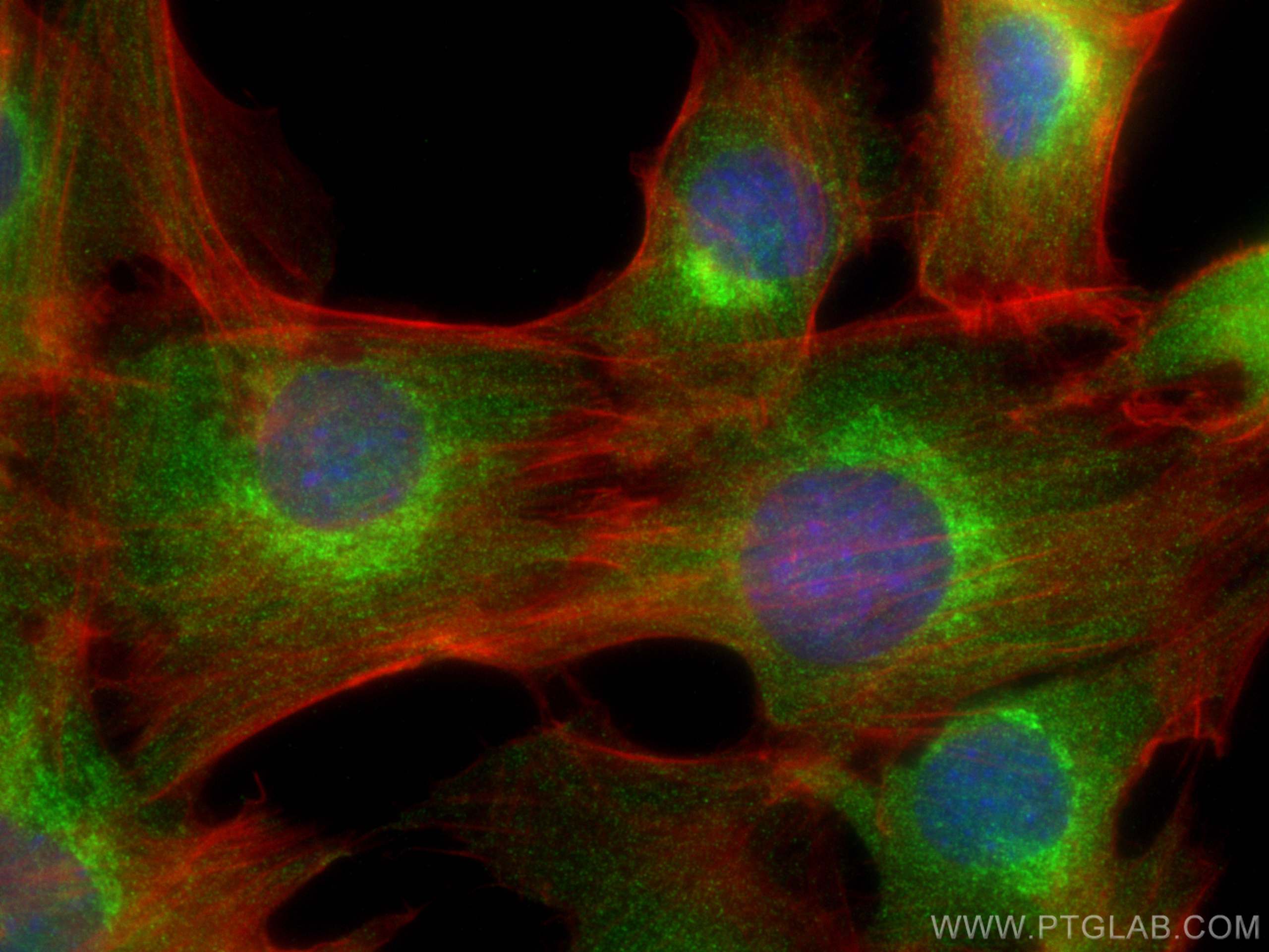
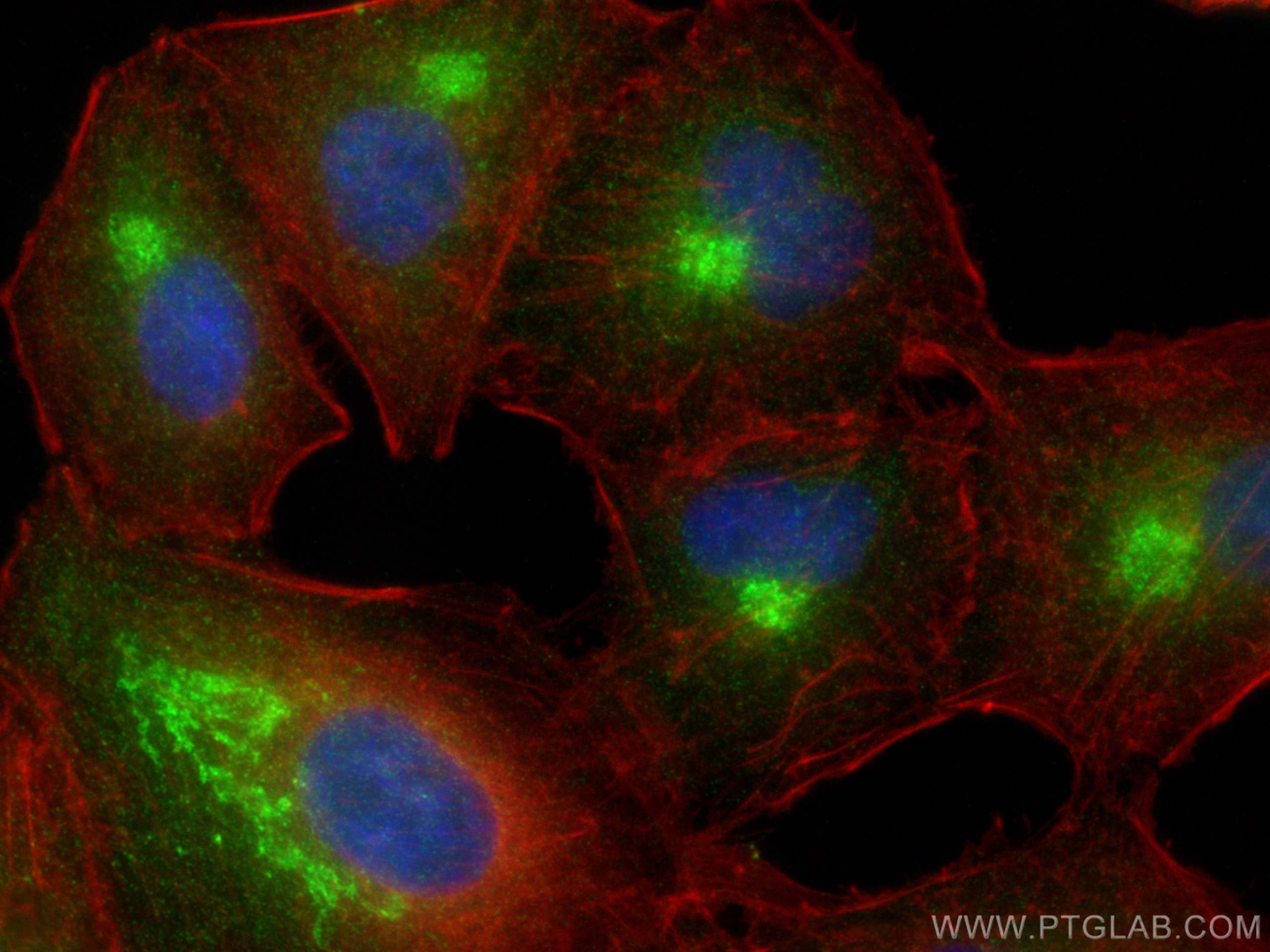
| Positive IF/ICC detected in | C2C12 cells, NIH/3T3 cells, A549 cells |
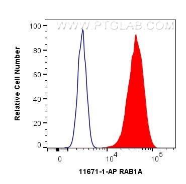
| Positive FC (Intra) detected in | C2C12 cells |
Recommended dilution
| Application | Dilution |
|---|---|
| Western Blot (WB) | WB : 1:2000-1:8000 |
| Immunoprecipitation (IP) | IP : 0.5-4.0 ug for 1.0-3.0 mg of total protein lysate |
| Immunohistochemistry (IHC) | IHC : 1:50-1:500 |
| Immunofluorescence (IF)/ICC | IF/ICC : 1:200-1:800 |
| Flow Cytometry (FC) (INTRA) | FC (INTRA) : 0.40 ug per 10^6 cells in a 100 ul suspension |
| It is recommended that this reagent should be titrated in each testing system to obtain optimal results. | |
| Sample-dependent, Check data in validation data gallery. | |
Product Information
11671-1-AP targets RAB1A in WB, IHC, IF/ICC, FC (Intra), IP, CoIP, ELISA applications and shows reactivity with human, mouse, rat samples.
| Tested Reactivity | human, mouse, rat |
| Cited Reactivity | human, mouse, rat, pig |
| Host / Isotype | Rabbit / IgG |
| Class | Polyclonal |
| Type | Antibody |
| Immunogen |
CatNo: Ag2264 Product name: Recombinant human RAB1A protein Source: e coli.-derived, PGEX-4T Tag: GST Domain: 1-205 aa of BC000905 Sequence: MSSMNPEYDYLFKLLLIGDSGVGKSCLLLRFADDTYTESYISTIGVDFKIRTIELDGKTIKLQIWDTAGQERFRTITSSYYRGAHGIIVVYDVTDQESFNNVKQWLQEIDRYASENVNKLLVGNKCDLTTKKVVDYTTAKEFADSLGIPFLETSAKNATNVEQSFMTMAAEIKKRMGPGATAGGAEKSNVKIQSTPVKQSGGGCC Predict reactive species |
| Full Name | RAB1A, member RAS oncogene family |
| Calculated Molecular Weight | 205 aa, 23 kDa |
| Observed Molecular Weight | 23 kDa |
| GenBank Accession Number | BC000905 |
| Gene Symbol | RAB1A |
| Gene ID (NCBI) | 5861 |
| RRID | AB_2173437 |
| Conjugate | Unconjugated |
| Form | Liquid |
| Purification Method | Antigen affinity purification |
| UNIPROT ID | P62820 |
| Storage Buffer | PBS with 0.02% sodium azide and 50% glycerol, pH 7.3. |
| Storage Conditions | Store at -20°C. Stable for one year after shipment. Aliquoting is unnecessary for -20oC storage. 20ul sizes contain 0.1% BSA. |
Background Information
Rab1A is a member of the Rab family of small GTPases with a well characterized function in the regulation of vesicle trafficking from the endoplasmic reticulum to the Golgi apparatus and within Golgi compartments. Rab1A has recently been reported an mTORC1 activator and a colorectal oncogene (Janice D. Thomas. et al. 2014. Cancer cell). RAB1A may be cross-reactions with both RAB1B and RAB1C.
Protocols
| Product Specific Protocols | |
|---|---|
| FC protocol for RAB1A antibody 11671-1-AP | Download protocol |
| IF protocol for RAB1A antibody 11671-1-AP | Download protocol |
| IHC protocol for RAB1A antibody 11671-1-AP | Download protocol |
| IP protocol for RAB1A antibody 11671-1-AP | Download protocol |
| WB protocol for RAB1A antibody 11671-1-AP | Download protocol |
| Standard Protocols | |
|---|---|
| Click here to view our Standard Protocols |
Publications
What published studies show
Able is busy answering your previous question. Please try again later.
| Species | Application | Title |
|---|---|---|
Autophagy RAB2 regulates the formation of autophagosome and autolysosome in mammalian cells.
| ||
Adv Sci (Weinh) ARL5B Drives Esophageal Squamous Cell Carcinoma Progression via ROCK1-SREBP1-Mediated Lipid Metabolic Reprogramming | ||
Cancer Lett Rab1A promotes IL-4R/JAK1/STAT6-dependent metastasis and determines JAK1 inhibitor sensitivity in non-small cell lung cancer.
| ||
Cell Commun Signal Salmonella Typhimurium hijacks a host glucose transporter for intravacuolar proliferation. | ||
JACS Au Targeting GDP-Dissociation Inhibitor Beta (GDI2) with a Benzo[a]quinolizidine Library to Induce Paraptosis for Cancer Therapy |
Reviews
What customers say
Users report strong performance for this antibody in Western Blot and Immunofluorescence. Two reviewers (5/5) confirmed clear, expected bands and fluorescence in HEK293 cells and pancreatic beta cells using datasheet-recommended dilutions (e.g., 1:1000 for WB), and both highly recommend the product. One reviewer (4/5) noted the antibody recognized the expected molecular weight band but observed a dirty background. Overall, the antibody is well-received for its specificity and reliable signal, with minor background issues in one case.
The reviews below have been submitted by verified Proteintech customers who received an incentive for providing their feedback.
Victoria (Verified Customer) (10-09-2025) | Great antibody
|
Laura (Verified Customer) (08-15-2025) | Antibody worked great in WB and IF. Bands and fluorescence were expected. Dilutions as per data sheet. I totally recommend.
|
Boyan (Verified Customer) (02-22-2024) | recognized a band around the expected MW, but this antibody gave a dirty background.
|




















