"Beta Tubulin Antibodies" Comparison
View side-by-side comparison of Beta Tubulin antibodies from other vendors to find the one that best suits your research needs.
Tested Applications
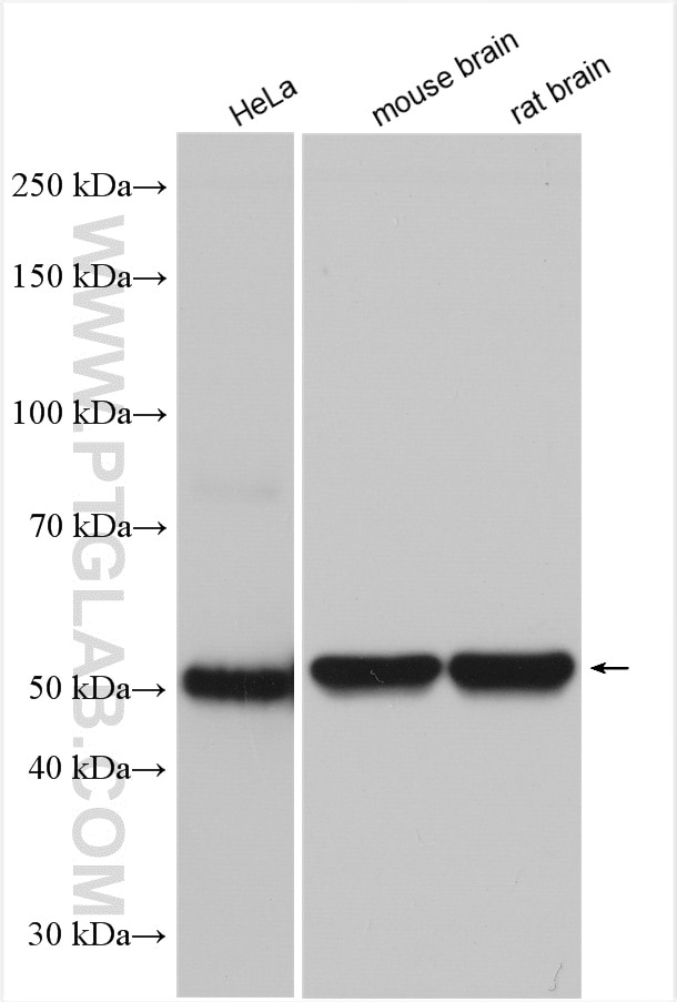
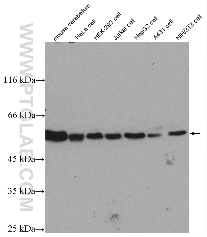
| Positive WB detected in | HeLa cells, mouse cerebellum tissue, mouse brain tissue, rat brain tissue, HEK-293 cells, Jurkat cells, HepG2 cells, A431 cells, NIH/3T3 cells |
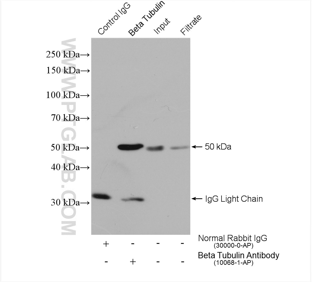
| Positive IP detected in | HEK-293 cells |
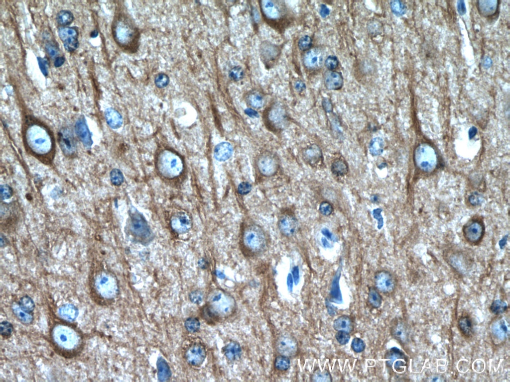
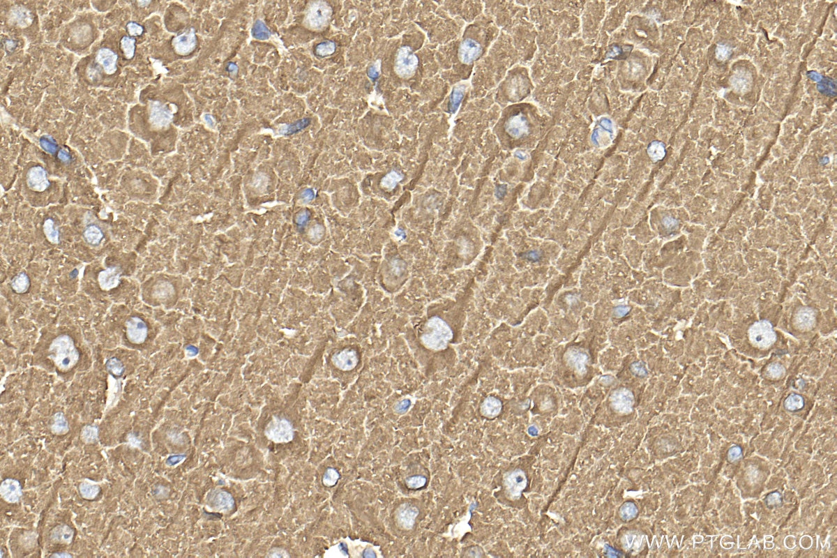
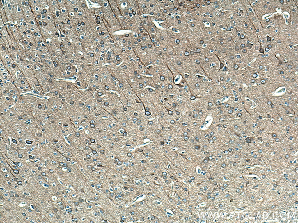
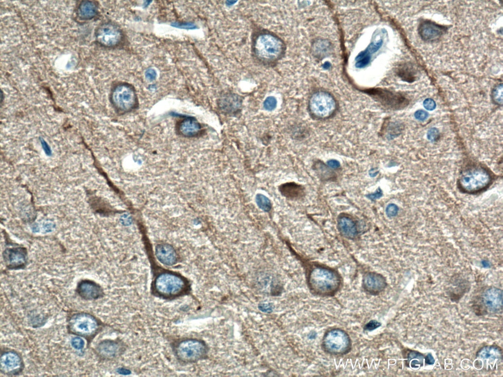
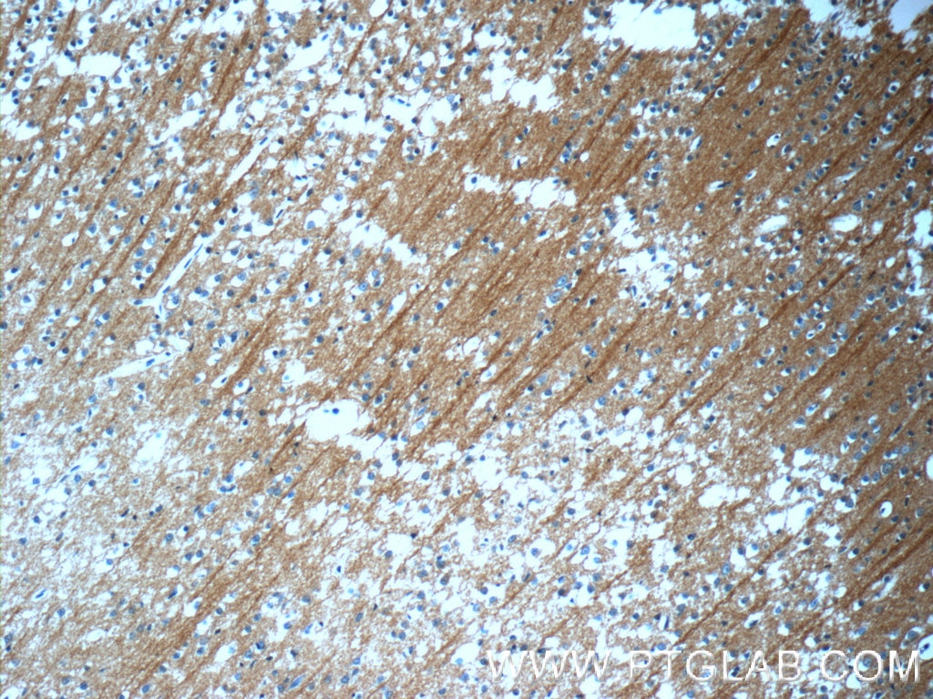
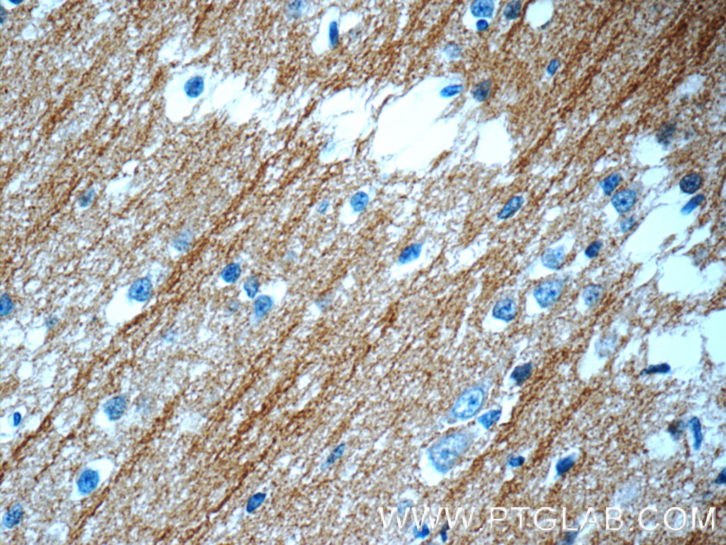
| Positive IHC detected in | rat brain tissue, human brain tissue, mouse brain tissue Note: suggested antigen retrieval with TE buffer pH 9.0; (*) Alternatively, antigen retrieval may be performed with citrate buffer pH 6.0 |
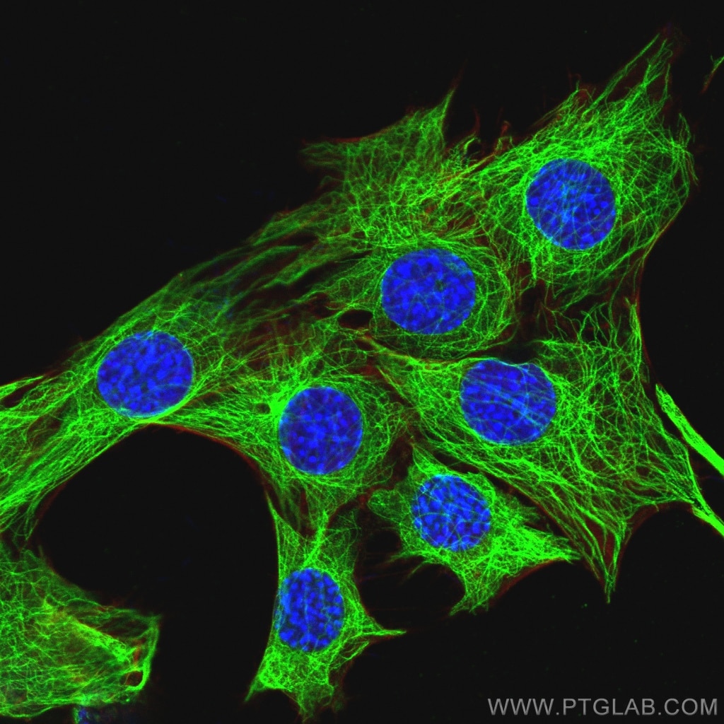
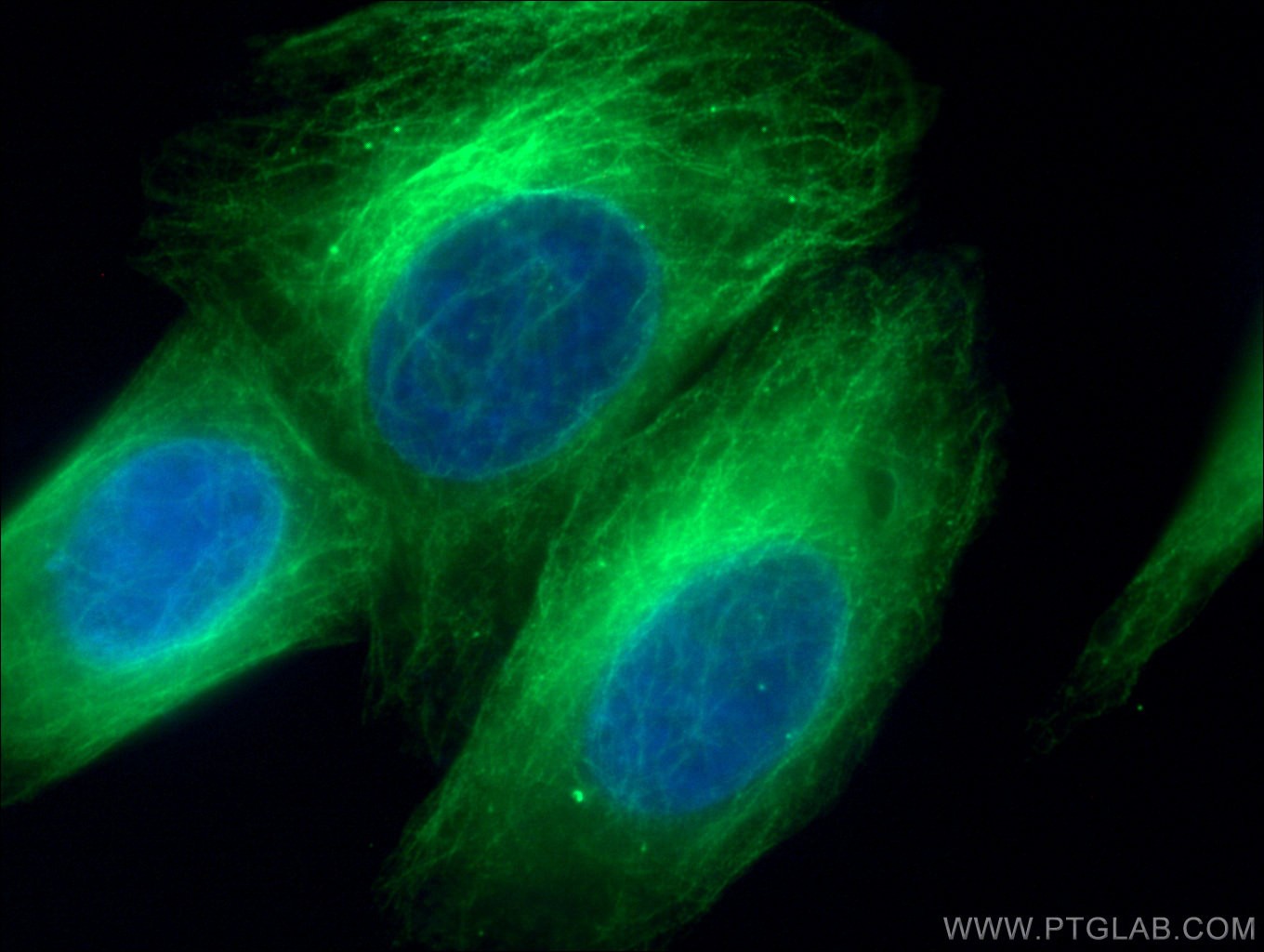
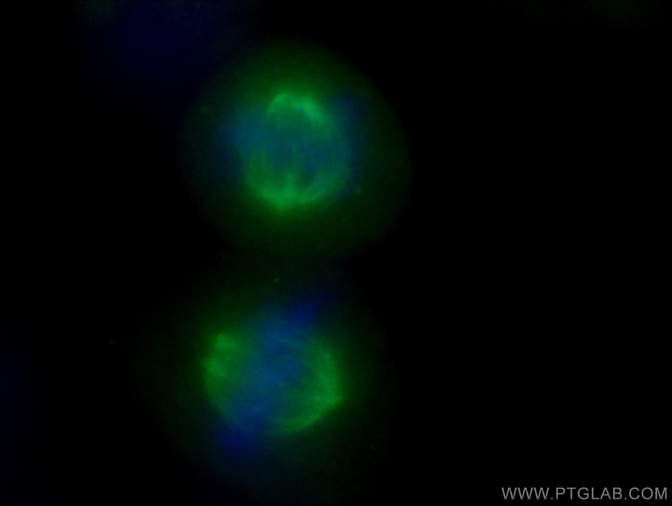
| Positive IF/ICC detected in | C2C12 cells, HepG2 cells |
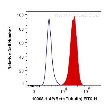
| Positive FC (Intra) detected in | HeLa cells |
Recommended dilution
| Application | Dilution |
|---|---|
| Western Blot (WB) | WB : 1:5000-1:16000 |
| Immunoprecipitation (IP) | IP : 0.5-4.0 ug for 1.0-3.0 mg of total protein lysate |
| Immunohistochemistry (IHC) | IHC : 1:200-1:800 |
| Immunofluorescence (IF)/ICC | IF/ICC : 1:200-1:800 |
| Flow Cytometry (FC) (INTRA) | FC (INTRA) : 0.40 ug per 10^6 cells in a 100 µl suspension |
| It is recommended that this reagent should be titrated in each testing system to obtain optimal results. | |
| Sample-dependent, Check data in validation data gallery. | |
Published Applications
| KD/KO | See 1 publications below |
| WB | See 567 publications below |
| IHC | See 5 publications below |
| IF | See 18 publications below |
| IP | See 3 publications below |
Product Information
10068-1-AP targets Beta Tubulin in WB, IHC, IF/ICC, FC (Intra), IP, ELISA applications and shows reactivity with human, mouse, rat samples.
| Tested Reactivity | human, mouse, rat |
| Cited Reactivity | human, mouse, rat, pig, canine, zebrafish, c.elegans, silkworm, caenorhabditis elegans |
| Host / Isotype | Rabbit / IgG |
| Class | Polyclonal |
| Type | Antibody |
| Immunogen |
CatNo: Ag0117 Product name: Recombinant human Tubulin-beta protein Source: e coli.-derived, PGEX-4T Tag: GST Domain: 44-259 aa of BC000748 Sequence: LERISVYYNEASSHKYVPRAILVDLEPGTMDSVRSGAFGHLFRPDNFIFGQSGAGNNWAKGHYTEGAELVDSVLDVVRKECENCDCLQGFQLTHSLGGGTGSGMGTLLISKVREEYPDRIMNTFSVVPSPKVSDTVVEPYNATLSIHQLVENTDETYCIDNEALYDICFRTLKLATPTYGDLNHLVSATMSGVTTSLRFPGQLNADLRKLAVNMVP Predict reactive species |
| Full Name | tubulin, beta 3 |
| Calculated Molecular Weight | 450 aa, 50 kDa |
| Observed Molecular Weight | 50-55 kDa |
| GenBank Accession Number | BC000748 |
| Gene Symbol | TUBB3 |
| Gene ID (NCBI) | 10381 |
| RRID | AB_2303998 |
| Conjugate | Unconjugated |
| Form | Liquid |
| Purification Method | Antigen affinity purification |
| UNIPROT ID | Q13509 |
| Storage Buffer | PBS with 0.02% sodium azide and 50% glycerol, pH 7.3. |
| Storage Conditions | Store at -20°C. Stable for one year after shipment. Aliquoting is unnecessary for -20oC storage. 20ul sizes contain 0.1% BSA. |
Background Information
There are five tubulins in human cells: alpha, beta, gamma, delta, and epsilon. Tubulins are conserved across species. They form heterodimers, which multimerize to form a microtubule filament. An alpha and beta tubulin heterodimer is the basic structural unit of microtubules. The heterodimer does not come apart, once formed. The alpha and beta tubulins, which are each about 55 kDa MW, are homologous but not identical. Alpha, beta, and gamma tubulins have all been used as loading controls. Tubulin expression may vary according to resistance to antimicrobial and antimitotic drugs.
Protocols
| Product Specific Protocols | |
|---|---|
| FC protocol for Beta Tubulin antibody 10068-1-AP | Download protocol |
| IF protocol for Beta Tubulin antibody 10068-1-AP | Download protocol |
| IHC protocol for Beta Tubulin antibody 10068-1-AP | Download protocol |
| IP protocol for Beta Tubulin antibody 10068-1-AP | Download protocol |
| WB protocol for Beta Tubulin antibody 10068-1-AP | Download protocol |
| Standard Protocols | |
|---|---|
| Click here to view our Standard Protocols |
Publications
| Species | Application | Title |
|---|---|---|
Cell Complement Signals Determine Opposite Effects of B Cells in Chemotherapy-Induced Immunity. | ||
Adv Mater Noninvasive Optogenetics Realized by iPSC-Derived Tentacled Carrier in Alzheimer's Disease Treatment | ||
Bioact Mater Delivering LINE1 antisense oligonucleotides via endothelial targeting extracellular vesicles to ameliorate myocardial infarction-induced cardiac senescence | ||
Nat Struct Mol Biol CRISPR-Cas9-based functional interrogation of unconventional translatome reveals human cancer dependency on cryptic non-canonical open reading frames | ||
Nat Commun MYG1 drives glycolysis and colorectal cancer development through nuclear-mitochondrial collaboration | ||