
Antibodies for ER Stress
ER Stress Research Focus
|
Introduction to the Endoplasmic Reticulum Popular Endoplasmic Reticulum Markers ER Stress In Neurodegenerative Diseases |
Introduction
The endoplasmic reticulum (ER) is a cellular organelle responsible for lipid or steroid synthesis, folding or maturation of proteins, calcium storage, and detoxification.
The ER consists of a membrane network of cisternae that goes through the cytoplasma and is in continuous connection with the nuclear envelope. The ER consists of two different regions that differ in their structure and function. The rough ER contains ribosomes attached to the cytoplasmic side of the membrane. The rough ER is mainly responsible for protein synthesis, while the smooth ER is lacking in ribosomes and functions as a storage for key enzymes and their products.
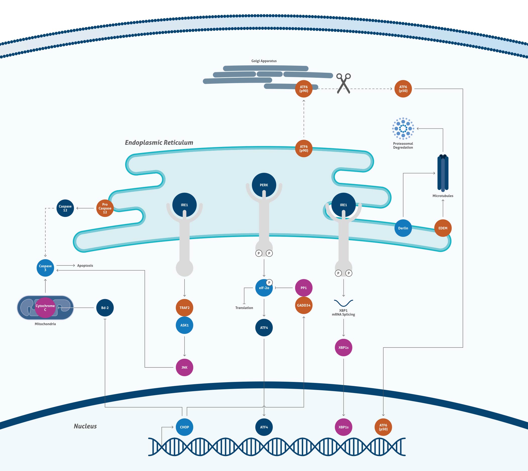
Cellular processes that lead to misfolding of proteins in the endoplasmic reticulum lead to ER stress and consequently to the activation of several signaling pathways named the unfolded protein response (UPR). ER stress has been shown to be connected to different neurodegenerative diseases, such as Parkinson’s or Alzheimer’s disease, inflammation, and cancer
Endoplasmic Reticulum

Most Popular Endoplasmic Reticulum Markers
| Antibody Name | Catalog number | Type |
| ERp57/ERp60 | 15967-1-AP | Rabbit Poly |
| ERp72 | 14712-1-AP | Rabbit Poly |
| GRP94 | 14700-1-AP | Rabbit Poly |
| PDI | 11245-1-AP | Rabbit Poly |
| TAP1 | 11114-1-AP | Rabbit Poly |
 |
|
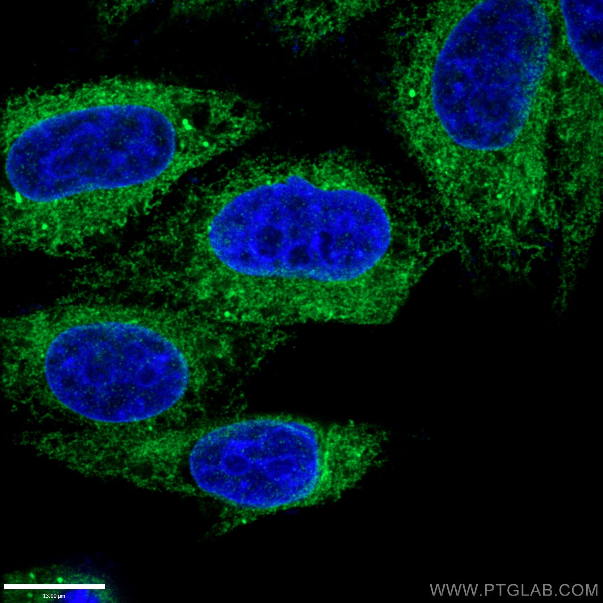
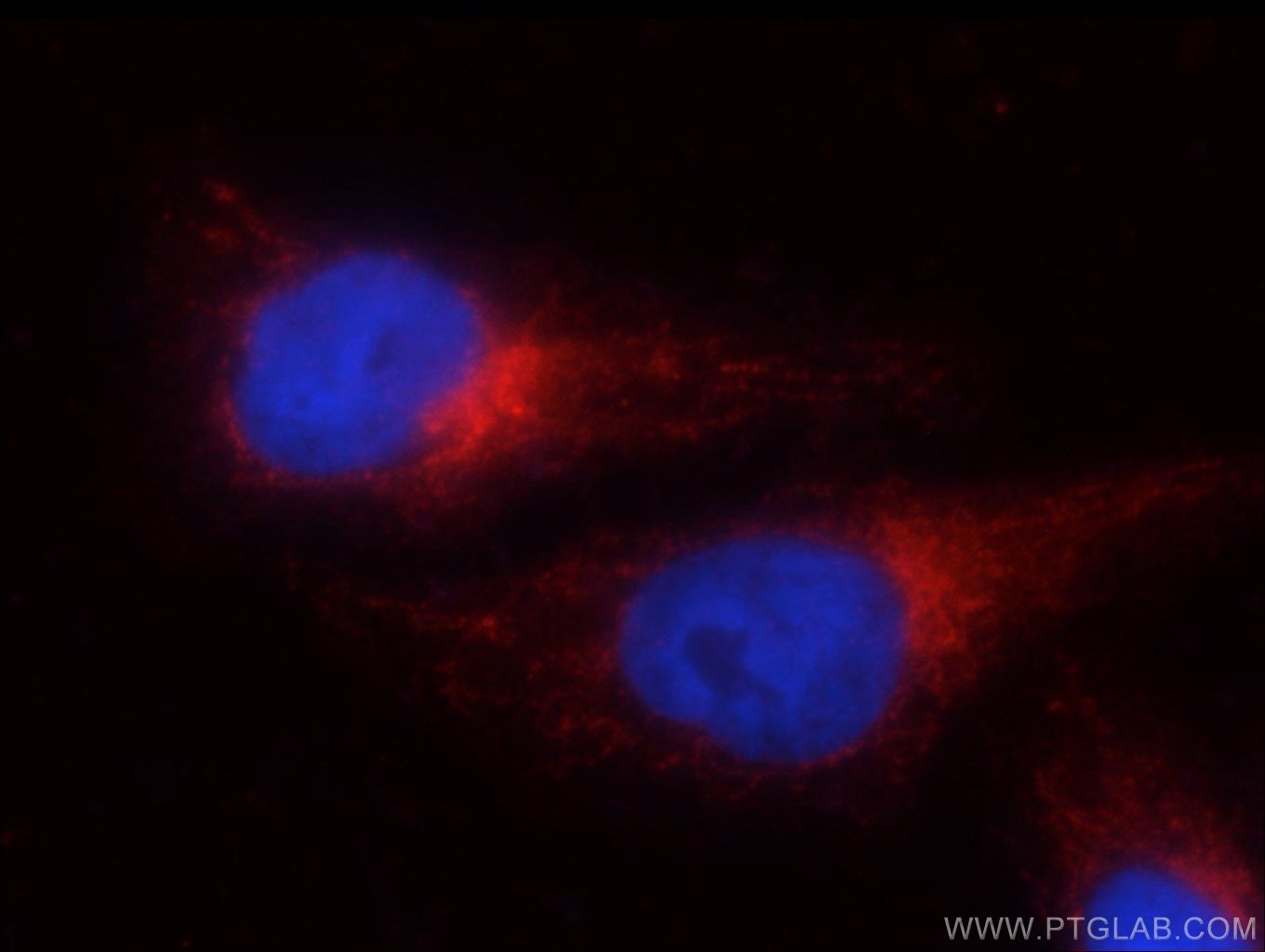
Immunofluorescence staining of fixed HepG2 cells (10% Formaldehyde) using ERp57/ERp60 antibody (15967-1 AP) at a dilution of 1:50 and goat anti-rabbit IgG(H+L) Alexa Fluor 488-conjugated secondary antibody. |
 |
|
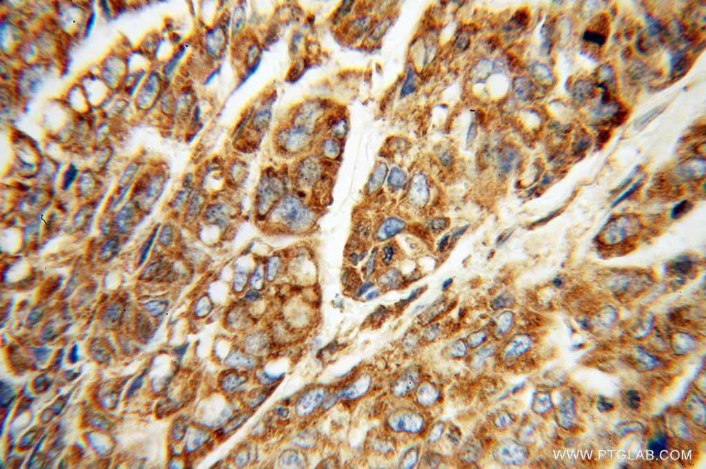
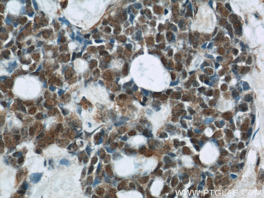
Immunohistochemical staining of paraffinembedded human lung cancer using ERp72 antibody (14712-1-AP) at a dilution of 1:100 (40x objective). |
ER stress is mainly caused by disturbance of the calcium homeostasis and accumulation of misfolded proteins. The ER responds by changing its protein profile, altering its cellular signalling and degradation of misfolded proteins. ER stress has been reported to be involved in different neurodegenerative diseases such as Alzheimer’s or Parkinson’s. To date, the exact mechanism and contribution of ER stress to these diseases remain unknown. Elucidation of the involvement of ER stress in neurodegeneration might help in the development of new neuroprotective therapies.
Most Popular Neurodegenerative Related Markers
| Antibody name | Catalog number | Type |
| AKT1 | 10176-2-AP | Rabbit Poly |
| alpha-synuclein | 10842-1-AP | Rabbit Poly |
| Amyloid beta | 60342-1-Ig | Mouse Mono |
| Tau | 10274-1-AP | Rabbit Poly |
| TDP-43 | 10782-2-AP | Rabbit Poly |
| PARK7, DJ-1 | 11681-1-AP | Rabbit Poly |
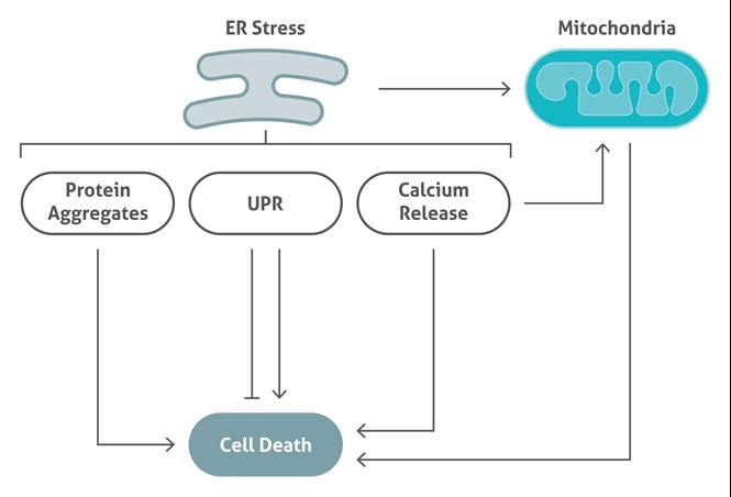
Disruption of the endoplasmic reticulum homeostasis leads to an accumulation of misfolded or unfolded proteins. This stage is called ER stress. ER stress is responsible for the activation of multiple signaling pathways, named the unfolded protein response (UPR). ER stress mainly induces cell dysfunction and cell death. Better understanding of ER stress and related UPR will help to elucidate new targets for ER stress-related diseases.
Related Antibodies
| Antibody Name | Catalog Number | Type |
| ASK1 | 14385-1-AP | Rabbit Poly |
| ATF4 | 10835-1-AP | Rabbit Poly |
| BCL-XL | 12789-1-AP | Rabbit Poly |
| Calnexin | 10427-2-AP | Rabbit Poly |
| Caspase3 | 19677-1-AP | Rabbit Poly |
| Caspase12 | 55238-1-AP | Rabbit Poly |
| Cytochrome C | 66264-1-Ig | Mouse Mono |
| JNK | 51151-1-AP | Rabbit Poly |
| XBP1 | 25997-1-AP | Rabbit Poly |
CHOP |
|
|
|
Catalog Number Type Rabbit Polyclonal Applications ELISA, FC, IHC, WB 27 Publications
|
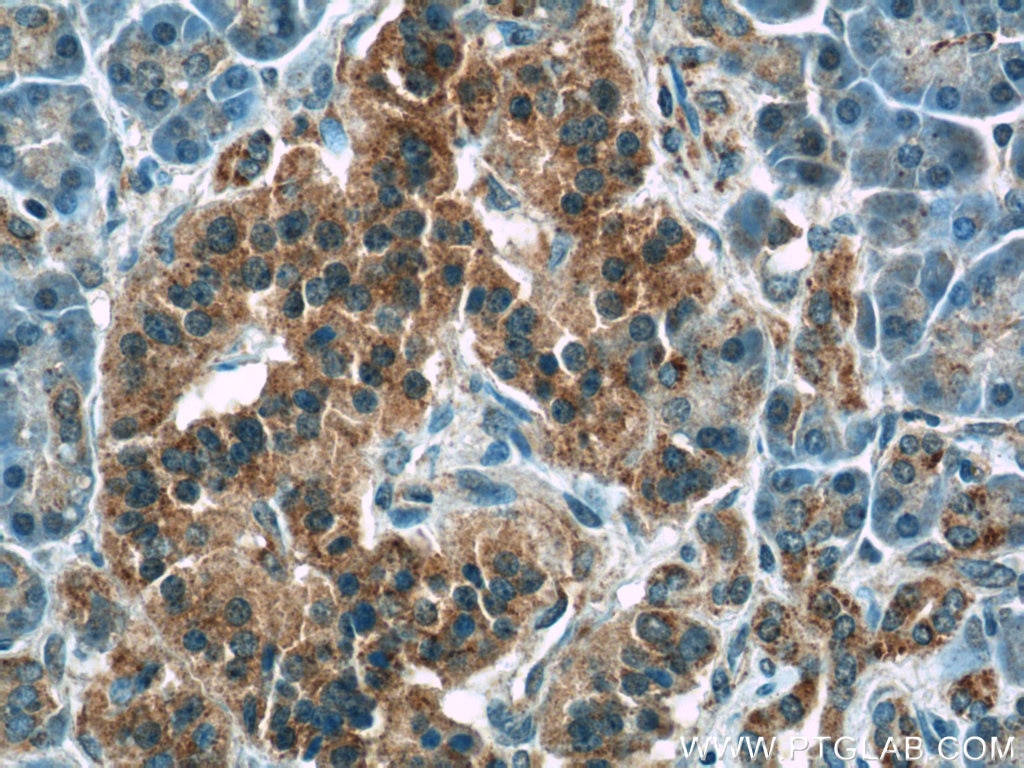
| Immunohistochemical staining of paraffin-embedded human cervical cancer using CHOP antibody(15204-1-AP) at a dilution of 1:50 (40x objective) | |
|
CHOP, also known as GADD153 or DDIT3, is a highly conserved gene in both the structural and regulatory regions of the hamster gene. Activated in response to unfolded and misfolded proteins, CHOP is significantly induced by ER stress. CHOP deficiency prevents ER stress in cells. CHOP is considered a pro-apoptotic marker of ER stress-dependent cell death. It acts as a dominant-negative inhibitor of the transcription factor C/EBP and LAP by forming a heterodimer. It may also play an important role in the malignant transformation of nevi to melanoma. |
|

GRP78 |
|
 |
Catalog number Type Rabbit Polyclonal Applications ELISA, FC, IF, IHC, IP, WB 38 Publications |
| Immunofluorescence staining of HepG2 cells using GRP78 antibody (11587-1-AP) at a dilution of 1:25 and goat anti-rabbit IgG rhodamine-labelled secondary antibody. |
|
|
GRP78 (HSPA5), also referred to as ‘immunoglobulin heavy chain-binding protein’ (BiP), is a member of the heat-shock protein-70 (HSP70) family and is involved in the folding and assembly of proteins in the endoplasmic reticulum (ER). It is a constitutively expressed resident protein of the ER in all eukaryotic cells. Recently it has been reported that GRP78 is associated with apoptosis or inhibition of cancer cell growth. |
|
PERK |
|
 |
Catalog number Type Rabbit Polyclonal Applications ELISA, IF, IHC, WB 1 Publication |
| Immunohistochemical staining of paraffin-embedded human placenta slide using PERK antibody (24390-1-AP) at a dilution of 1:50 | |
|
PERK is also known as PEK, EIF2AK3 (Eukaryotic translation initiation factor 2-alpha kinase 3), and belongs to the GCN2 subfamily. It potentially acts as a metabolic sensor in the insulin-secreting beta-cells to modulate the trafficking and quality control of proinsulin in the ER relative to the physiological demands for circulating insulin. PERK and EIF2AK3 also have a functional role in regulating translation under non-stressed conditions, in addition to their long-established roles as stress kinases. |
|
ATF6 |
|
 |
Catalog number Type Rabbit Polyclonal Applications ELISA, IF, IHC, IP, WB 6 Publications
|
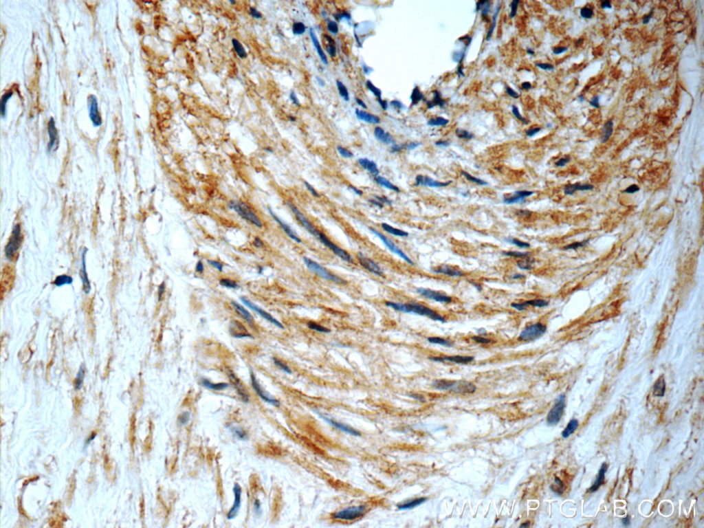
| Immunohistochemical staining of paraffin-embedded human pancreas tissue slide using ATF6 antibody (24169-1-AP) at a dilution of 1:50 (40x objective). | |
|
Activating Transcription Factor 6 (ATF6), as the name suggests, is a transcription factor. It increases the expression of unfolded protein response target genes in response to ER stress. It binds DNA on the 5’-CCAC[GA]-3’ half of the ER stress response element (ERSE) (5’-CCAAT-N(9)-CCAC[GA]-3’) and of ERSE II (5’-ATTGG-N-CCACG-3’). During unfolded protein response an approximative 50 kDa fragment containing the cytoplasmic transcription factor domain is released by proteolysis. The cleavage seemso be performed sequentially by site-1 and site-2 proteases. The fully glycosylated form of ATF6, a 670 amino acid protein, exhibits an electrophoretic mobility of ~90 kDa in denaturing SDS-gels, in part because of the glycosylated modifications. ATF6 has 3 consensus sites for N-linked glycosylation and exists constitutively as a glycosylated protein. Differentially glycosylated ATF6 forms may result from mutations or experimental treatment. |
|
More Endoplasmic Reticulum Related Markers
Antibodies specific for proteins of the endoplasmic reticulum serve to provide more detailed understanding of the molecular mechanisms regulating ER stress and to elucidate the roles of proteins related to several diseases.
| Antibody Name | Catalog Number | Type |
| APP | 60342-1-Ig | Mouse Mono |
| BCHE | 23854-1-AP | Rabbit Poly |
| CALR | 10292-1-AP | Rabbit Poly |
| DLG4 | 20665-1-AP | Rabbit Poly |
| HMOX1 | 10701-1-AP | Rabbit Poly |
| HMOX2 | 14817-1-AP | Rabbit Poly |
| HSPA1A | 10995-1-AP | Rabbit Poly |
| IFNG | 15365-1-AP | Rabbit Poly |
| P4HB | 11245-1-AP | Rabbit Poly |
| PD1A4 | 14712-1-AP | Rabbit Poly |
| SERCA2 | 13985-1-AP | Rabbit Poly |
| TAP1 | 11114-1-AP | Rabbit Poly |
| UGGT1 | 14170-1-AP | Rabbit Poly |
| VCP | 10736-1-AP | Rabbit Poly |
Loading control antibodies
| GAPDH Antibody |  |
| Catalog no.: 60004-1-Ig | |
|
GAPDH is commonly used as a protein loading control in western blot due to its consistently high expression in most cell types. This enzyme participates in several cellular events such as glycolysis, DNA repair, and apoptosis. Proteintech monoclonal GAPDH antibodies are raised against a whole-protein antigen of human origin and have over 4,960 citations. |
| Beta Actin Antibody (KD/KO validated) |  |
| Catalog no.: 66009-1-Ig | |
|
Beta-actin is usually used as a loading control due to its broad and consistent expression across all eukaryotic cell types and the fact that expression levels of this protein are not affected by most experimental treatments. 66009-1-Ig has been cited in over 2,460 publications and has wide species reactivity. |
Able™