- Featured Product
- KD/KO Validated
Lamin B1 Polyklonaler Antikörper
Lamin B1 Polyklonal Antikörper für WB, IHC, IF/ICC, IF-P, FC (Intra), IP, ELISA
Wirt / Isotyp
Kaninchen / IgG
Getestete Reaktivität
human, Maus, Ratte und mehr (6)
Anwendung
WB, IHC, IF/ICC, IF-P, FC (Intra), IP, CoIP, ChIP, ELISA
Konjugation
Unkonjugiert
Kat-Nr. : 12987-1-AP
Synonyme
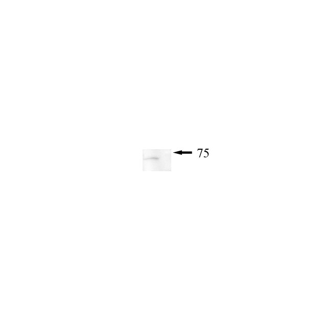
 at dilution of 1:20000 incubated at room temperature for 1.5 hours.")
 at dilution of 1:20000 incubated at room temperature for 1.5 hours.")
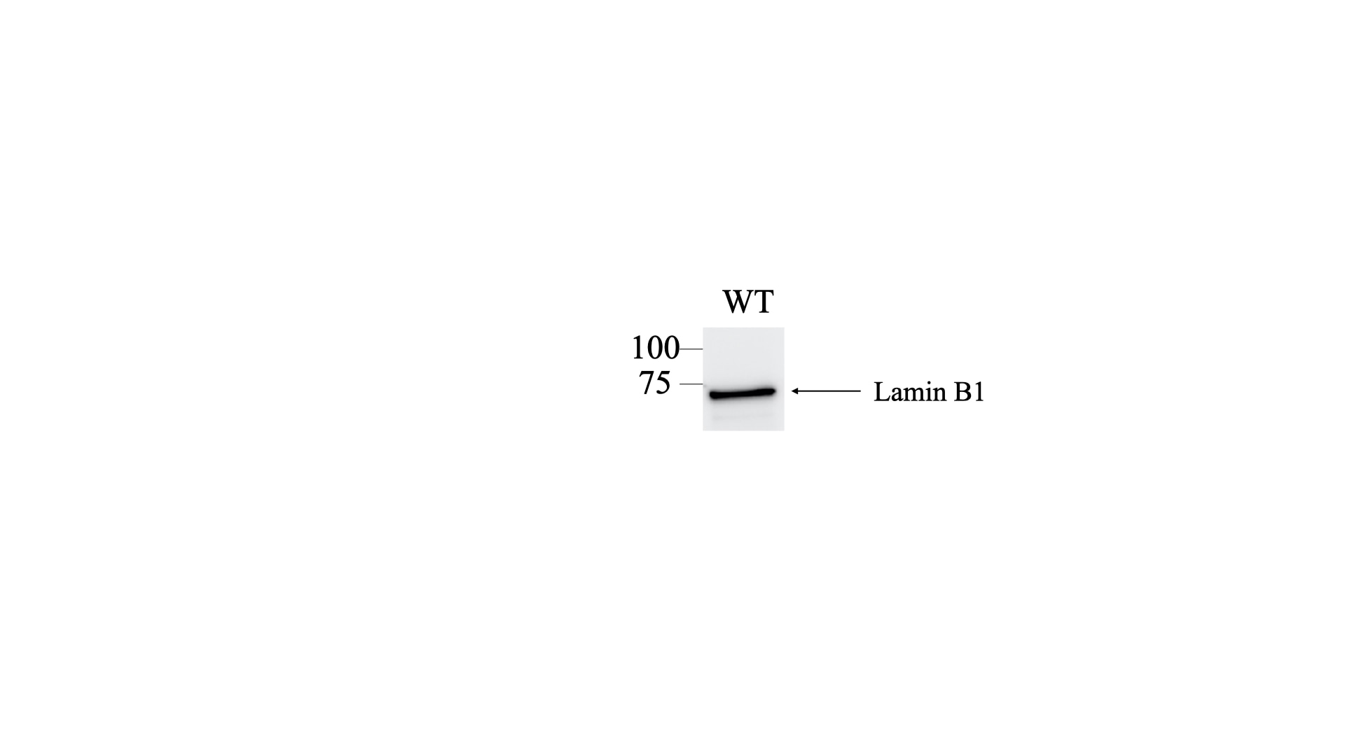
 at dilution of 1:10000 incubated at room temperature for 1.5 hours.")
 with HeLa cells lysate 1320 ug.")
 at dilution of 1:100 (under 10x lens). Heat mediated antigen retrieval with Tris-EDTA buffer (pH 9.0).")
 at dilution of 1:1000 (under 10x lens). Heat mediated antigen retrieval with Tris-EDTA buffer (pH 9.0).")
 at dilution of 1:2000 (under 10x lens). Heat mediated antigen retrieval with Tris-EDTA buffer (pH 9.0).")
 at dilution of 1:2000 (under 40x lens). Heat mediated antigen retrieval with Tris-EDTA buffer (pH 9.0).")
 at dilution of 1:4000 (under 20x lens). Heat mediated antigen retrieval with Tris-EDTA buffer (pH 9.0).")
 at dilution of 1:1200 (under 20x lens). Heat mediated antigen retrieval with Tris-EDTA buffer (pH 9.0).")
 at dilution of 1:4000 (under 20x lens). Heat mediated antigen retrieval with Tris-EDTA buffer (pH 9.0).")
 at dilution of 1:50.")
 at dilution of 1:50.")
 fixed human skin cancer tissue using 12987-1-AP (Lamin B1 antibody) at dilution of 1:50 and Alexa Fluor 488-conjugated AffiniPure Goat Anti-Rabbit IgG(H+L).")
 fixed HepG2 cells using 12987-1-AP (Lamin B1 antibody) at dilution of 1:200 and Alexa Fluor 488-conjugated AffiniPure Goat Anti-Rabbit IgG(H+L).")
 fixed HepG2 cells using Lamin B1 antibody (12987-1-AP) at dilution of 1:400 and CoraLite®488-Conjugated AffiniPure Goat Anti-Rabbit IgG(H+L), CL594-Phalloidin (red).")
 fixed NIH/3T3 cells using 12987-1-AP (Lamin B1 antibody) at dilution of 1:100 and Alexa Fluor 488-conjugated AffiniPure Goat Anti-Rabbit IgG(H+L).")
.")
 and CoraLite®488-Conjugated AffiniPure Goat Anti-Rabbit IgG(H+L) at dilution 1:1000 (red), or 0.4 ug Isotype Control. Cells were fixed with 4% PFA and permeabilized with Flow Cytometry Perm Buffer (PF00011-C).")
"Lamin B1 Antibodies" Comparison
View side-by-side comparison of Lamin B1 antibodies from other vendors to find the one that best suits your research needs.
Geprüfte Anwendungen
| Erfolgreiche Detektion in WB | HeLa-Zellen, HepG2-Zellen, Jurkat-Zellen, K-562-Zellen, MCF-7-Zellen, NIH/3T3-Zellen, RAW 264.7-Zellen |
| Erfolgreiche IP | HeLa-Zellen |
| Erfolgreiche Detektion in IHC | Mausherzgewebe, humanes Kolonkarzinomgewebe, humanes Kolongewebe, humanes Nierengewebe, humanes Leberkarzinomgewebe, Mausnierengewebe Hinweis: Antigendemaskierung mit TE-Puffer pH 9,0 empfohlen. (*) Wahlweise kann die Antigendemaskierung auch mit Citratpuffer pH 6,0 erfolgen. |
| Erfolgreiche Detektion in IF-P | humanes Hautkrebsgewebe |
| Erfolgreiche Detektion in IF/ICC | HepG2-Zellen, humanes Hautkrebsgewebe, NIH/3T3-Zellen |
| Erfolgreiche Detektion in FC (Intra) | HEK-293-Zellen |
Empfohlene Verdünnung
| Anwendung | Verdünnung |
|---|---|
| Western Blot (WB) | WB : 1:5000-1:50000 |
| Immunpräzipitation (IP) | IP : 0.5-4.0 ug for 1.0-3.0 mg of total protein lysate |
| Immunhistochemie (IHC) | IHC : 1:1000-1:4000 |
| Immunfluoreszenz (IF)-P | IF-P : 1:50-1:500 |
| Immunfluoreszenz (IF)/ICC | IF/ICC : 1:200-1:800 |
| Durchflusszytometrie (FC) (INTRA) | FC (INTRA) : 0.40 ug per 10^6 cells in a 100 µl suspension |
| It is recommended that this reagent should be titrated in each testing system to obtain optimal results. | |
| Sample-dependent, check data in validation data gallery | |
Veröffentlichte Anwendungen
| KD/KO | See 5 publications below |
| WB | See 1244 publications below |
| IHC | See 11 publications below |
| IF | See 69 publications below |
| IP | See 2 publications below |
| CoIP | See 1 publications below |
| ChIP | See 4 publications below |
Produktinformation
12987-1-AP bindet in WB, IHC, IF/ICC, IF-P, FC (Intra), IP, CoIP, ChIP, ELISA Lamin B1 und zeigt Reaktivität mit human, Maus, Ratten
| Getestete Reaktivität | human, Maus, Ratte |
| In Publikationen genannte Reaktivität | human, Affe, hamster, Hausschwein, Huhn, Hund, Maus, Ratte, Bombyx Mori |
| Wirt / Isotyp | Kaninchen / IgG |
| Klonalität | Polyklonal |
| Typ | Antikörper |
| Immunogen | Lamin B1 fusion protein Ag3631 |
| Vollständiger Name | lamin B1 |
| Berechnetes Molekulargewicht | 66 kDa |
| Beobachtetes Molekulargewicht | 66-70 kDa |
| GenBank-Zugangsnummer | BC012295 |
| Gene symbol | Lamin B1 |
| Gene ID (NCBI) | 4001 |
| Konjugation | Unkonjugiert |
| Form | Liquid |
| Reinigungsmethode | Antigen-Affinitätsreinigung |
| Lagerungspuffer | PBS with 0.02% sodium azide and 50% glycerol |
| Lagerungsbedingungen | Bei -20°C lagern. Nach dem Versand ein Jahr lang stabil Aliquotieren ist bei -20oC Lagerung nicht notwendig. 20ul Größen enthalten 0,1% BSA. |
Hintergrundinformationen
Lamins are components of the nuclear lamina, a fibrous layer on the nucleoplasmic side of the inner nuclear membrane, which is thought to provide a framework for the nuclear envelope and may also interact with chromatin. The nuclear lamina consists of a two-dimensional matrix of proteins located next to the inner nuclear membrane. The lamin family of proteins make up the matrix and are highly conserved in evolution. During mitosis, the lamina matrix is reversibly disassembled as the lamin proteins are phosphorylated. Vertebrate lamins consist of two types, A and B. This gene encodes one of the two B type proteins, B1. Expression of uncleavable mutant lamin A or B caused significant delays in the onset of chromatin condensation and nuclear shrinkage during apoptosis (PMID:11953316). This protein is not suitable for samples where the nuclear envelope has been removed.
Protokolle
| PRODUKTSPEZIFISCHE PROTOKOLLE | |
|---|---|
| WB protocol for Lamin B1 antibody 12987-1-AP | Protokoll herunterladen |
| IHC protocol for Lamin B1 antibody 12987-1-AP | Protokoll herunterladenl |
| IF protocol for Lamin B1 antibody 12987-1-AP | Protokoll herunterladen |
| IP protocol for Lamin B1 antibody 12987-1-AP | Protokoll herunterladen |
| FC protocol for Lamin B1 antibody 12987-1-AP | Download protocol |
| STANDARD-PROTOKOLLE | |
|---|---|
| Klicken Sie hier, um unsere Standardprotokolle anzuzeigen |
Publikationen
| Species | Application | Title |
|---|---|---|
Nat Biotechnol Drag-and-drop genome insertion of large sequences without double-strand DNA cleavage using CRISPR-directed integrases | ||
Cell Res Disruption of ER ion homeostasis maintained by an ER anion channel CLCC1 contributes to ALS-like pathologies | ||
Nature Stella safeguards the oocyte methylome by preventing de novo methylation mediated by DNMT1. | ||
Mol Cancer Diversifying the anthracycline class of anti-cancer drugs identifies aclarubicin for superior survival of acute myeloid leukemia patients | ||
Immunity 25-Hydroxycholesterol regulates lysosome AMP kinase activation and metabolic reprogramming to educate immunosuppressive macrophages |
Rezensionen
The reviews below have been submitted by verified Proteintech customers who received an incentive for providing their feedback.
FH Rajkumar (Verified Customer) (04-18-2025) | I used this antibody for HRP-based detection and it worked very well at the suggested dilution. The signal was strong. Highly recommend for similar applications.
|
FH Mi (Verified Customer) (02-21-2023) | Works great in human adipocytes.
|
FH Ning (Verified Customer) (01-28-2023) | Excellent antibody to probe lamin B1.
 |
FH Tsimafei (Verified Customer) (12-04-2022) | 20µg of total protein were loaded. Incubation for 2h at room temperature. Excellent antibody
 |
FH Alejandro (Verified Customer) (08-07-2022) | Works fine for flow cytometry
|
FH Iram (Verified Customer) (09-04-2020) | Very sharp bands for nuclear protein
|
FH Chun (Verified Customer) (12-05-2019) | Excellent antibody
|
FH Marco (Verified Customer) (09-02-2019) | good staining in conjunction with Alexa Fluor Plus 488 secondary
|

