Tested Applications
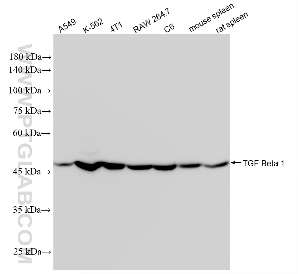
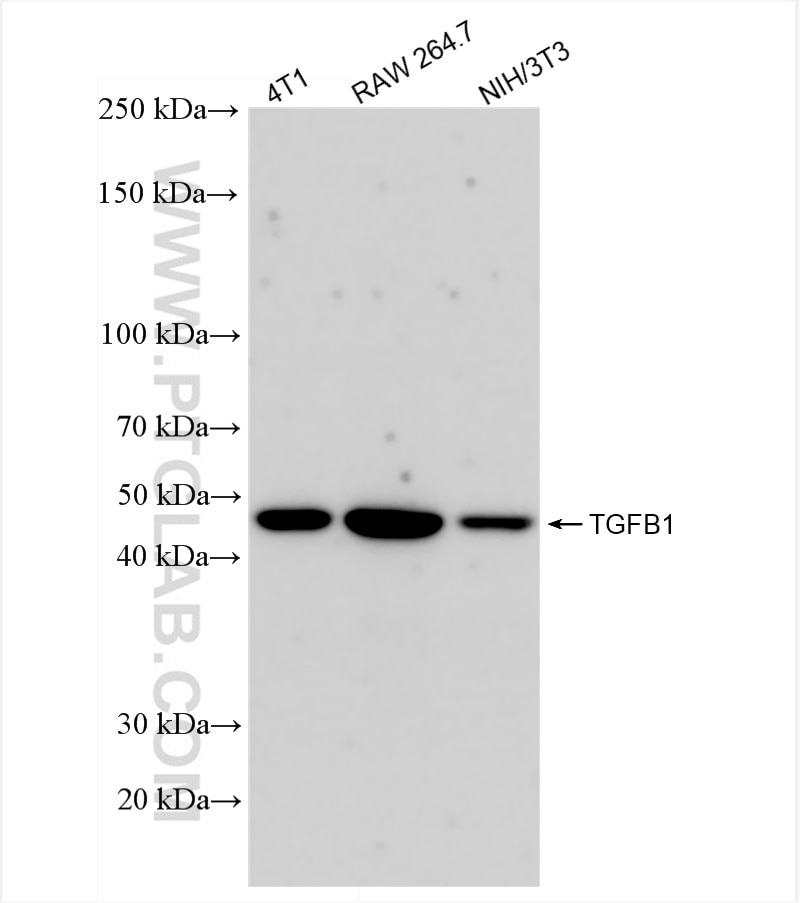
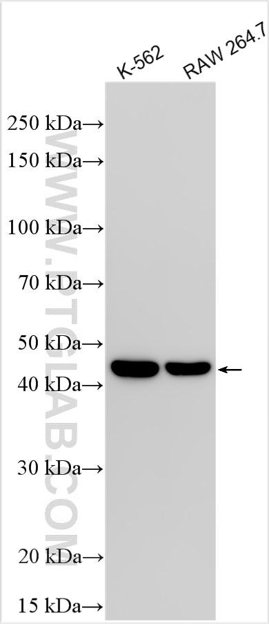
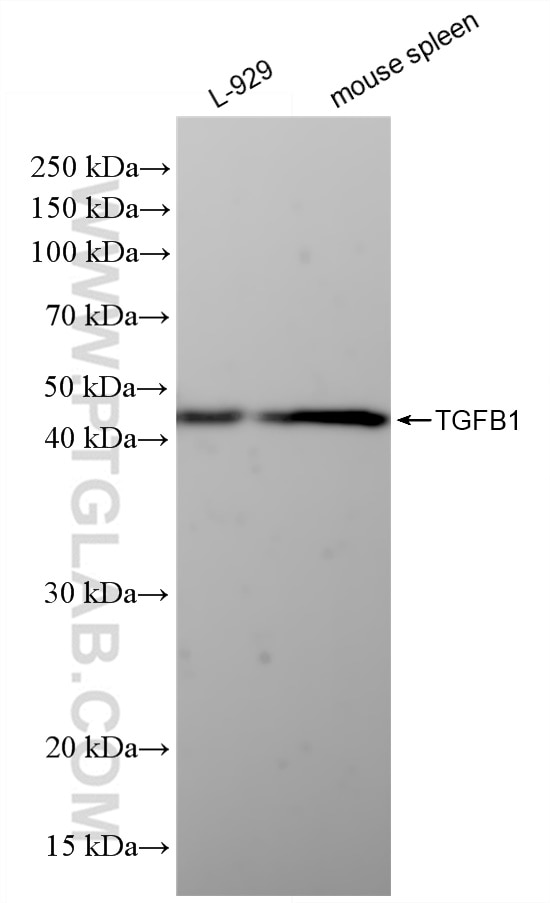
| Positive WB detected in | A549 cells, 4T1 cells, HepG2 cells, K-562 cells, L-929 cells, RAW 264.7 cells, C6 cells, mouse spleen tissue, rat spleen tissue |
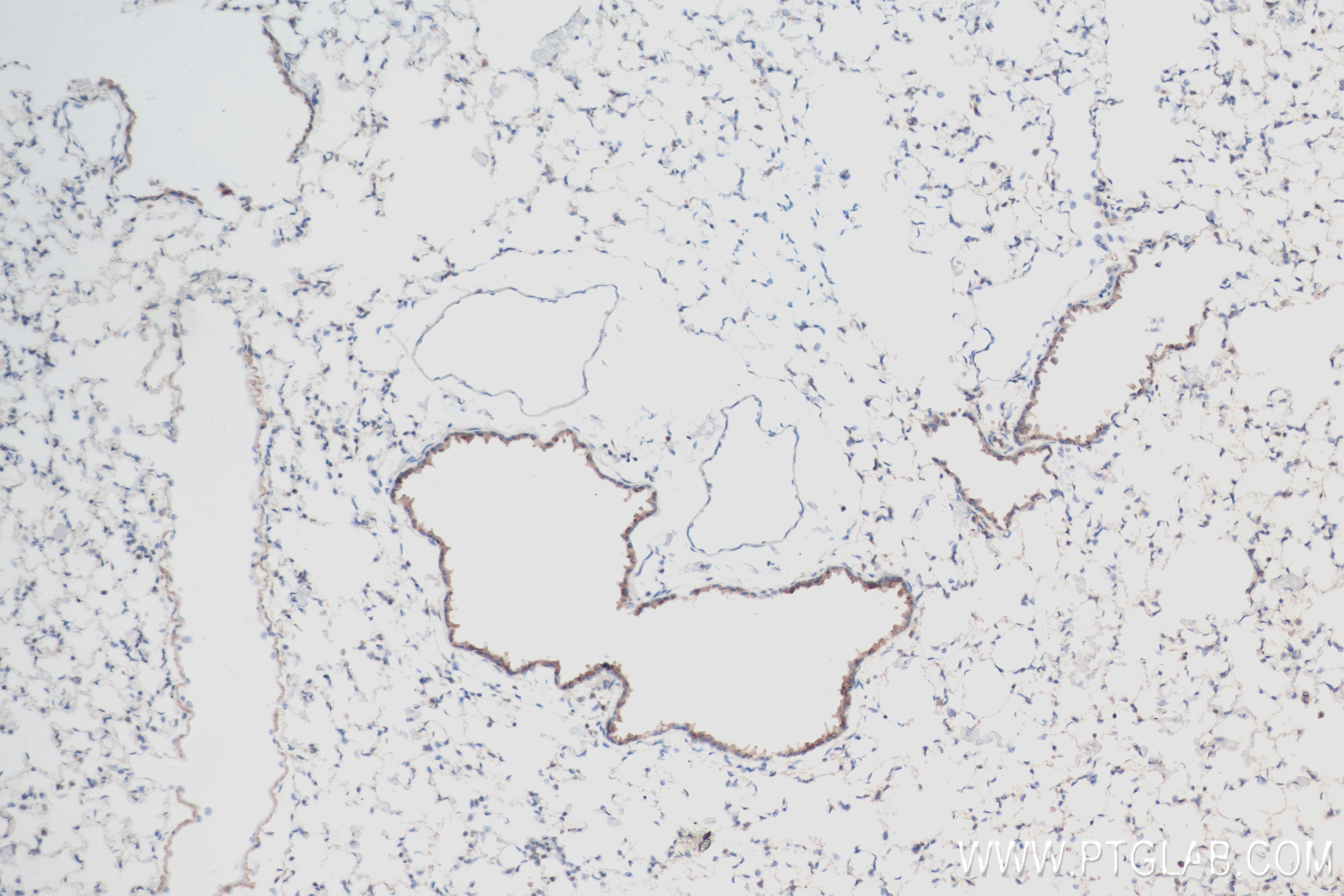
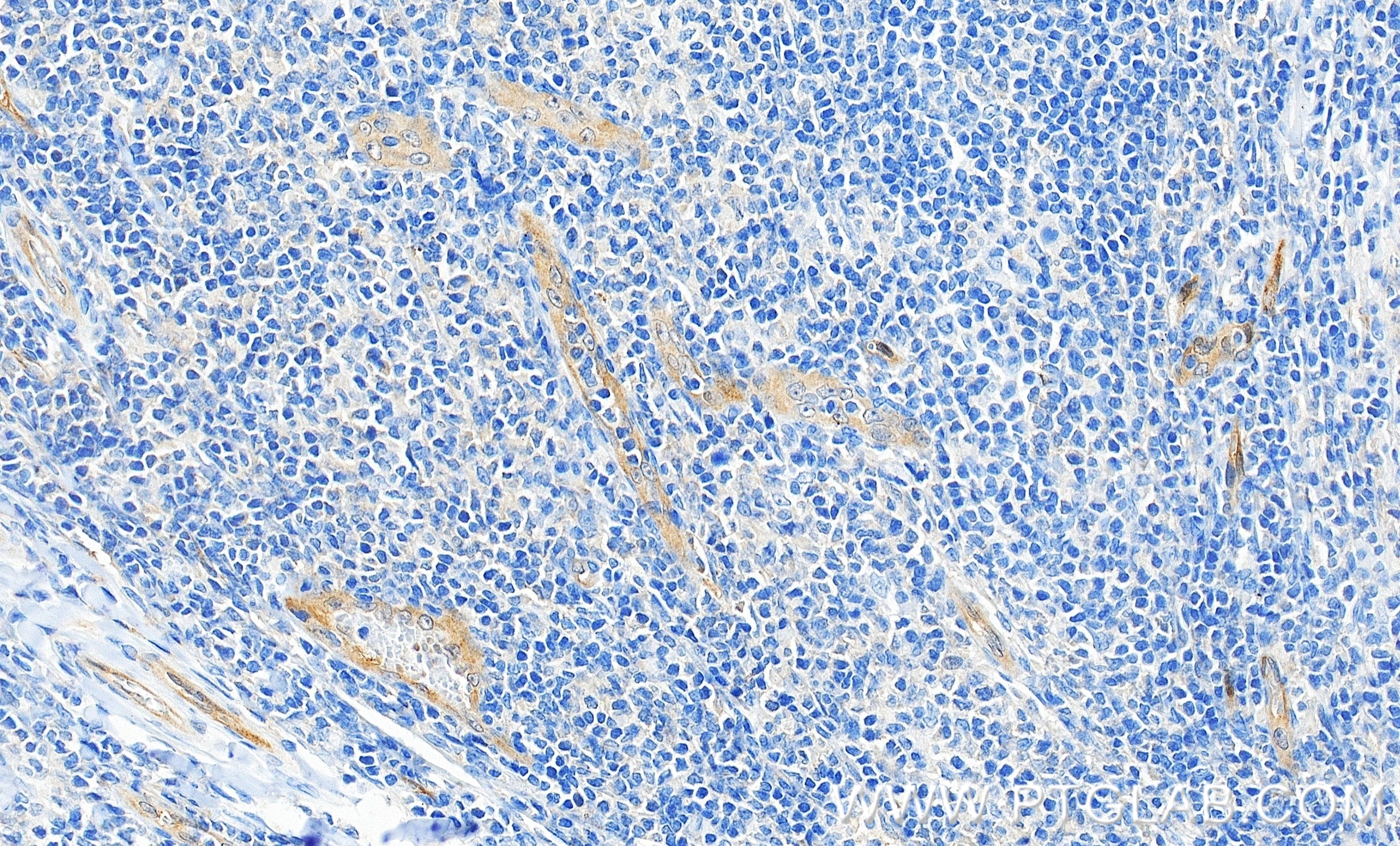
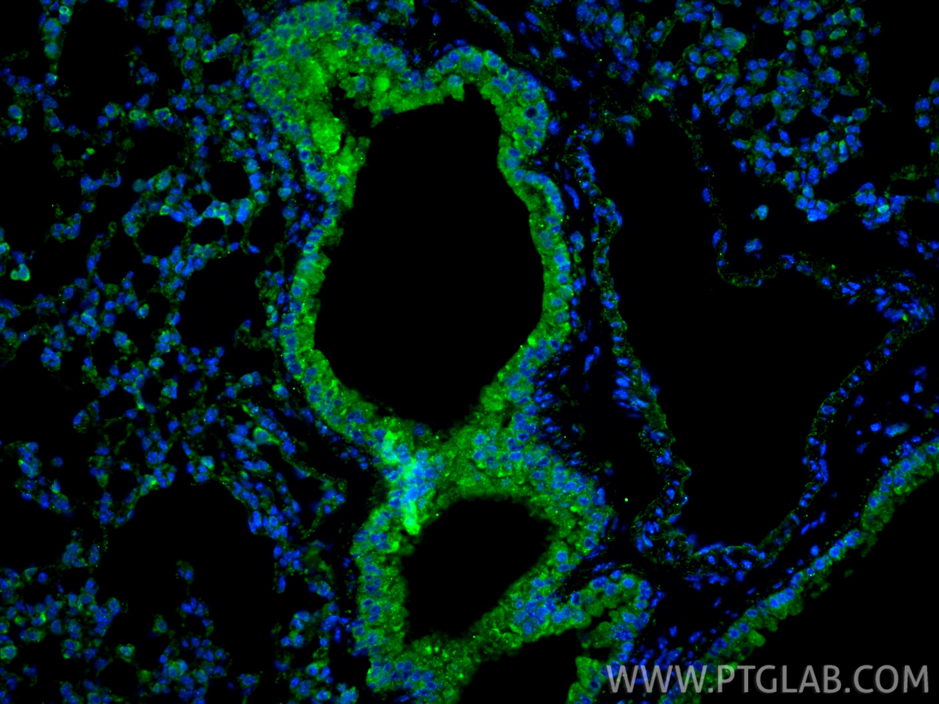
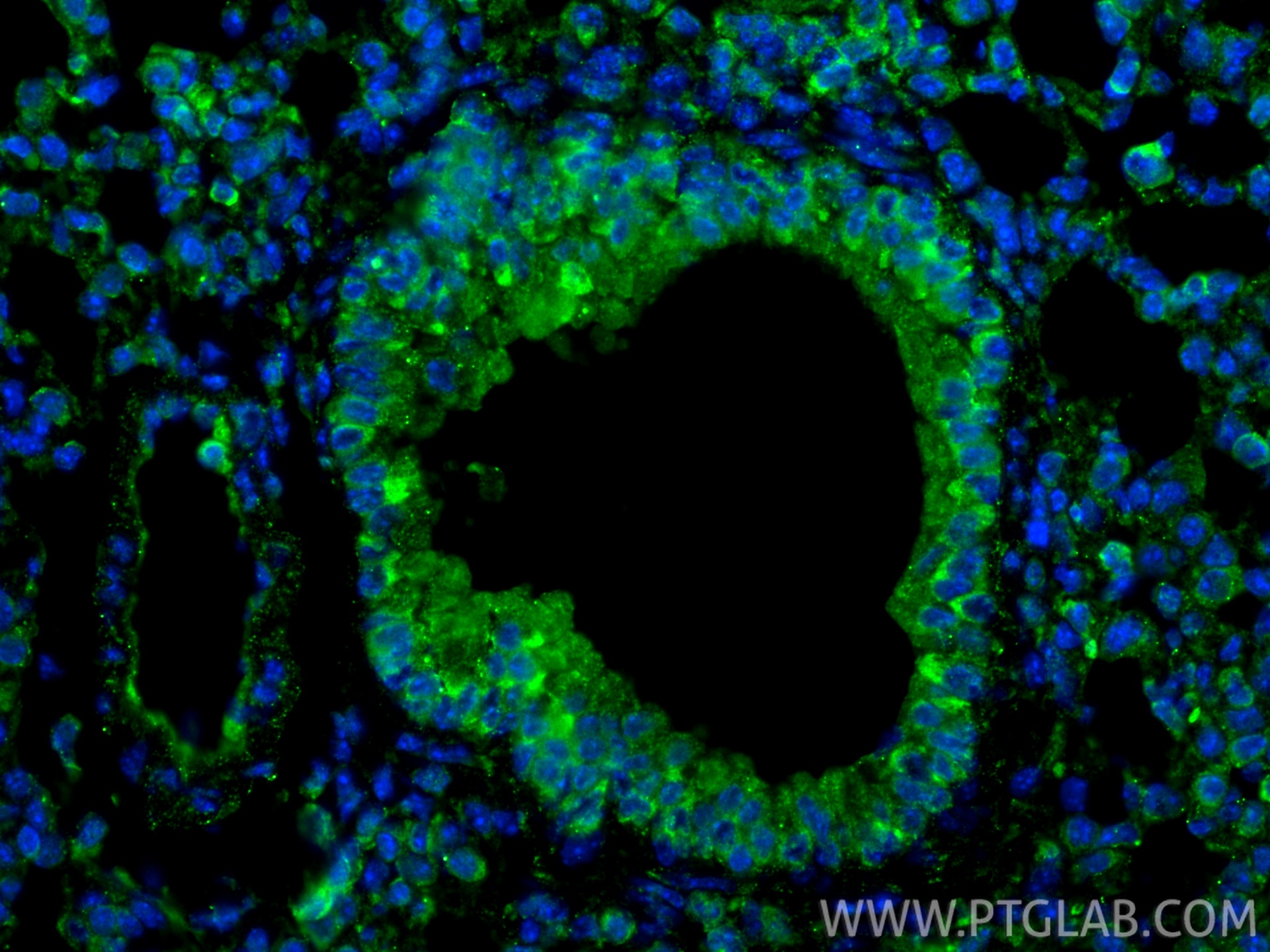
| Positive IHC detected in | human tonsillitis tissue, mouse lung tissue Note: suggested antigen retrieval with TE buffer pH 9.0; (*) Alternatively, antigen retrieval may be performed with citrate buffer pH 6.0 |
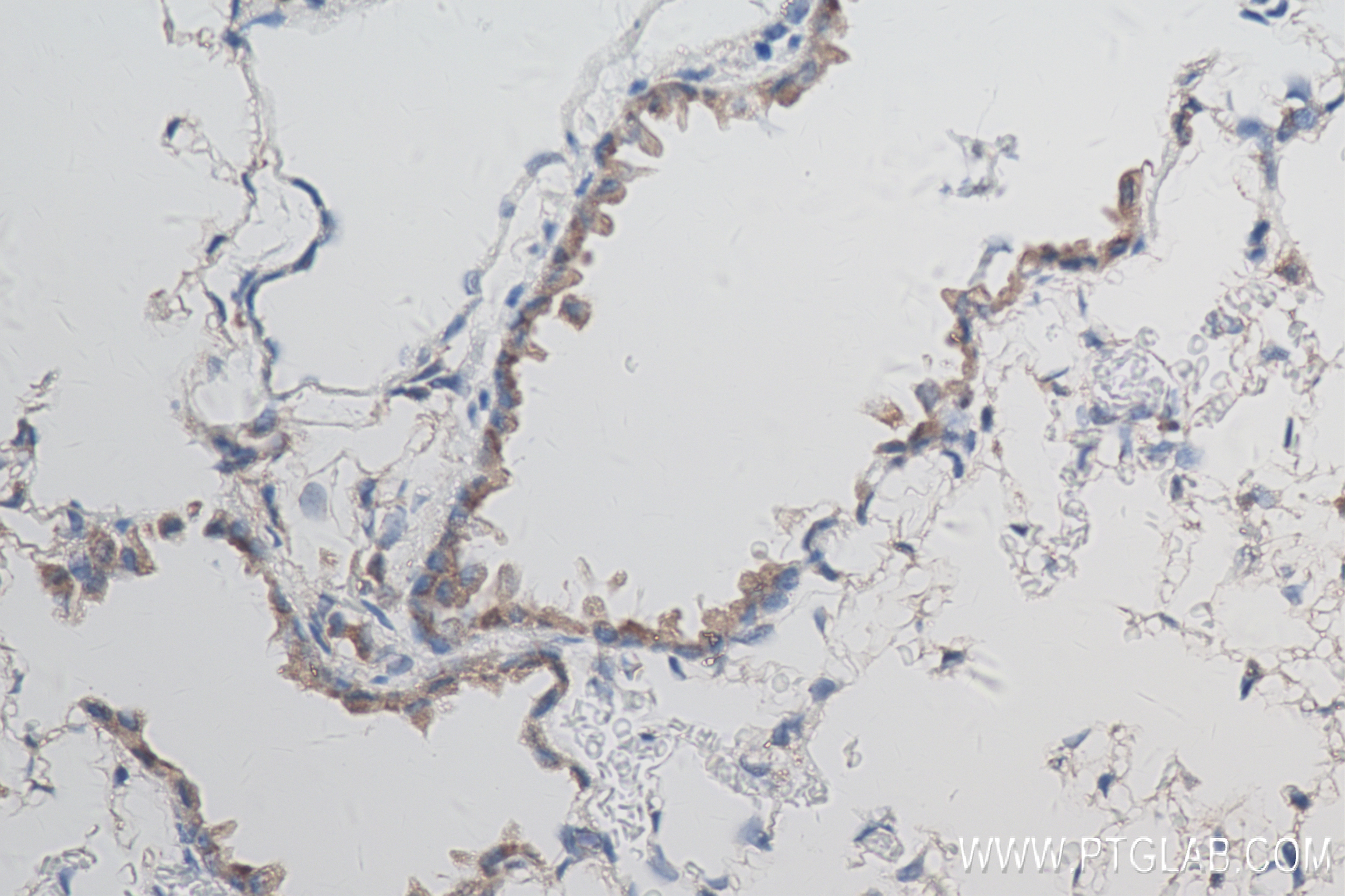
| Positive IF-P detected in | mouse lung tissue |
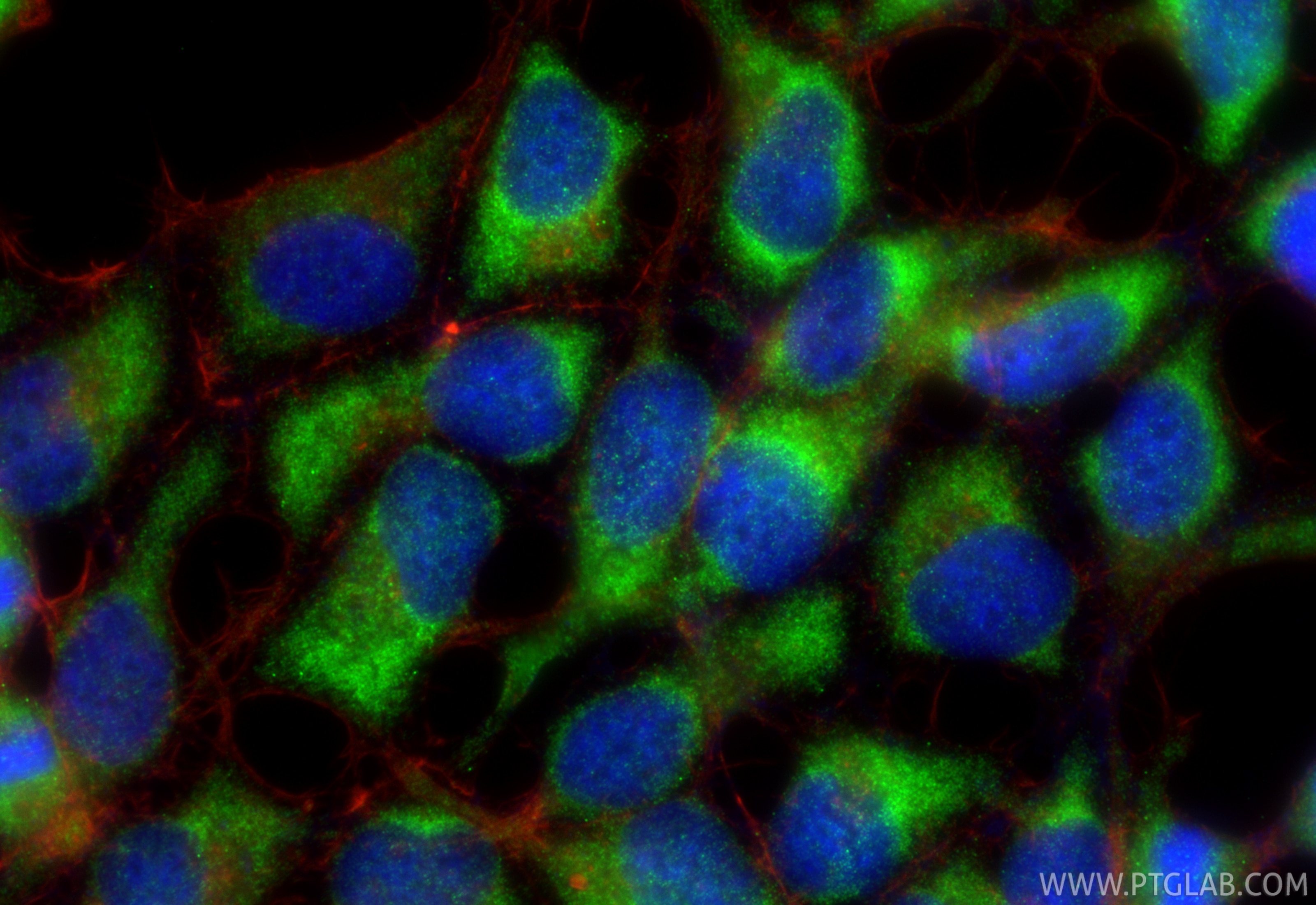
| Positive IF/ICC detected in | HEK-293 cells |
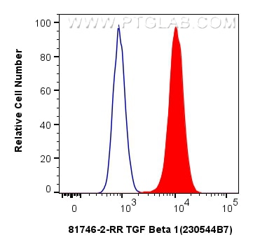
| Positive FC (Intra) detected in | HEK-293 cells |
Recommended dilution
| Application | Dilution |
|---|---|
| Western Blot (WB) | WB : 1:2000-1:12000 |
| Immunohistochemistry (IHC) | IHC : 1:500-1:2000 |
| Immunofluorescence (IF)-P | IF-P : 1:50-1:500 |
| Immunofluorescence (IF)/ICC | IF/ICC : 1:50-1:500 |
| Flow Cytometry (FC) (INTRA) | FC (INTRA) : 0.25 ug per 10^6 cells in a 100 µl suspension |
| It is recommended that this reagent should be titrated in each testing system to obtain optimal results. | |
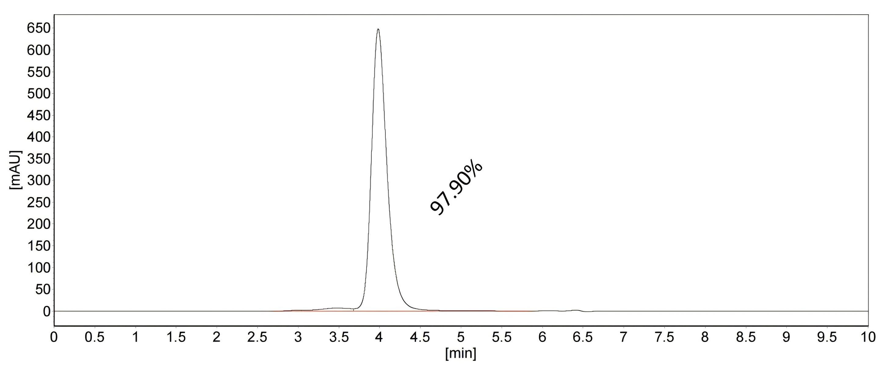
| Sample-dependent, Check data in validation data gallery. | |
Published Applications
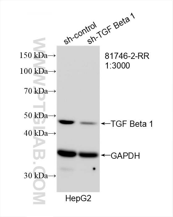
| KD/KO | See 2 publications below |
| WB | See 70 publications below |
| IHC | See 23 publications below |
| IF | See 19 publications below |
| ELISA | See 2 publications below |
| FC | See 1 publications below |
| CoIP | See 2 publications below |
Product Information
81746-2-RR targets TGF Beta 1 in WB, IHC, IF/ICC, IF-P, FC (Intra), CoIP, ELISA applications and shows reactivity with human, mouse, rat samples.
| Tested Reactivity | human, mouse, rat |
| Cited Reactivity | human, mouse, rat, rabbit, canine, chicken |
| Host / Isotype | Rabbit / IgG |
| Class | Recombinant |
| Type | Antibody |
| Immunogen |
CatNo: Ag13591 Product name: Recombinant human TGFB1 protein Source: e coli.-derived, PET28a Tag: 6*His Domain: 14-258 aa of BC000125 Sequence: PLLWLLVLTPGRPAAGLSTCKTIDMELVKRKRIEAIRGQILSKLRLASPPSQGEVPPGPLPEAVLALYNSTRDRVAGESAEPEPEPEADYYAKEVTRVLMVETHNEIYDKFKQSTHSIYMFFNTSELREAVPEPVLLSRAELRLLRLKLKVEQHVELYQKYSNNSWRYLSNRLLAPSDSPEWLSFDVTGVVRQWLSRGGEIEGFRLSAHCSCDSRDNTLQVDINGFTTGRRGDLATIHGMNRPFL Predict reactive species |
| Full Name | transforming growth factor, beta 1 |
| Calculated Molecular Weight | 44 kDa |
| Observed Molecular Weight | 44 kDa |
| GenBank Accession Number | BC000125 |
| Gene Symbol | TGFB1 |
| Gene ID (NCBI) | 7040 |
| RRID | AB_3670503 |
| Conjugate | Unconjugated |
| Form | Liquid |
| Purification Method | Protein A purfication |
| UNIPROT ID | P01137 |
| Storage Buffer | PBS with 0.02% sodium azide and 50% glycerol, pH 7.3. |
| Storage Conditions | Store at -20°C. Stable for one year after shipment. Aliquoting is unnecessary for -20oC storage. 20ul sizes contain 0.1% BSA. |
Background Information
TGF Beta 1 (TGF-β1) is a pleiotropic cytokine and a member of the TGF-β superfamily (including TGF-β1, TGF-β2, and TGF-β3). It is encoded by the TGFB1 gene and acts as a secreted protein involved in controlling cell growth, proliferation, differentiation, and apoptosis. TGF-β1 is produced by various cell types (e.g., platelets, lymphocytes, macrophages, epithelial cells, and inflammatory cells) and is secreted as a precursor requiring proteolytic activation to yield the mature, biologically active peptide. TGF-β1 plays crucial roles in immune regulation, wound healing, fibrosis, and tissue remodeling (including cardiac injury and fibrosis). It also influences cancer progression by promoting epithelial-to-mesenchymal transition (EMT) and metastasis in tumors.
Protocols
| Product Specific Protocols | |
|---|---|
| FC protocol for TGF Beta 1 antibody 81746-2-RR | Download protocol |
| IF protocol for TGF Beta 1 antibody 81746-2-RR | Download protocol |
| IHC protocol for TGF Beta 1 antibody 81746-2-RR | Download protocol |
| WB protocol for TGF Beta 1 antibody 81746-2-RR | Download protocol |
| Standard Protocols | |
|---|---|
| Click here to view our Standard Protocols |
Publications
| Species | Application | Title |
|---|---|---|
Pharmacol Res Lipocalin 2 (LCN2) confers acquired resistance to almonertinib in NSCLC through LCN2-MMP-9 signaling pathway | ||
Free Radic Biol Med N6-methyladenosine modification of SPOP relieves ferroptosis and diabetic cardiomyopathy by enhancing ubiquitination of VDAC3 | ||
J Biomed Mater Res B Appl Biomater A Sandwiched Patch for Prevention of Pelvic Adhesion After Uterine Operation | ||
J Orthop Surg Res Regeneration process of severed rabbit common calcanean tendons influenced by external compression | ||
Adv Sci (Weinh) Decoding the Cardiac Immune Microenvironment and Fibroblast Crosstalk in Radiotherapy Combined with Immunotherapy-Induced Cardiac Fibrosis Based on Single-Cell Transcriptomic Analysis. | ||
Mol Nutr Food Res Curcumin Inhibits Renal Fibrosis by Suppressing S100A8/A9-TLR4 Signaling via Gut Microbiota-Derived Short-Chain Fatty Acid in Macrophages. |
Reviews
The reviews below have been submitted by verified Proteintech customers who received an incentive for providing their feedback.
FH Sonam (Verified Customer) (10-16-2025) | AB worked well
|