
Scientist Stories: Dr Joe McKellar on Imaging the Cytoskeletal Architecture of LUHMES-Derived Neurons
Dr Joe McKellar shows us what a neuron's skeleton really looks like.
This story is part of #InTheLabWithProteintech, highlighting how Proteintech reagents support researchers in advancing their work.
This feature marks the last of a three-part spotlight on Joe McKellar, PhD, Scientific Director at Viroscope Imaging and winner of CiteAb Image of the Year and Nikon Small World, sharing insights from his work in advanced microscopy and his PhD research in Health Biology.
Read Part 1: https://www.ptglab.com/news/blog/scientist-stories-dr-joe-mckellar-on-imaging-influenza-a/
What is the biological significance of the image?
"During my postdoc, I studied deltaviruses, which we discovered package themselves inside other viruses’ particles. We termed this spreading through a “viral Trojan Horse”. To prove that a deltavirus could use a herpesvirus as a viral Trojan Horse to infect human neurons, we turned to the LUHMES cell model. Since I hadn't worked with these before, I spent some time optimizing a 4-color multiplexing protocol to visualize the infection. Before adding the viruses, I wanted to understand the cellular architecture I would be working with. This image is the result of that optimization, capturing the DNA and three distinct components of the neuronal cytoskeleton, actin, tubulin, and vimentin, simultaneously."
How do you design a multiplex experiment at that scale without signal interference?
"At a 4-color scale, preventing signal interference comes down to careful fluorophore selection and leveraging your microscope's hardware. First, you have to ensure there is minimal overlap in the emission spectra of your chosen fluorophores. But the real technical advantage comes from the imaging system itself. I shot this on a ZEISS LSM980 equipped with an Airyscan 2 detector. A standard confocal microscope uses a physical pinhole to block out-of-focus light. The Airyscan detector replaces that single pinhole with an array of 32 distinct detector elements. This allows you to collect more light while simultaneously increasing the optical resolution beyond the traditional diffraction limit. Combined with the spectral tuning capabilities of the LSM series, you can cleanly separate four dense channels without any optical bleed-through, Airyscan or not."
What are the technical aspects you considered in the protocol?
"While the microscope handles the back-end resolution, the wet-lab protocol has to be flawless at the front-end to make a 4-color multiplex work. The blocking step is critical for example. You must meticulously saturate any non-specific binding sites to eliminate background noise. When combining different primary and secondary antibodies, you must carefully select the host species of your antibodies and make your blocking buffer accordingly. If your protocol isn't optimized at the bench, the microscope won’t be able to save the final image."
View the Vimentin antibody Joe used. https://www.ptglab.com/products/VIM-Antibody-10366-1-AP.htm
How does this work contribute to the broader landscape of the field?
"This imaging work allowed us to successfully demonstrate that human neurons can be infected by these deltavirus/herpesvirus Trojan Horse particles, and that both viruses are capable of replicating within them. Because we were initially studying animal deltaviruses, proving that they can hitchhike inside a helper virus capable of infecting human neurons is a significant leap. It suggests that these satellite viruses have the potential to cross species barriers and potentially contribute to neurological pathologies in the human population."
What are you planning to tackle next?
"One of my focuses at Viroscope Imaging is to participate in tackling the reproducibility crisis through visual validation. For academic and industry researchers, knowing with absolute certainty that an antibody is specific to its target in your chosen cell line or species is invaluable. Far too much time and funding are wasted on testing reagents that are not fully validated. I want to participate in this industry-wide effort by validating antibodies across various biological systems and species. By providing definitive, high-resolution visual proof of specificity, my goal is to give researchers the confidence they need to push their discoveries forward at a much faster pace."
Featured Product
The following Proteintech reagent was used in this study:
- Vimentin Polyclonal Antibody (Cat No. 10366-1-AP)
Used to detect and visualise vimentin distribution across the cytoskeletal network of LUHMES-derived neurons, contributing to a multicolour mapping of cytoskeletal architecture alongside tubulin, actin, and DNA.
View product: https://www.ptglab.com/products/VIM-Antibody-10366-1-AP.htm
About the Scientist
Joe McKellar is a virologist and cell biologist with a PhD in Health Biology, specializing in advanced microscopy. As the Scientific Director of Viroscope Imaging, he leads an agency dedicated to highlighting biotechnologies and cutting-edge research through high-resolution visual narratives. His academic work has consistently relied on the lens of a microscope to answer complex biological questions, from understanding cellular defense mechanisms to characterizing novel forms of viral transmission. This dedication to visual science has earned him recognition in international competitions, such as the CiteAb Image of the Year and Nikon Small World. When he isn't exploring cellular landscapes under a confocal microscope, you can usually find him strategizing over a game of Magic: The Gathering.
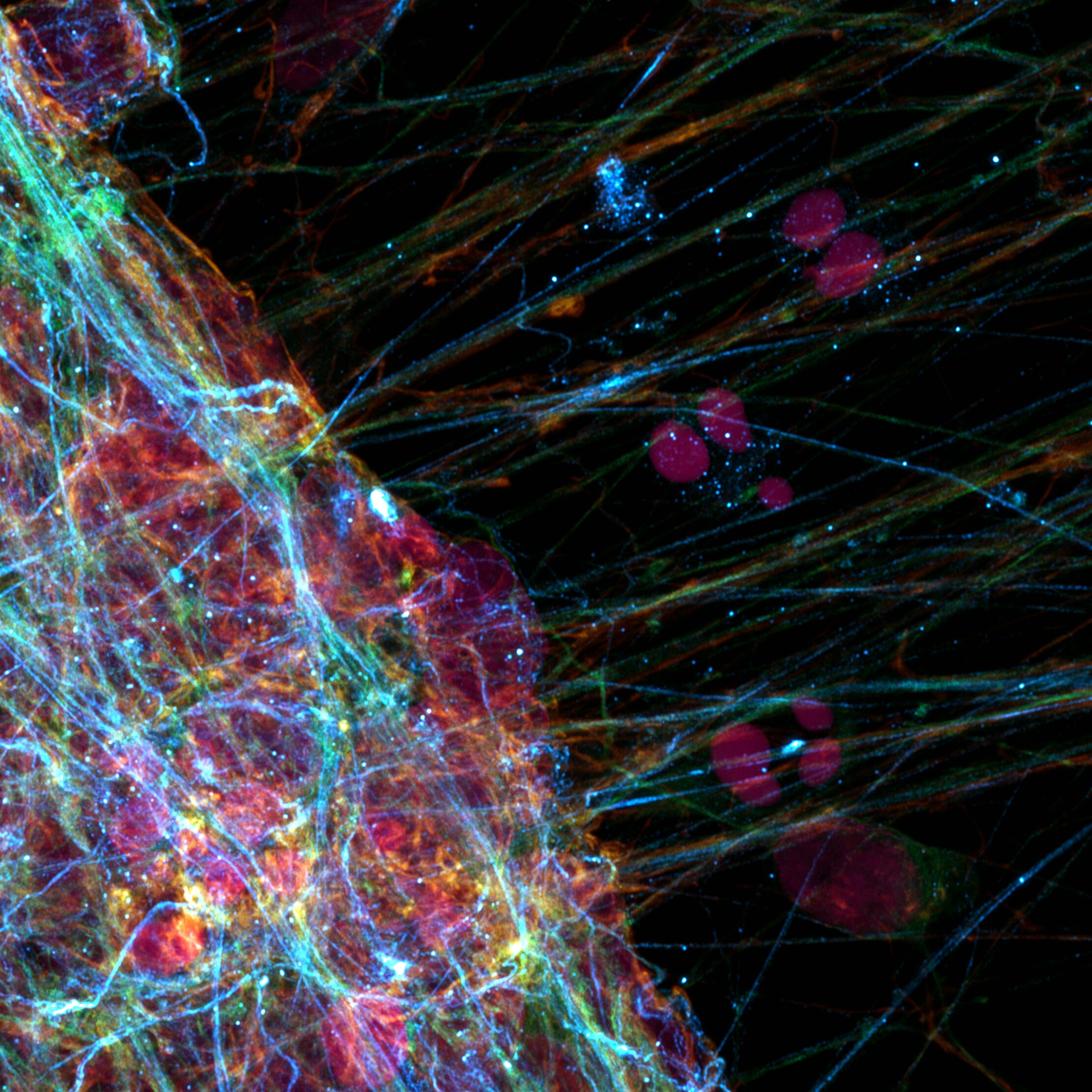
Airyscan fluorescence image (Maximum Intensity Projection) of LUHMES-derived neurons, showing a mass of cells on the left of the image extending prolongations outward. Vimentin was detected using a Proteintech anti-Vimentin antibody (Cat. No. 10366-1-AP) and is shown in cyan. Tubulin is shown in green, Actin in gold, and DNA in pink, together mapping the full cytoskeletal landscape of the neuronal extensions. Image acquired using a ZEISS LSM980 with Airyscan 2.
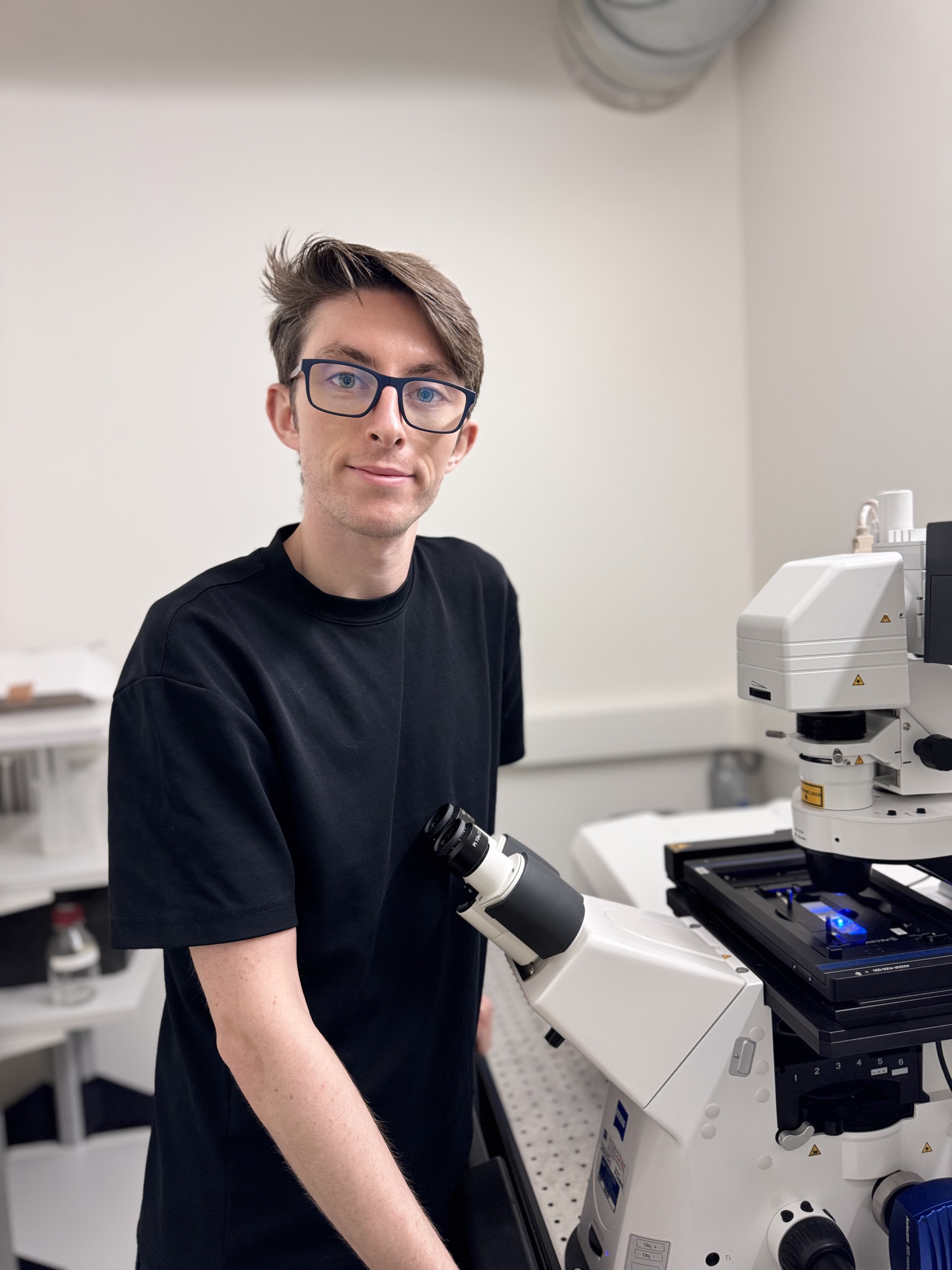
Joe McKellar, PhD
Able™