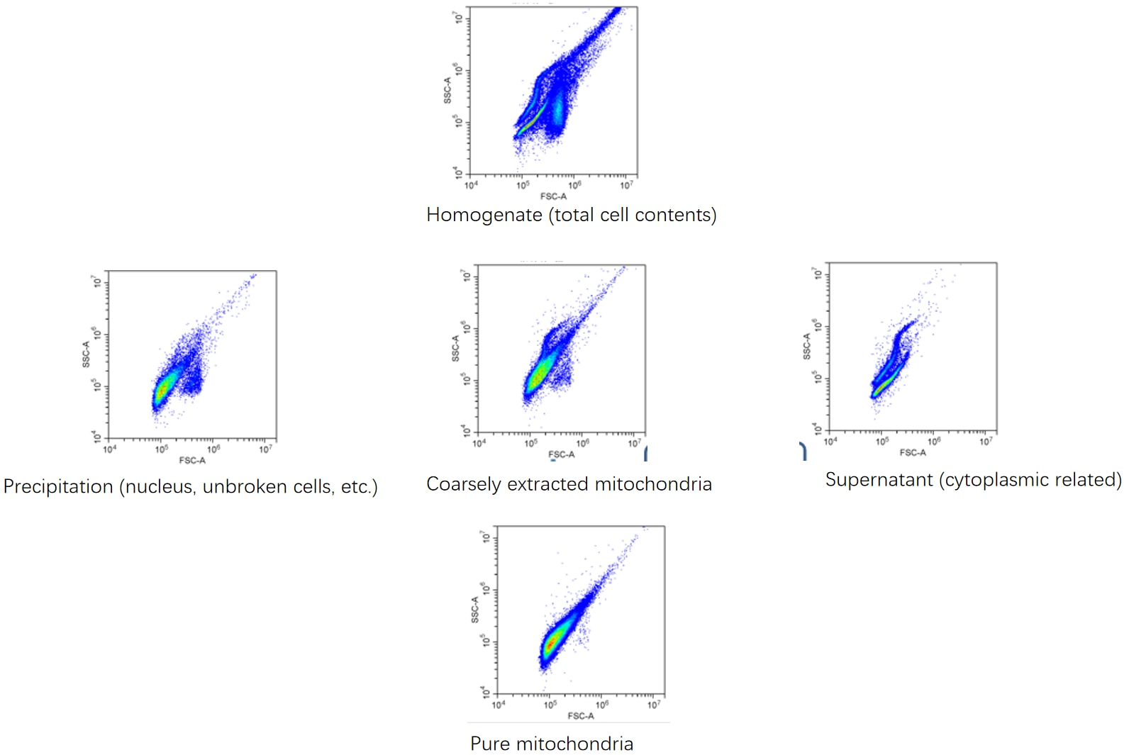
. The pellet and supernatant obtained during the isolation process were each diluted 100-fold and analysed by flow cytometry.")
Product Information
This mitochondrial protein isolation and extraction kit, developed by Proteintech, efficiently separates high-purity mitochondria from animal tissues and cultured cells. The isolated mitochondria retain intact inner and outer membranes with fully functional mitochondrial capabilities, making them suitable for studies on mitochondrial physiological functions. Additionally, the isolated mitochondria can be lysed using the provided lysis buffer for subsequent protein analysis experiments such as Western blotting and immunoprecipitation.
About the Kit:
● This kit enables the isolation of high-purity mitochondria suitable for flow cytometric analyses.
● The kit provides a mitochondrial preservation solution for preserving isolated mitochondria for subsequent experiments.
● The kit provides a mitochondrial lysis buffer for efficient disruption of the isolated high-purity mitochondria.
● The kit provides the mitochondrial dye Janus Green B for rapid assessment of extraction efficiency.
● The 0.4% trypan blue solution enables evaluation of homogenisation efficacy. Cease homogenisation once 60% cell disruption is achieved, avoiding excessive processing.
● This kit is exclusively for fresh tissue samples. Do not use cryopreserved tissue.
● For preparing mitochondrial proteins, add the protease inhibitor mixture in advance. Otherwise, no addition is required.
● A minimum sample size of 20 million cells or 100 mg tissue is recommended. With this sample quantity, the kit can process 30 samples.
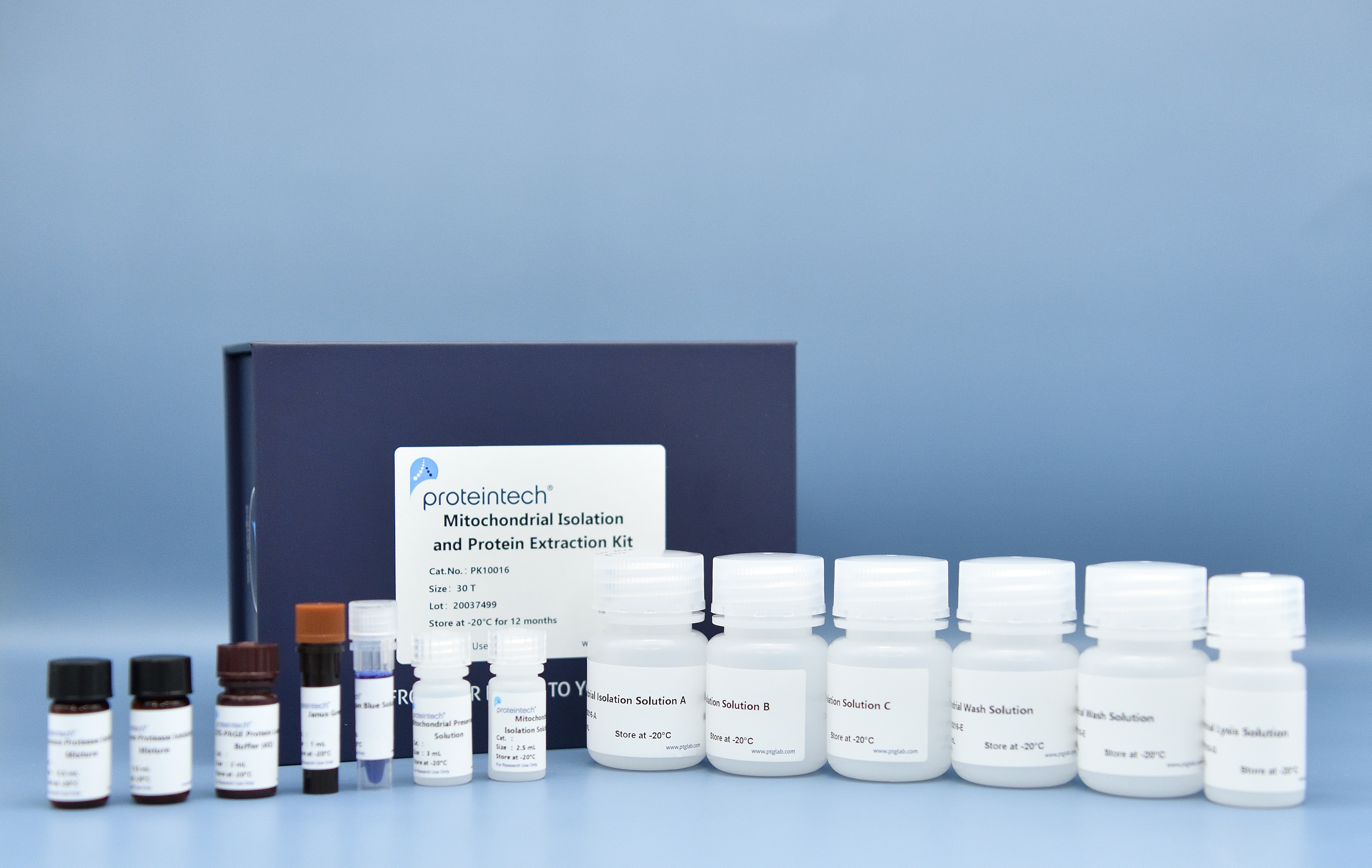
Product Component
Component | Catalog No. | Size |
Mitochondrial Isolation Solution A | PK10016-A | 30 mL |
Mitochondrial Isolation Solution B | PK10016-B | 30 mL |
Mitochondrial Isolation Solution C | PK10016-C | 15 mL |
Mitochondrial Isolation Solution D | PK10016-D | 2.5 mL |
Mitochondrial Wash Solution | PK10016-E | 30 mL*2 |
Mitochondrial Preservation Solution | PK10016-F | 3 mL |
Mitochondrial Lysis Solution | PK10016-G | 10 mL |
Janus Green B | PK10016-H | 1 mL |
SDS-PAGE Protein Loading Buffer (4X) | PK10016-I | 2 mL |
Common Protease Inhibitor Mixture | PK10016-J | 0.5 mL*2 |
0.4% Trypan Blue Solution | PK10016-K | 1 mL |
Storage
Store at -20°C for 12 months or 4°C for up to one month.
Usage
1. Pre‑Extraction Preparation
Thaw all solutions in the kit at room temperature, then place them on ice. Pre‑cool the centrifuge to 4 °C in advance. If preparing mitochondrial protein samples, take appropriate volumes of Mitochondria Isolation Solution A, Mitochondria Isolation Solution B, Mitochondria Isolation Solution C, Mitochondria Wash Solution, and Mitochondria Lysis Solution according to the sample amount. Add protease inhibitor cocktail (100×) at a ratio of 100:1 before use.
2. Sample Pre‑Treatment
a. For Cells
Collect cells and centrifuge at 500 g for 5 min at 4 °C. Wash twice with pre‑cooled PBS. On ice, add 1 mL of pre‑cooled Mitochondria Isolation Solution A directly per 20 million cells.
b. For Tissues
Obtain fresh animal tissue, wash away impurities such as fat and blood from the tissue with ice‑cold PBS, blot dry with filter paper, and weigh. Mince the tissue in a centrifuge tube. On ice, add 1 mL of pre‑cooled Mitochondria Isolation Solution A per 100 mg tissue.
3. Sample Homogenization
Transfer the sample suspension to a pre‑cooled glass homogenizer of appropriate size and homogenize the sample under ice‑bath conditions.
Note: Homogenization cycles vary by sample type. Cultured cells are recommended to be homogenized 3–5 times; tissue samples 5–10 times.
Identification method: Take 20 uL of cell homogenate after 3 cycles or tissue homogenate after 5 cycles, mix with an equal volume of 0.4% trypan blue solution, and observe under a microscope. Stop homogenization when the proportion of trypan blue‑positive (blue) ruptured cells reaches 60%. Avoid over‑homogenization. If the proportion of positive (blue) cells is below 60%, increase homogenization by 1–2 cycles and re‑assess using trypan blue as described.
4. Crude Mitochondria Isolation
Based on the volume of the sample homogenate from Step 3, add an equal volume of Mitochondria Isolation Solution B to the bottom of the centrifuge tube, then slowly layer the sample homogenate on top along the tube wall.
For cultured cell samples: centrifuge at 600 g for 10 min at 4 °C and collect the uppermost fraction.
For tissue samples: centrifuge at 700 g for 10 min at 4 °C and collect the uppermost fraction.
Notes: (1) Layer the homogenate carefully along the tube wall onto Mitochondria Isolation Solution B and centrifuge promptly to prevent particles in the homogenate from settling naturally, which impairs subsequent centrifugal separation. (2) The supernatant will separate into two layers after centrifugation. Collect the uppermost layer. If layering is indistinct, collect the entire supernatant.
5. Transfer the collected supernatant to new 1.5 mL EP tubes at 1 mL per tube. Centrifuge at 10 000 g for 10 min at 4 °C and collect the pellet. The obtained pellet is crude mitochondria.
6. High‑Purity Mitochondria Isolation
a. Preparation of High‑Purity Mitochondria Isolation Solution
Prepare fresh by mixing Mitochondria Isolation Solution C and Mitochondria Isolation Solution D at a ratio of 17:3. Add 0.5 mL to new EP tubes (1.5 mL) in advance.
b. Resuspend the crude mitochondrial pellet from Step 5 in 0.5 mL of Mitochondria Wash Solution per 1.5 mL EP tube.
c. Slowly layer the resuspended crude mitochondria onto the high‑purity mitochondria isolation solution. Centrifuge at 22 000 g for 10 min at 4 °C and collect the pellet.
d. Resuspend each pellet in 1 mL of Mitochondria Wash Solution, centrifuge at 16 000 g for 5 min at 4 °C, and collect the pellet. The final pellet is high‑purity mitochondria.
7. Mitochondria Staining and Identification
Smear an appropriate amount of the pellet, stain with Janus Green B solution for 20 min, cover with a coverslip, and examine under a microscope. Mitochondria appear as small blue‑green rod‑shaped or dumbbell‑shaped structures.
8. Use of Mitochondria
a. For Functional or Enzymatic Activity Assays of Intact Mitochondria
Resuspend mitochondria from an initial 20 million cells or 100 mg tissue in 100 uL of Mitochondria Preservation Solution and use promptly. If not used immediately, store at −80 °C. Frozen‑thawed mitochondria are not recommended for intact mitochondrial functional studies, but can be used for mitochondrial protein analysis, Western blotting, immunoprecipitation, etc.
b. For Mitochondrial Protein Analysis
Add 100 uL of Mitochondria Lysis Solution (supplemented with general protease inhibitor cocktail at 100:1 before use) to mitochondria from an initial 20 million cells or 100 mg tissue. Incubate on ice for 30 min, then centrifuge at 10 000 g for 5 min at 4 °C. The supernatant is the mitochondrial sample, which can be used for PAGE, Western blotting, immunoprecipitation, and other downstream experiments.
c. For Western Blotting
Mix the lysed mitochondrial sample with SDS‑PAGE loading buffer at a ratio of 3:1. Heat in a 100 °C boiling water bath for 5 min to fully denature proteins. After cooling to room temperature, load directly into the wells of an SDS‑PAGE gel.
Notes
1. All steps for mitochondrial isolation must be conducted at low temperature.
2. Mitochondrial content and size vary across different samples. Adjust the volumes of mitochondrial isolation buffer and lysis buffer according to experimental conditions.
3. This kit is suitable only for fresh tissue samples. Do not use cryopreserved tissue.
4. The coarse or high-purity mitochondrial separation may be performed according to experimental requirements.
5. Mitochondria stored in mitochondrial preservation buffer should be used promptly.
6. When preparing mitochondrial protein samples, add Common Protease Inhibitor Cocktail (Catalog No. PR20032) to Mitochondrial Isolation Buffer A, Mitochondrial Isolation Buffer B, Mitochondrial Isolation Buffer C, Mitochondrial Wash Buffer, and Mitochondrial Lysis Buffer in advance. For phospho-antibody detection, add Phosphatase Inhibitor Cocktail (Catalog No. PR20015) in advance.
7. The common protease inhibitor mixture in this kit is suitable for normal protein extraction. For proteins prone to degradation, the Enhanced Protease Inhibitor Cocktail (Catalog No. PR20016) is recommended.
8. The SDS-PAGE Protein Loading Buffer contains a slight amount of DTT, which may emit a mildly pungent odour, but does not contain highly toxic Dithiothreitol.
9. Mitochondrial proteins extracted using this kit can be quantified by the BCA method. Do not use the Bradford method.
10. All reagents in this kit should be dissolved at room temperature. Do not heat.
11. For your safety and health, wear a lab coat and disposable gloves during operation.
FAQ
|
Problem |
Possible Cause |
Solution |
|
No mitochondria detected |
Excessive homogenisation leads to mitochondrial fragmentation |
Reduce the number of homogenisation cycles |
|
The sample is not fresh |
Isolate from fresh samples |
|
|
The sample size is too small |
Increase the volume of the sample extracted |
|
|
The purity of the mitochondria is not high |
Improper handling resulted in the coarse mitochondria remaining in the upper layer of the high-purity mitochondrial separation solution, preventing the two from separating into distinct layers. |
Carefully add the coarse mitochondria along the wall of the tube to the supernatant of the high-purity mitochondrial isolation solution, and centrifuge rapidly. |
Cited in Article as
PK10016, Mitochondrial Isolation and Protein Extraction Kit, Proteintech, IL, USA
Publications
| Application | Title |
|---|---|
Adv Sci (Weinh) Hierarchical Targeting Nanodrug with Holistic DNA Protection for Effective Treatment of Acute Kidney Injury | |
EMBO J Coronaviral ORF6 protein mediates inter-organelle contacts and modulates host cell lipid flux for virus production | |
Signal Transduct Target Ther DUSP6 ablation restores CAR T-cell fitness impaired by tumor CD58 loss through invigoration of AP-1 signaling. | |
Phytomedicine Kaempferol attenuated LPS-induced microglial neurotoxicity by promoting mitophagy to inhibit mtDNA-mediated NLRP3 inflammasome activation. | |
J Transl Med SIRT3 ameliorates diabetes-associated cognitive dysfunction via regulating mitochondria-associated ER membranes |