")
")
")
")
")


")
")
")
")
")
")
")
")
")
")
")
")
")
")
")
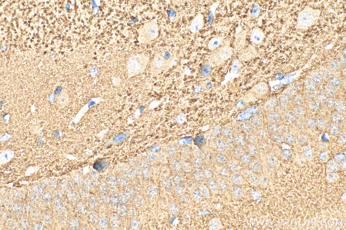
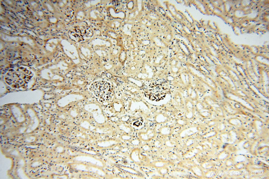
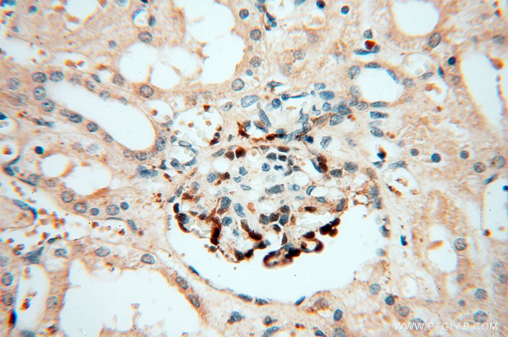
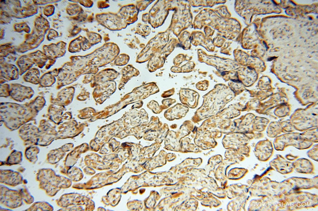
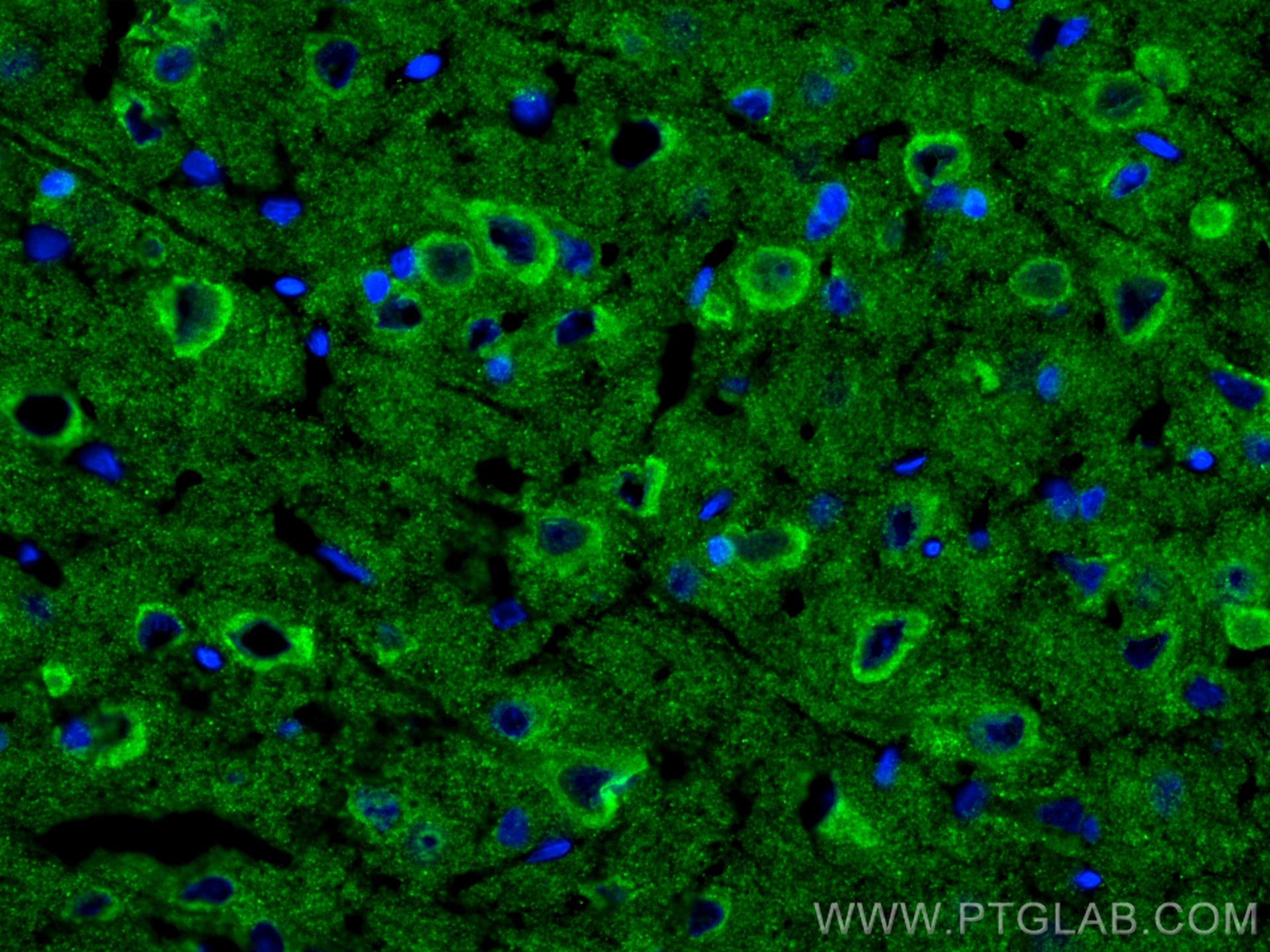
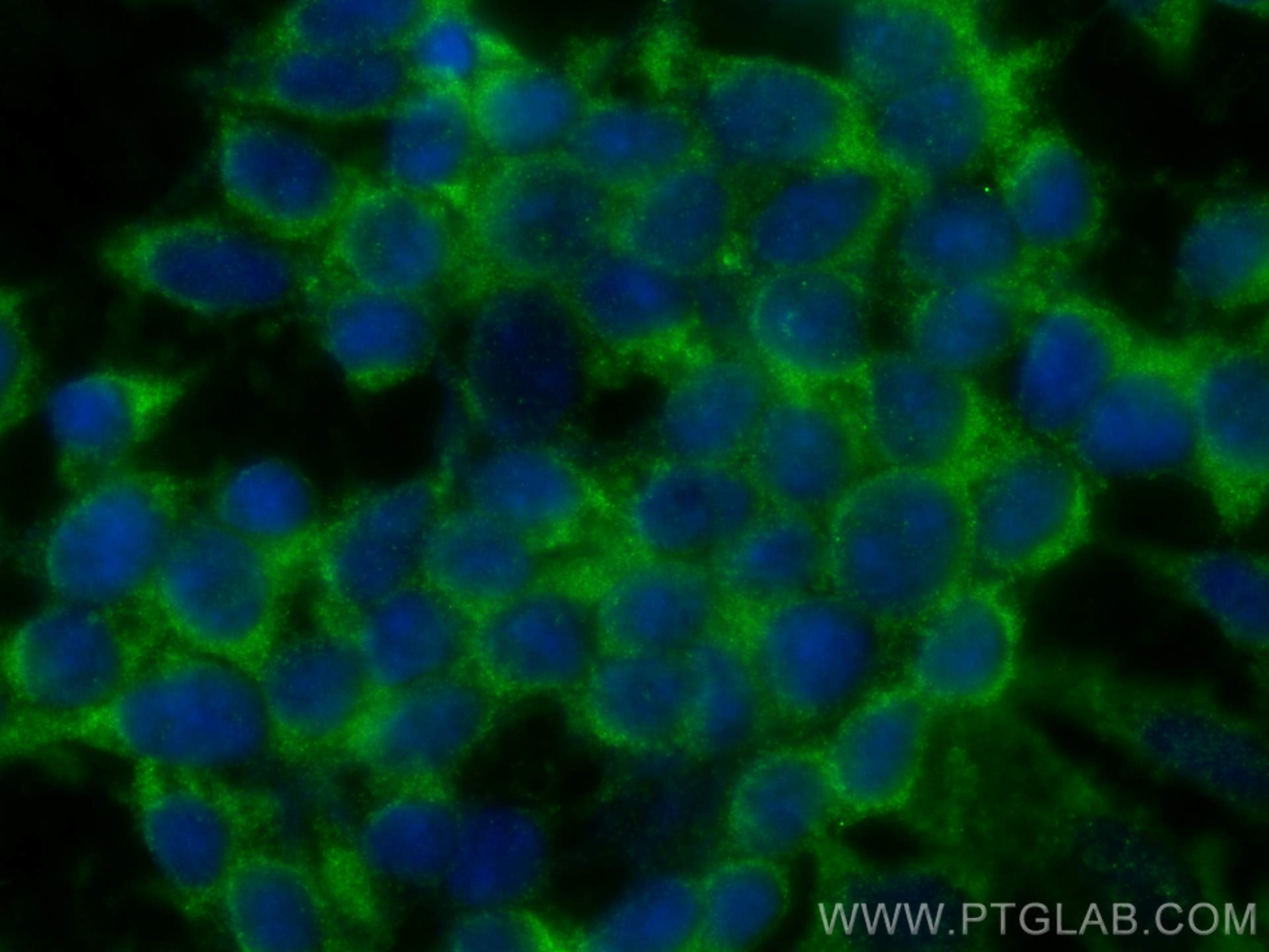
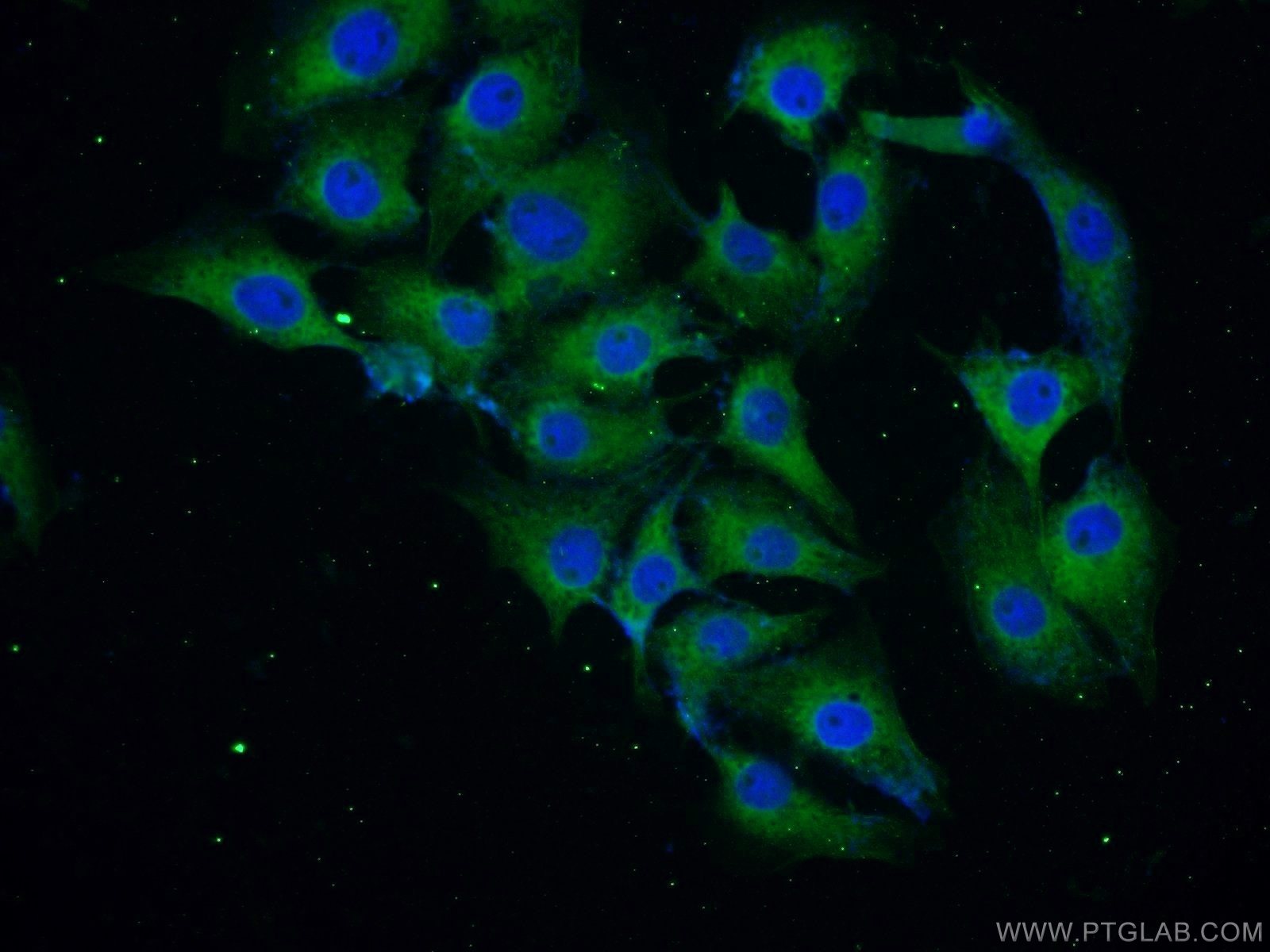
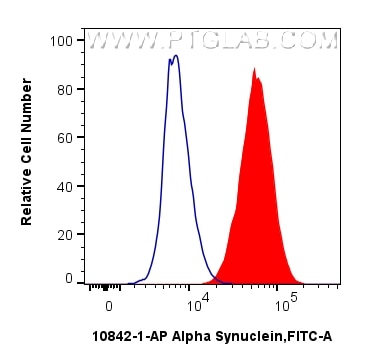
Product Information
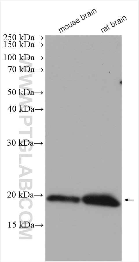
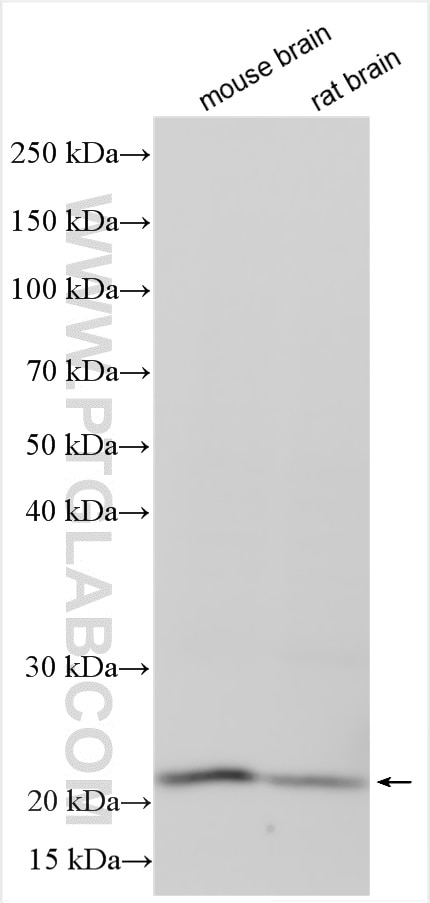
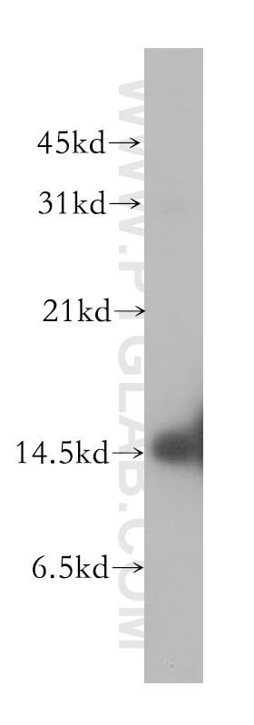
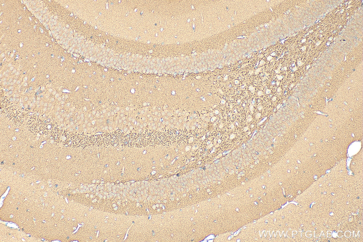
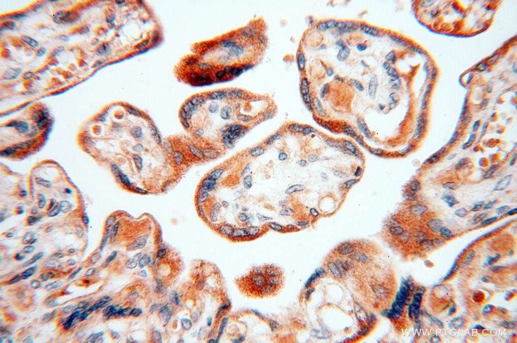
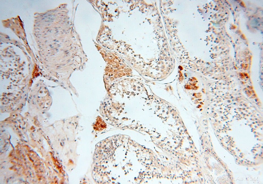
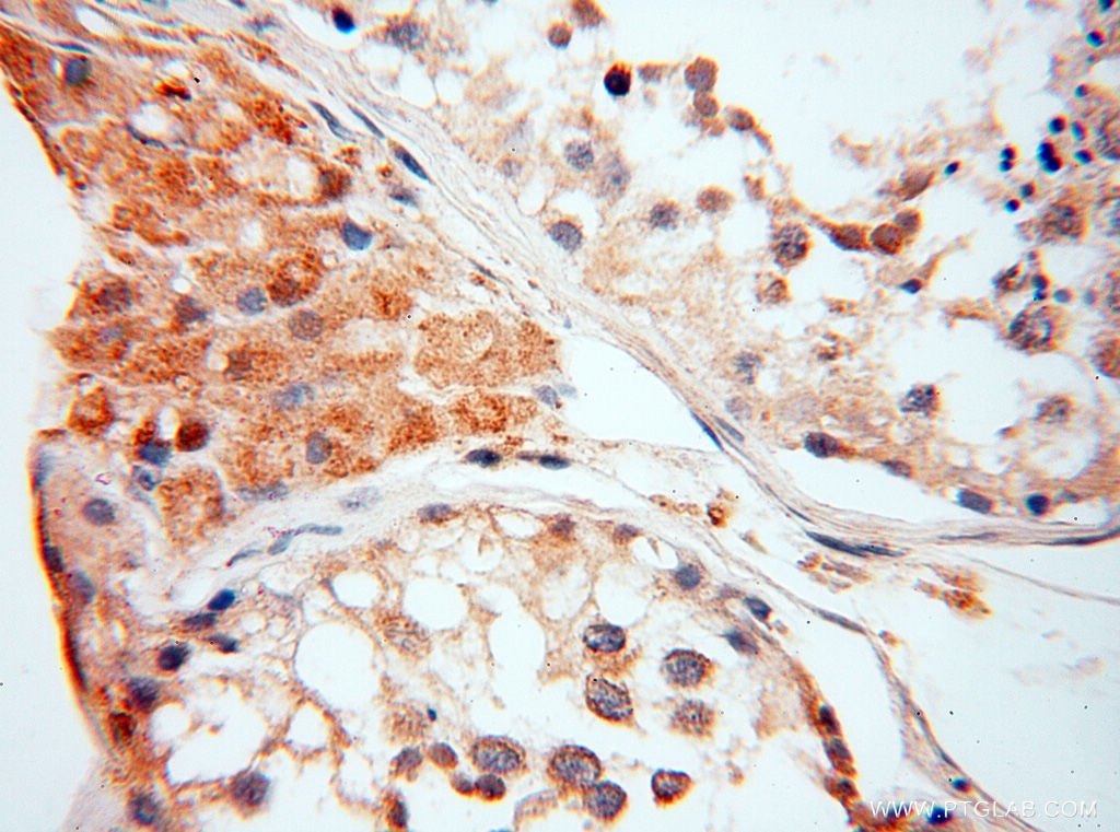
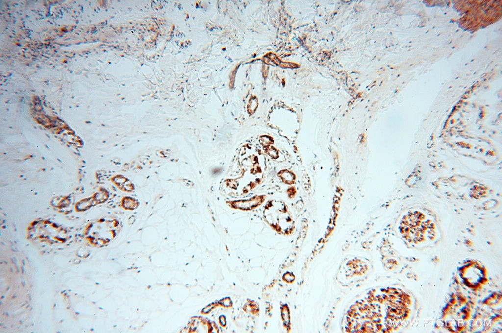
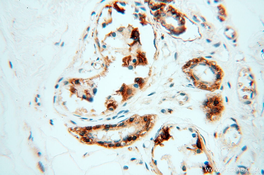
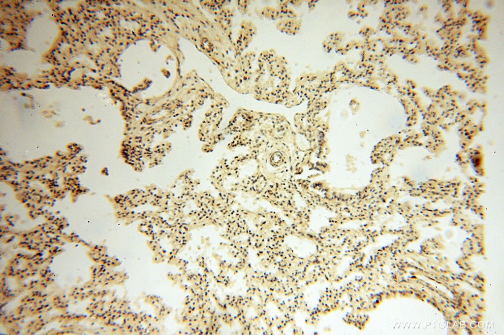
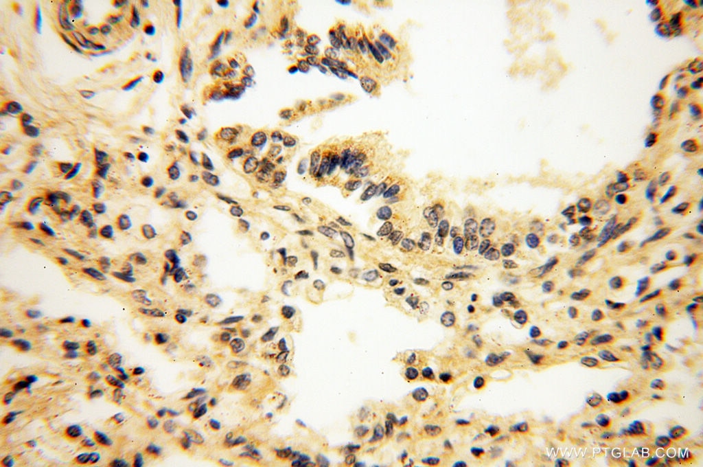
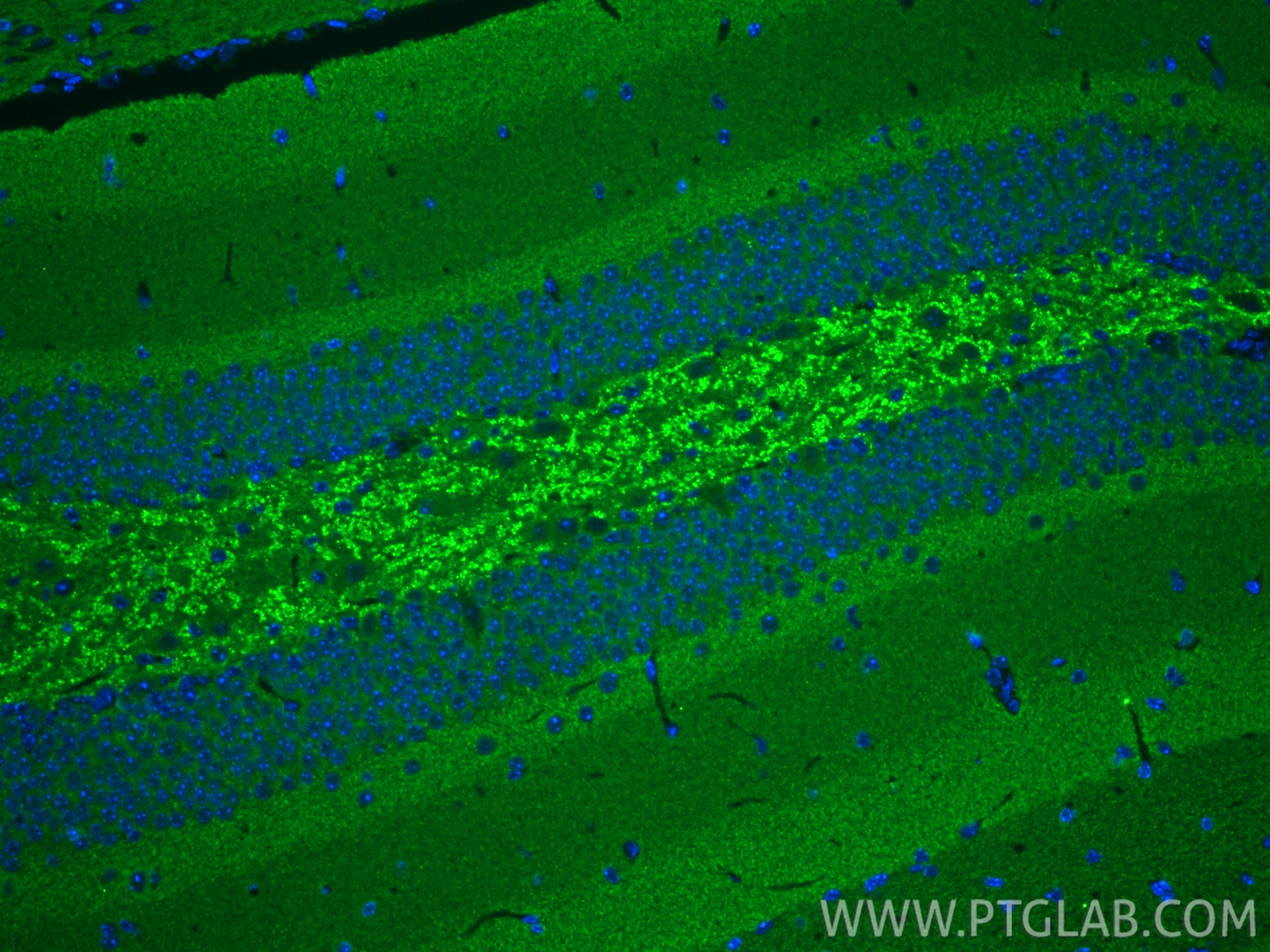
10842-1-PBS targets Alpha Synuclein in WB, IHC, IF/ICC, IF-P, IF-Fro, ELISA applications and shows reactivity with human, mouse, rat samples.
| Tested Reactivity | human, mouse, rat |
| Host / Isotype | Rabbit / IgG |
| Class | Polyclonal |
| Type | Antibody |
| Immunogen |
CatNo: Ag1285 Product name: Recombinant human a-Synuclein protein Source: e coli.-derived, PKG Tag: GST Domain: 1-140 aa of BC013293 Sequence: MDVFMKGLSKAKEGVVAAAEKTKQGVAEAAGKTKEGVLYVGSKTKEGVVHGVATVAEKTKEQVTNVGGAVVTGVTAVAQKTVEGAGSIAAATGFVKKDQLGKNEEGAPQEGILEDMPVDPDNEAYEMPSEEGYQDYEPEA Predict reactive species |
| Full Name | synuclein, alpha (non A4 component of amyloid precursor) |
| Calculated Molecular Weight | 14 kDa |
| Observed Molecular Weight | 15-19 kDa |
| GenBank Accession Number | BC013293 |
| Gene Symbol | Alpha Synuclein |
| Gene ID (NCBI) | 6622 |
| RRID | AB_2192672 |
| Conjugate | Unconjugated |
| Form | Liquid |
| Purification Method | Antigen affinity purification |
| UNIPROT ID | P37840 |
| Storage Buffer | PBS only, pH 7.3. |
| Storage Conditions | Store at -80°C. |
Background Information
Alpha Synuclein (α-syn) is a 14-19 kDa phosphoprotein that is primarily localize to the presynaptic terminals of mature neurons, where it is involved in synaptic function and plasticity. Α-syn has drawn intense interest ever since the late 1990s, when the first α-synuclein missense mutation was identified as a cause of familial Parkinson's disease (PD). Aggregated and hyper-phosphorylated forms of α-syn protein are the pathological hallmark of Lewy body disease, which includes Parkinson's disease (PD), diffuse Lewy body disease (DLBD), and Lewy body variant of Alzheimer's disease (LBV). This antibody can recognize all the isoforms of α-syn.
Protocols
| Product Specific Protocols | |
|---|---|
| IHC protocol for Alpha Synuclein antibody 10842-1-PBS | Download protocol |
| Standard Protocols | |
|---|---|
| Click here to view our Standard Protocols |