Tested Applications
| Positive FC (Intra) detected in | HeLa cells |
Recommended dilution
| Application | Dilution |
|---|---|
| Flow Cytometry (FC) (INTRA) | FC (INTRA) : 0.80 ug per 10^6 cells in a 100 µl suspension |
| It is recommended that this reagent should be titrated in each testing system to obtain optimal results. | |
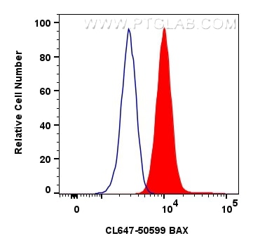
| Sample-dependent, Check data in validation data gallery. | |
Product Information
CL647-50599 targets BAX in FC (Intra) applications and shows reactivity with human, mouse, rat samples.
| Tested Reactivity | human, mouse, rat |
| Host / Isotype | Rabbit / IgG |
| Class | Polyclonal |
| Type | Antibody |
| Immunogen |
CatNo: Ag0576 Product name: Recombinant human BAX protein Source: e coli.-derived, PGEX-4T Tag: GST Domain: 1-192 aa of BC014175 Sequence: MDGSGEQPRGGGPTSSEQIMKTGALLLQGFIQDRAGRMGGEAPELALDPVPQDASTKKLSECLKRIGDELDSNMELQRMIAAVDTDSPREVFFRVAADMFSDGNFNWGRVVALFYFASKLVLKALCTKVPELIRTIMGWTLDFLRERLLGWIQDQGGWDGLLSYFGTPTWQTVTIFVAGVLTASLTIWKKMG Predict reactive species |
| Full Name | BCL2-associated X protein |
| Calculated Molecular Weight | 21 kDa |
| Observed Molecular Weight | 21 kDa |
| GenBank Accession Number | BC014175 |
| Gene Symbol | BAX |
| Gene ID (NCBI) | 581 |
| RRID | AB_2934970 |
| Conjugate | CoraLite® Plus 647 Fluorescent Dye |
| Excitation/Emission Maxima Wavelengths | 654 nm / 674 nm |
| Excitation Laser | Red Laser (633 nm) |
| Form | Liquid |
| Purification Method | Antigen affinity purification |
| UNIPROT ID | Q07812 |
| Storage Buffer | PBS with 50% glycerol, 0.05% Proclin300, 0.5% BSA, pH 7.3. |
| Storage Conditions | Store at -20°C. Avoid exposure to light. Stable for one year after shipment. Aliquoting is unnecessary for -20oC storage. |
Background Information
BAX (also known as BCL2 Associated X, Bcl-2-Like Protein 4, Bcl2-L-4, BCL2L4) is a member of the BCL2 family of proteins that play a key role in the regulation of apoptosis in higher eukaryotes (https://www.uniprot.org/uniprot/Q07812). BAX comprises 4 Bcl-2 homology domains (BH1-BH4) and a C-terminal transmembrane domain. In healthy mammalian cells, BAX is localized to the cytoplasm through its interaction with the anti-apoptotic BL-2 family members BCL2L1/Bcl-xL (PMIDs: 28755482, 21458670). In response to apoptotic stimuli, however, BAX undergoes a conformational change that causes it to translocate to the outer mitochondrial membrane where it initiates the mitochondrial pathway of apoptosis via two potential mechanisms. Firstly, upon translocation to the outer mitochondrial membrane, BAX interacts with the mitochondrial voltage-dependent anion channel (VDAC) leading to the opening of the channel, loss of membrane potential, and the release of cytochrome c from the mitochondrion (PMID:10766872). The release of cytochrome C into the cytoplasm leads to the activation of Caspase3, initiating apoptosis. Secondly, activated BAX forms homodimers, which then assemble into oligomers on the mitochondrial outer membrane to create pores that permeabilize the mitochondrion leading to the release of cytochrome C (PMID:25458844).
BAX has been shown to be involved in p53-mediated apoptosis. Expression of the human bax gene has been shown to be directly regulated by p53, and the bax promoter contains four motifs with homology to consensus p53-binding sites (PMID:7834749). Furthermore, p53 directly interacts with BAX to promote its activation (PMID:14963330).
What is the molecular weight of BAX?
BAX is a 192 amino acid protein that has a predicted molecular weight of 21.1 kDa.
What is the subcellular localization of BAX?
In healthy mammalian cells, BAX is localized to the cytoplasm. In response to apoptotic stimuli, BAX translocates to the outer mitochondrial membrane.
What is the tissue specificity of BAX?
BAX is ubiquitously expressed (PMID: 25613900).
What is the connection between BAX and cancer?
BAX appears to play an important role in suppressing cancer development, and decreased BAX levels are associated with chemo- and radioresistance in a number of cancers including lung cancer, chronic lymphocytic leukemia (CLL), and prostate cancer. Loss-of-function mutations of BAX have also been reported in hematopoietic malignancies in humans (PMID:9531611) and in gastrointestinal cancer of the microsatellite mutator phenotype (MMP), where BAX inactivation contributes to tumor progression by providing a survival advantage (PMID:9331106).
A number of anticancer drugs used clinically have been demonstrated to induce BAX activation indirectly to facilitate apoptosis of tumor cells. Recent efforts have demonstrated that BAX itself may be a promising direct target for small-molecule drug discovery for novel anticancer drugs and several direct BAX activators have been identified that may hold promise for cancer therapy, with the potential to overcome chemo- and radioresistance (PMID:25230299).
Protocols
| Product Specific Protocols | |
|---|---|
| FC protocol for CL Plus 647 BAX antibody CL647-50599 | Download protocol |
| Standard Protocols | |
|---|---|
| Click here to view our Standard Protocols |