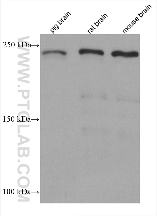
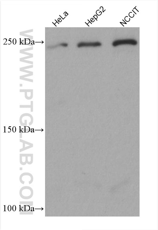
Various lysates were subjected to SDS PAGE followed by western blot with 67631-1-Ig (CKAP5 antibody) at dilution of 1:10000 incubated at room temperature for 1.5 hours.
WB analysis using 67631-1-Ig
Various lysates were subjected to SDS PAGE followed by western blot with 67631-1-Ig (CKAP5 antibody) at dilution of 1:10000 incubated at room temperature for 1.5 hours.
IHC staining of mouse cerebellum using 67631-1-Ig
Immunohistochemical analysis of paraffin-embedded mouse cerebellum tissue slide using 67631-1-Ig (Ch-TOG antibody) at dilution of 1:500 (under 10x lens). Heat mediated antigen retrieval with Tris-EDTA buffer (pH 9.0).
Immunohistochemical analysis of paraffin-embedded mouse cerebellum tissue slide using 67631-1-Ig (Ch-TOG antibody) at dilution of 1:500 (under 40x lens). Heat mediated antigen retrieval with Tris-EDTA buffer (pH 9.0).
IHC staining of human rectal cancer using 67631-1-Ig
Immunohistochemical analysis of paraffin-embedded human rectal cancer tissue slide using 67631-1-Ig (Ch-TOG antibody) at dilution of 1:1000 (under 10x lens). Heat mediated antigen retrieval with Tris-EDTA buffer (pH 9.0).
IHC staining of human rectal cancer using 67631-1-Ig
Immunohistochemical analysis of paraffin-embedded human rectal cancer tissue slide using 67631-1-Ig (Ch-TOG antibody) at dilution of 1:1000 (under 40x lens). Heat mediated antigen retrieval with Tris-EDTA buffer (pH 9.0).
IF Staining of HepG2 using 67631-1-Ig
Immunofluorescent analysis of (-20°C Ethanol) fixed HepG2 cells using Ch-TOG antibody (67631-1-Ig, Clone: 1H5G1 ) at dilution of 1:4000 and CoraLite®488-Conjugated AffiniPure Goat Anti-Mouse IgG(H+L), CL594-Phalloidin (red).
The Proteintech guarantee covers Proteintech antibodies in any species and any application, including those not listed on the datasheet. If the antibody doesn’t perform, you can receive a hassle-free refund or credit note.
pig brain tissue, HeLa cells, HepG2 cells, NCCIT cells, rat brain tissue, mouse brain tissue
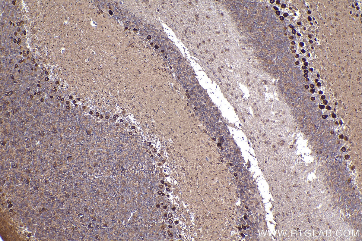
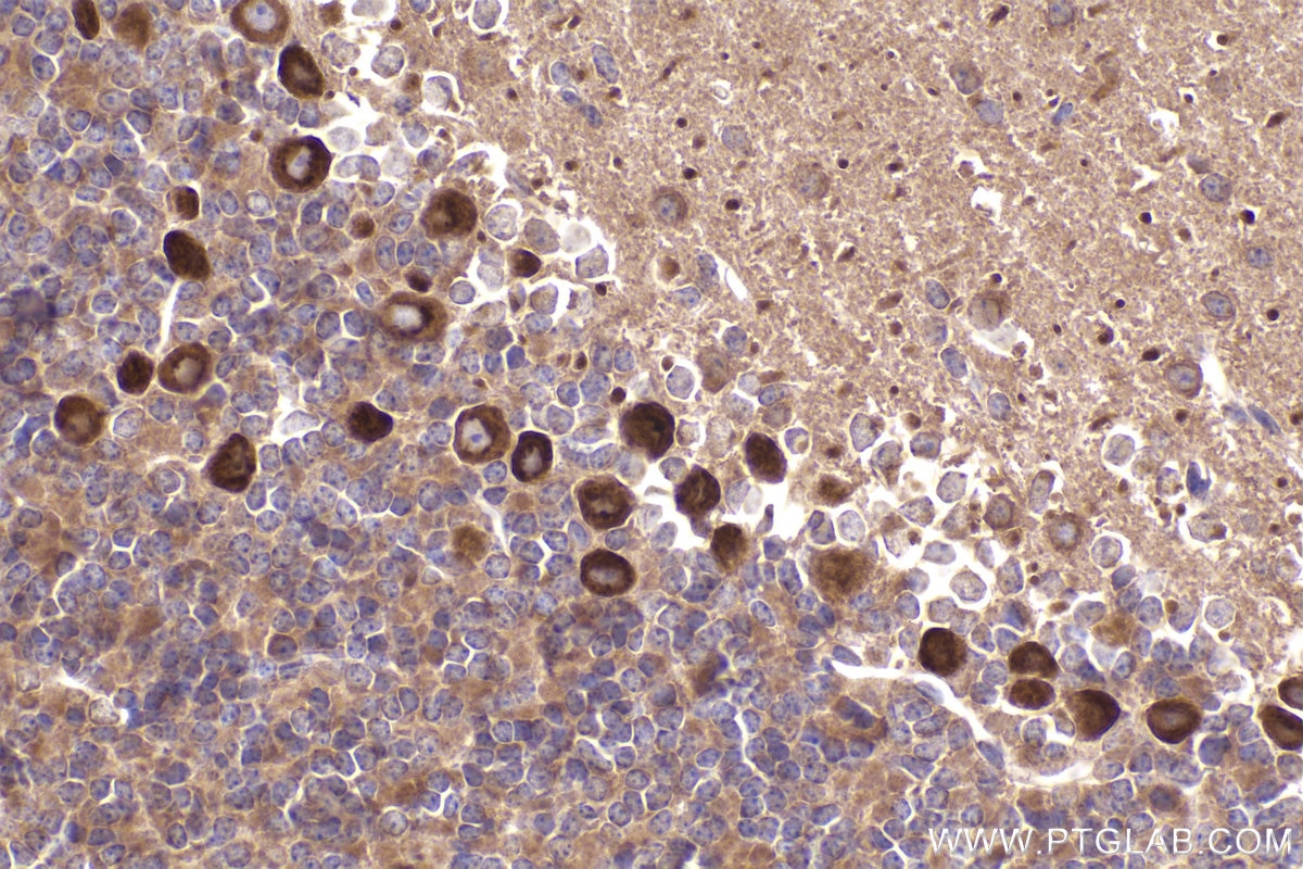
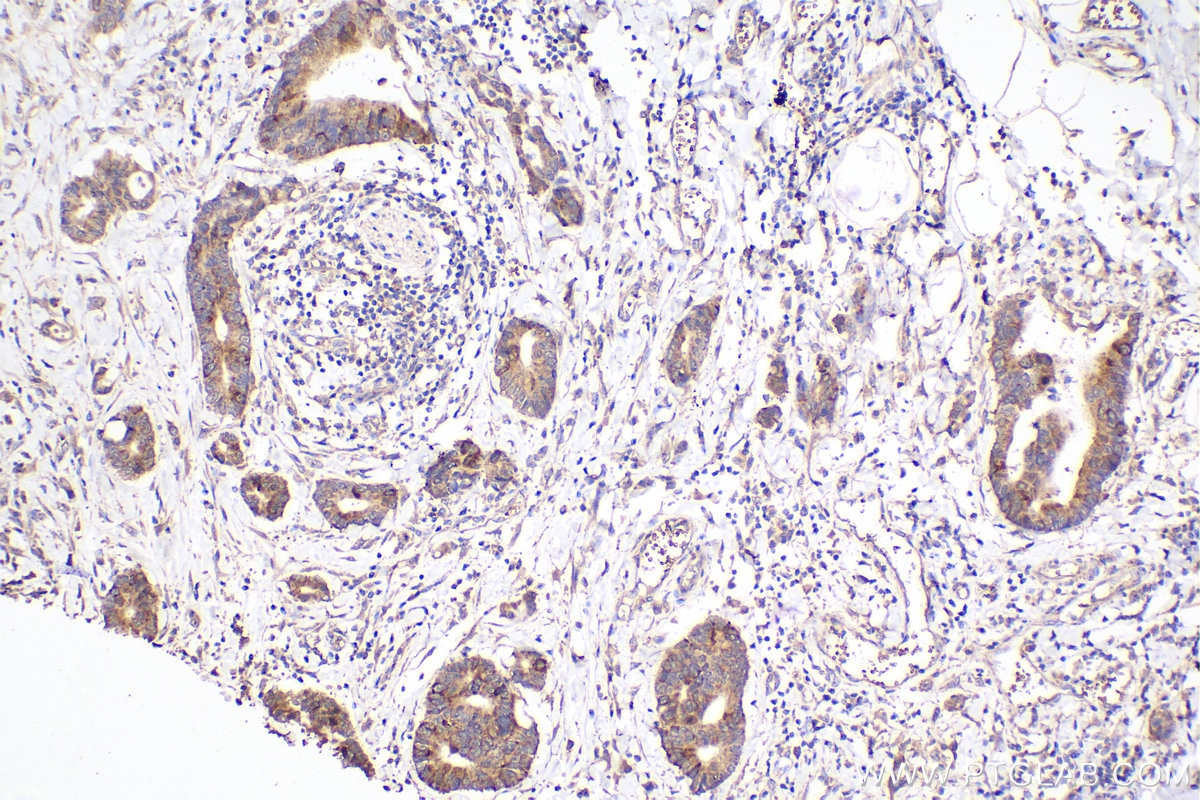
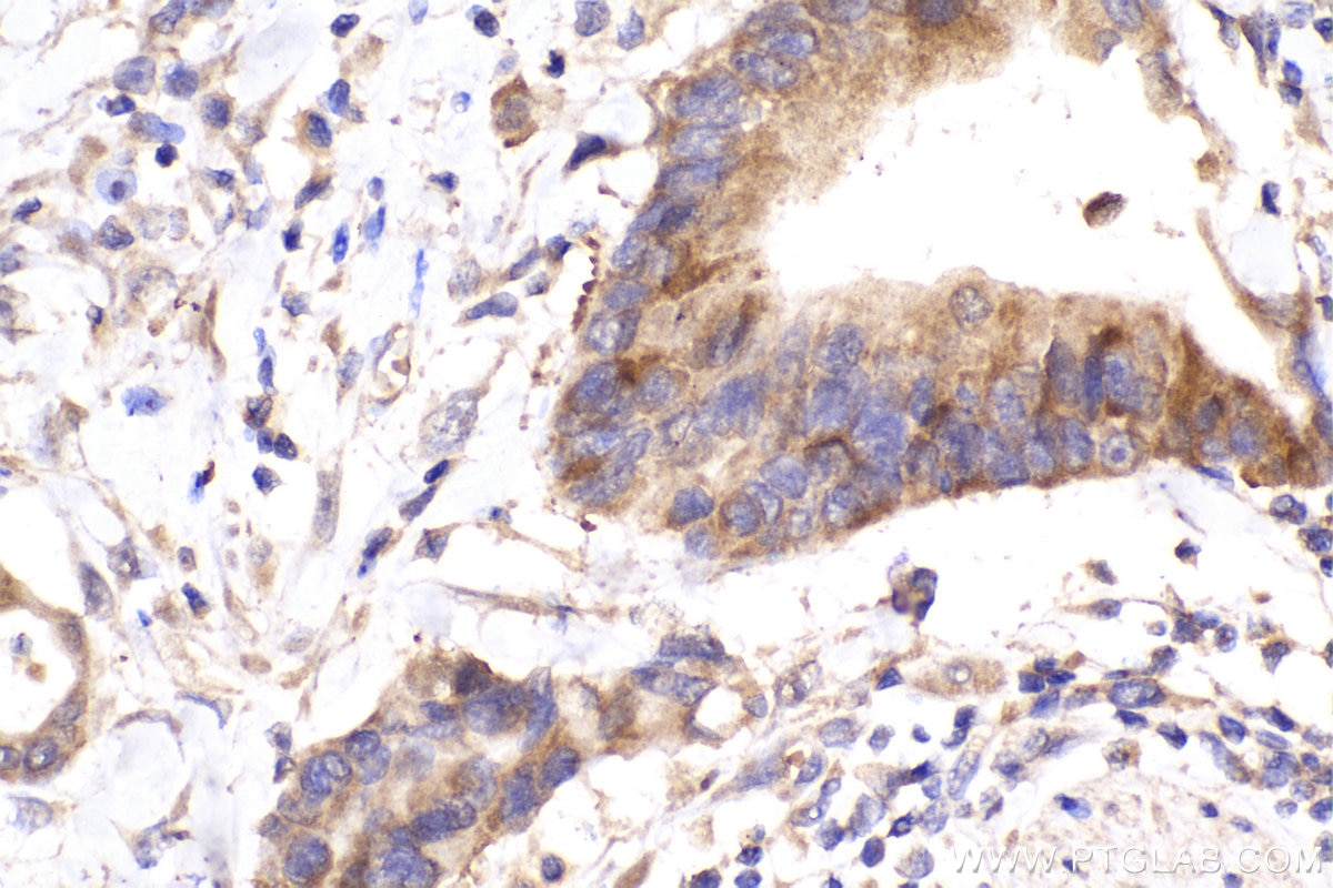
Positive IHC detected in
mouse cerebellum tissue, human rectal cancer tissue Note: suggested antigen retrieval with TE buffer pH 9.0; (*) Alternatively, antigen retrieval may be performed with citrate buffer pH 6.0
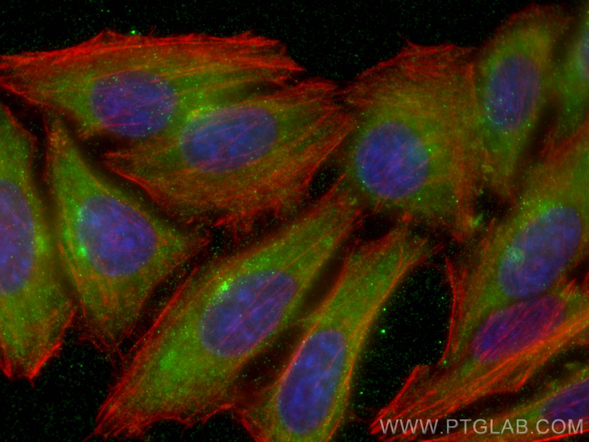
Positive IF/ICC detected in
HepG2 cells
Recommended dilution
Application
Dilution
Western Blot (WB)
WB : 1:5000-1:50000
Immunohistochemistry (IHC)
IHC : 1:250-1:1000
Immunofluorescence (IF)/ICC
IF/ICC : 1:2000-1:8000
It is recommended that this reagent should be titrated in each testing system to obtain optimal results.
Sample-dependent, Check data in validation data gallery.
Product Information
67631-1-Ig targets Ch-TOG in WB, IHC, IF/ICC, ELISA applications and shows reactivity with human, mouse, rat, pig samples.
PBS with 0.02% sodium azide and 50% glycerol pH 7.3.
Storage Conditions
Store at -20°C. Stable for one year after shipment. Aliquoting is unnecessary for -20oC storage. 20ul sizes contain 0.1% BSA.
Background Information
Ch-TOG (colonic hepatic tumor-overexpressed gene), also known as XMAP215 or CKAP5, is a microtubule polymerase which can promotes cytoplasmic microtubule nucleation and elongation. Through interacting with Aurora-A and TACC3, it plays a major role in organizing spindle poles.
Various lysates were subjected to SDS PAGE followed by western blot with 67631-1-Ig (CKAP5 antibody) at dilution of 1:10000 incubated at room temperature for 1.5 hours.
WB analysis using 67631-1-Ig
Various lysates were subjected to SDS PAGE followed by western blot with 67631-1-Ig (CKAP5 antibody) at dilution of 1:10000 incubated at room temperature for 1.5 hours.
IHC Figures
IHC staining of mouse cerebellum using 67631-1-Ig
Immunohistochemical analysis of paraffin-embedded mouse cerebellum tissue slide using 67631-1-Ig (Ch-TOG antibody) at dilution of 1:500 (under 10x lens). Heat mediated antigen retrieval with Tris-EDTA buffer (pH 9.0).
IHC staining of mouse cerebellum using 67631-1-Ig
Immunohistochemical analysis of paraffin-embedded mouse cerebellum tissue slide using 67631-1-Ig (Ch-TOG antibody) at dilution of 1:500 (under 40x lens). Heat mediated antigen retrieval with Tris-EDTA buffer (pH 9.0).
IHC staining of human rectal cancer using 67631-1-Ig
Immunohistochemical analysis of paraffin-embedded human rectal cancer tissue slide using 67631-1-Ig (Ch-TOG antibody) at dilution of 1:1000 (under 10x lens). Heat mediated antigen retrieval with Tris-EDTA buffer (pH 9.0).
IHC staining of human rectal cancer using 67631-1-Ig
Immunohistochemical analysis of paraffin-embedded human rectal cancer tissue slide using 67631-1-Ig (Ch-TOG antibody) at dilution of 1:1000 (under 40x lens). Heat mediated antigen retrieval with Tris-EDTA buffer (pH 9.0).
IF/ICC Figures
IF Staining of HepG2 using 67631-1-Ig
Immunofluorescent analysis of (-20°C Ethanol) fixed HepG2 cells using Ch-TOG antibody (67631-1-Ig, Clone: 1H5G1 ) at dilution of 1:4000 and CoraLite®488-Conjugated AffiniPure Goat Anti-Mouse IgG(H+L), CL594-Phalloidin (red).
The species listed in Tested Reactivity are in-house verified and applicable species. For unlisted species, please refer to the homology analysis of the immunogen sequence and related species. For rabbit polyclonal antibodies, homology >70% is recommended. For mouse monoclonal antibodies and rabbit recombinant antibodies, homology >90% is recommended. Generally, the higher the homology, the greater the applicability. However, there will be certain differences in protein expression in different species, tissues or cells. Therefore, the homology analysis results are for reference only and do not serve as a guarantee.
At Proteintech, we pride ourselves on our antibody quality, customer service and transparency. As such, we are comparing our antibodies with other vendors, enabling easy identification and comparisons of key data to help you choose the suitable antibody for your needs.
We have selected the top cited antibodies from these vendors for you to compare.