Tested Applications
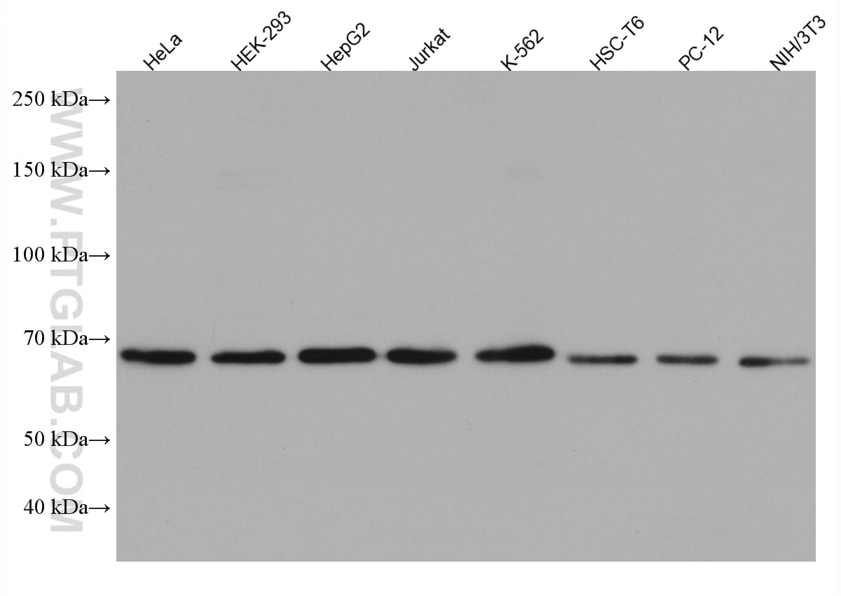
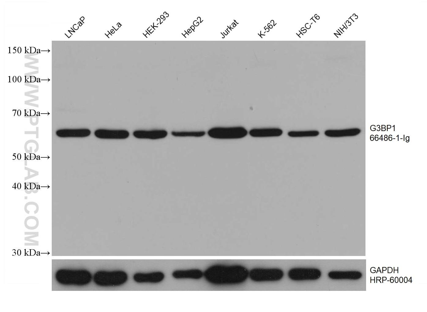
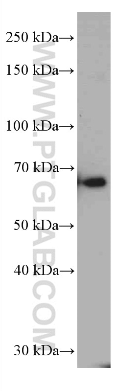
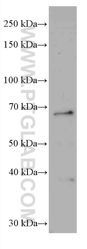
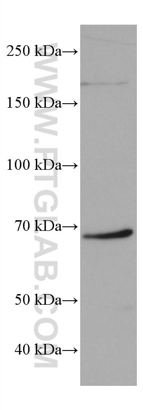
| Positive WB detected in | LNCaP cells, RAW 264.7 cells, pig brain tissue, HeLa cells, mouse brain tissue, HEK-293 cells, HepG2 cells, Jurkat cells, K-562 cells, HSC-T6 cells, PC-12 cells, NIH/3T3 cells, 4T1 cells |
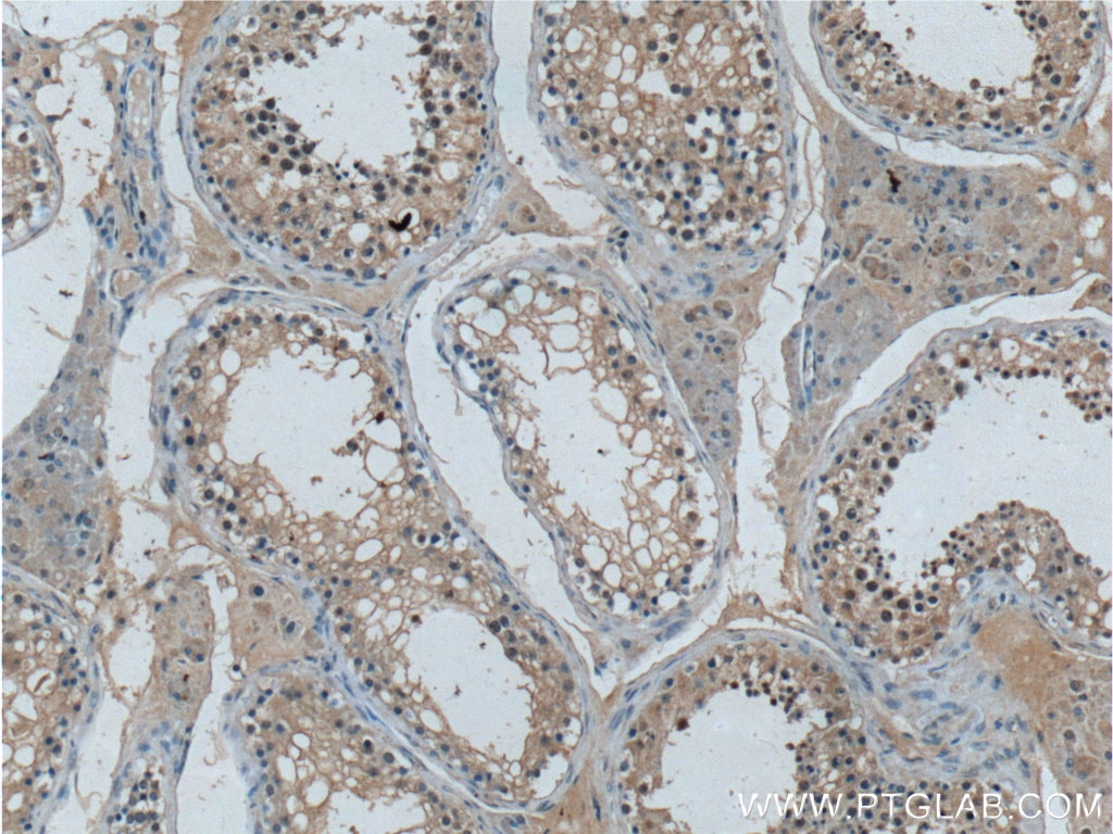
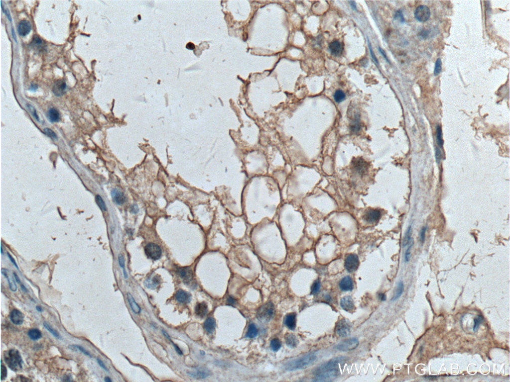
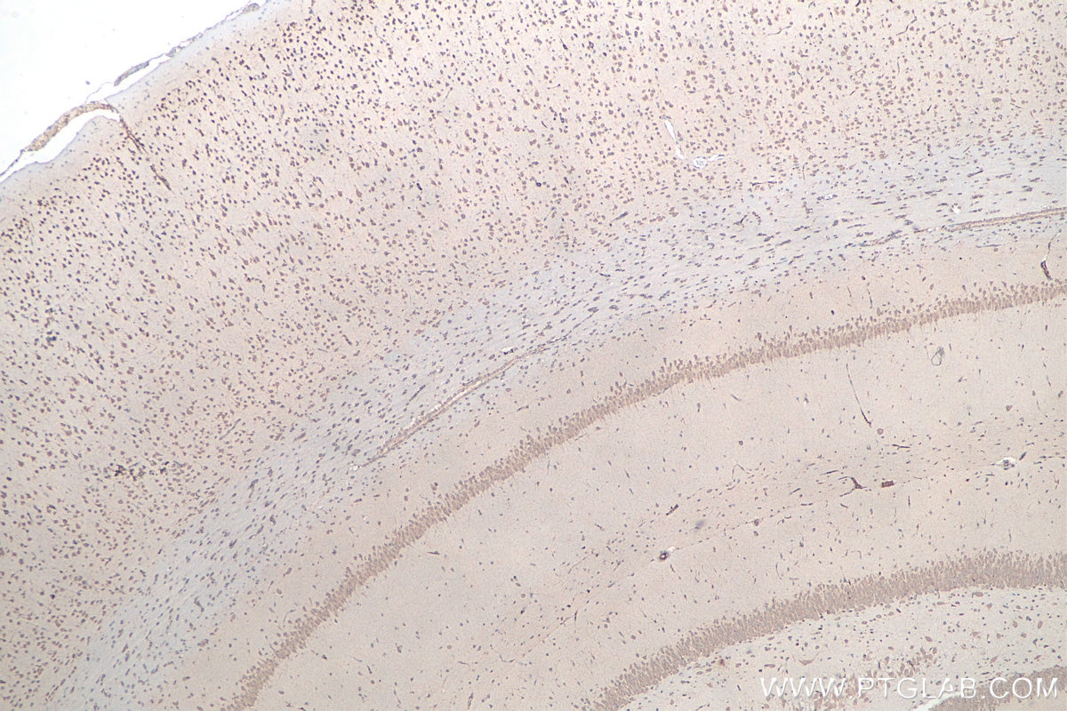
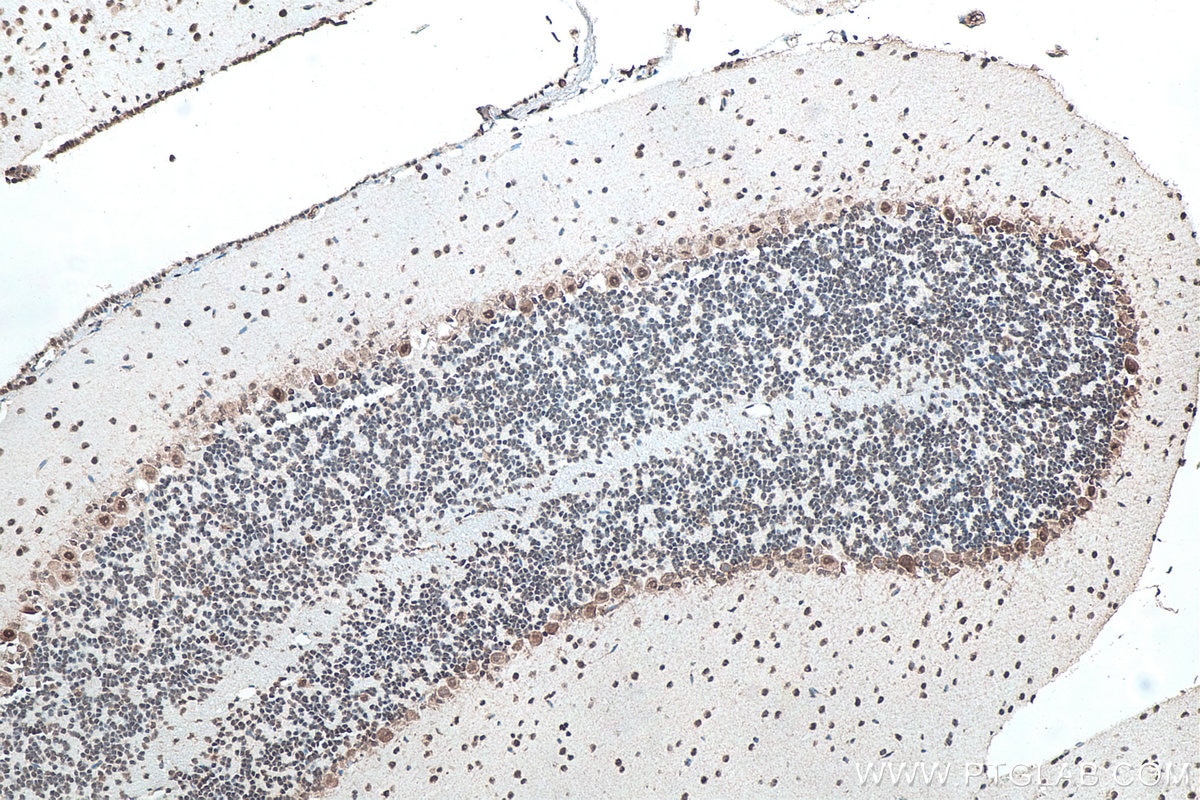
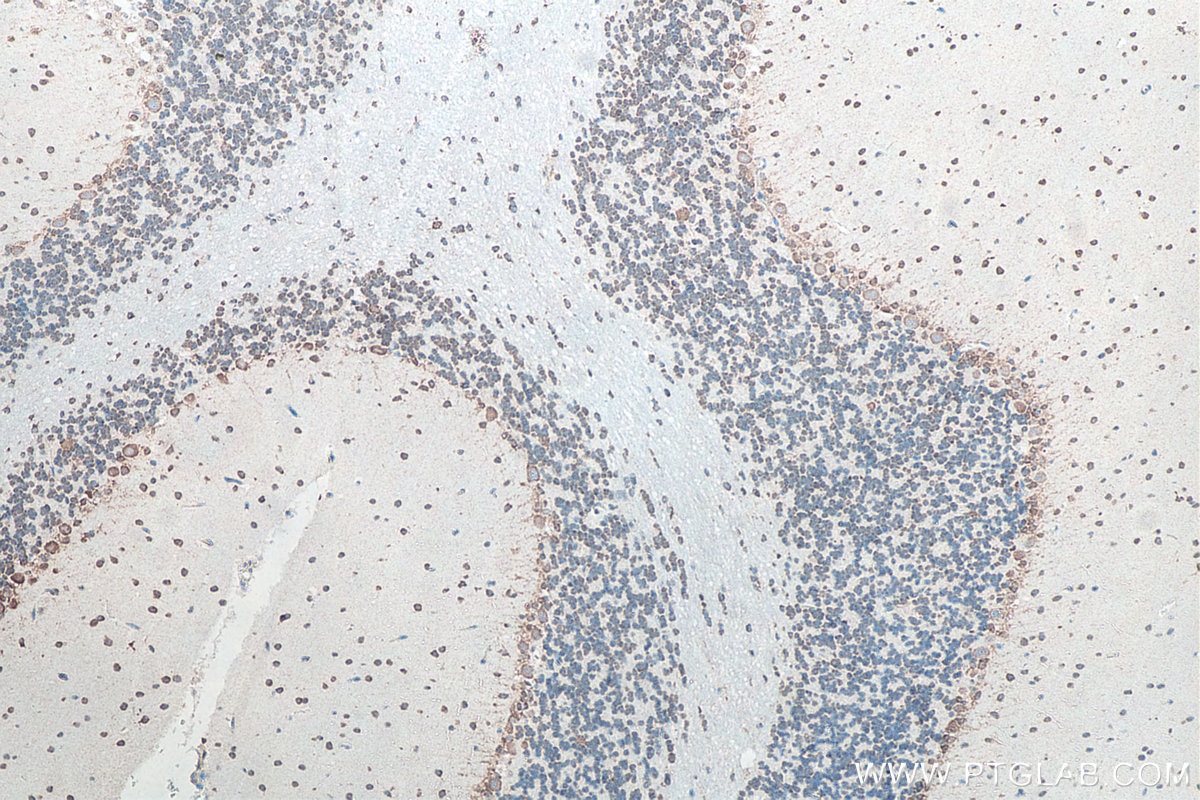
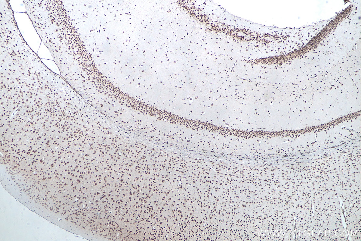
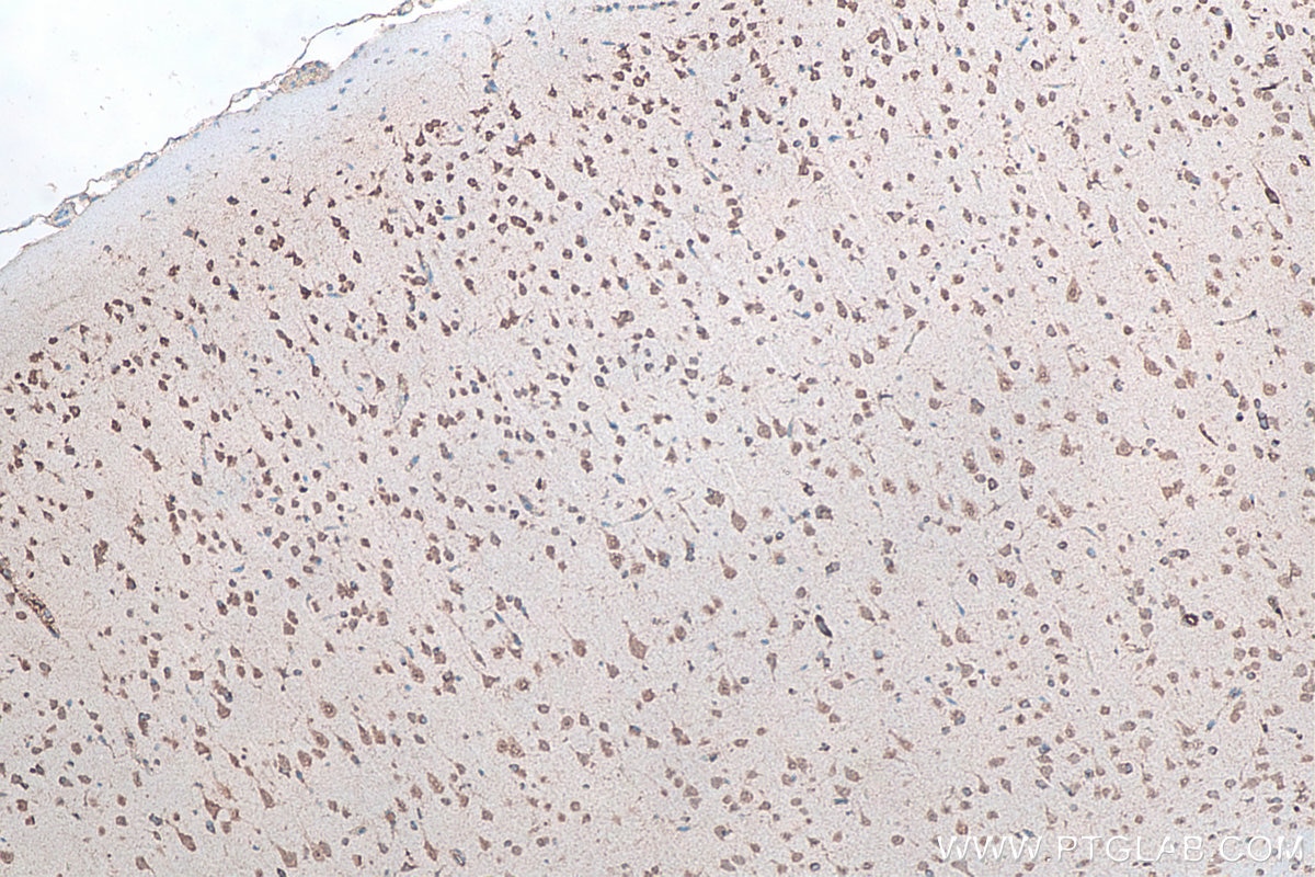
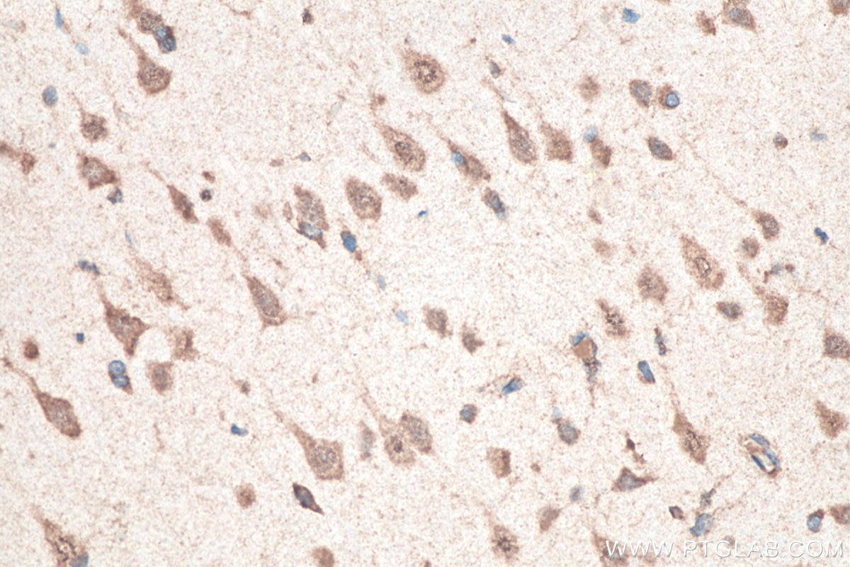
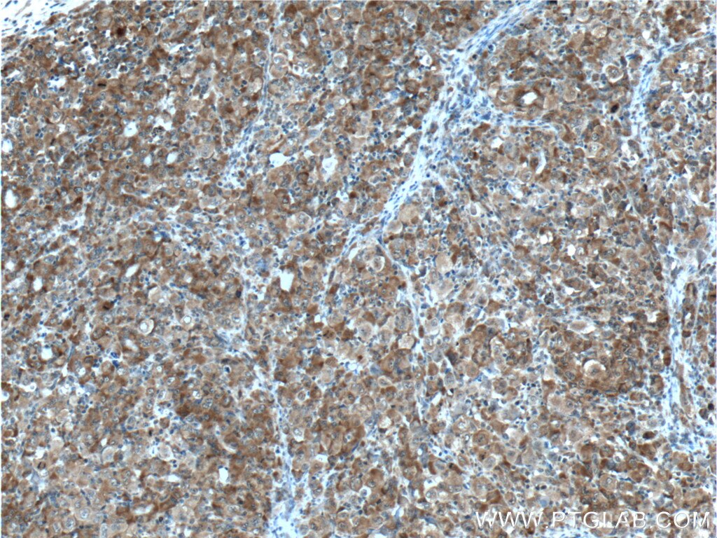
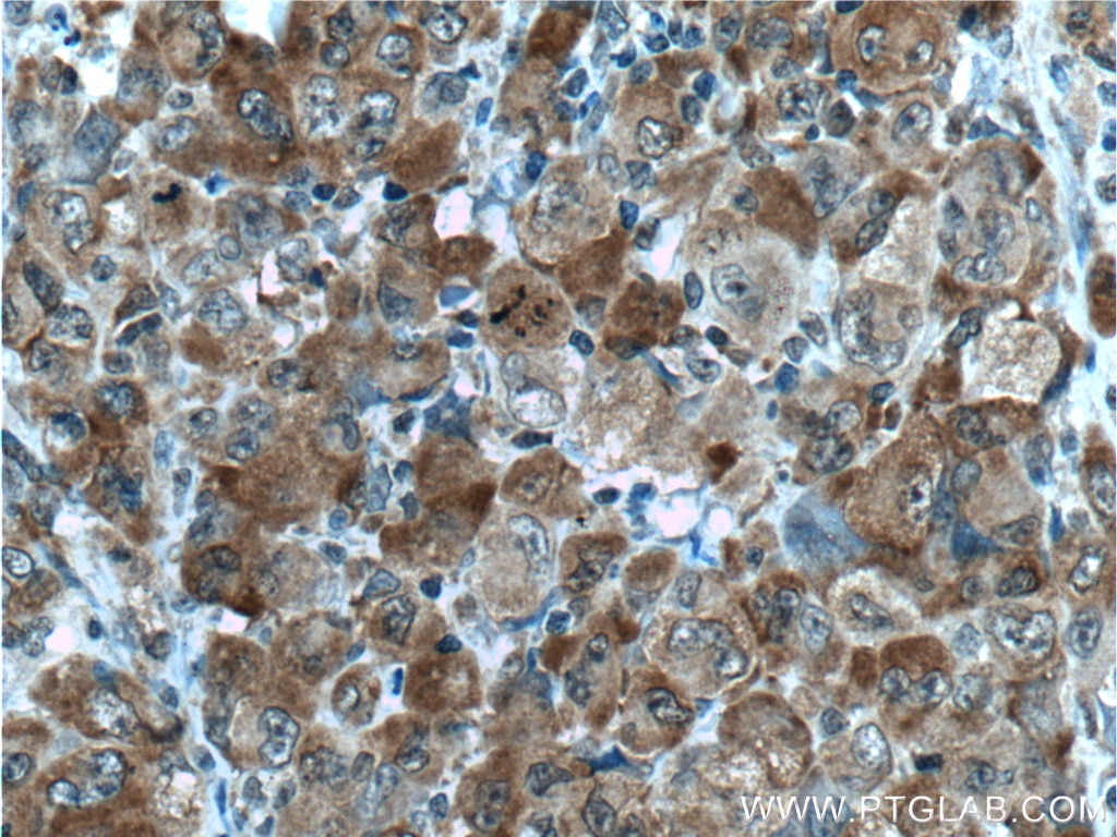
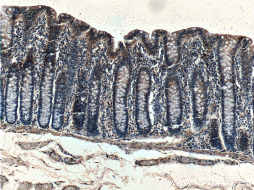
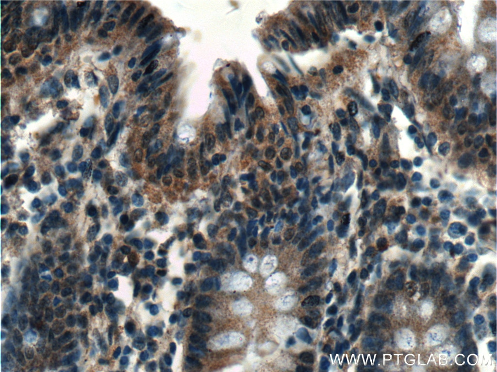
| Positive IHC detected in | human testis tissue, human colon tissue, human lymphoma tissue, rat brain tissue Note: suggested antigen retrieval with TE buffer pH 9.0; (*) Alternatively, antigen retrieval may be performed with citrate buffer pH 6.0 |
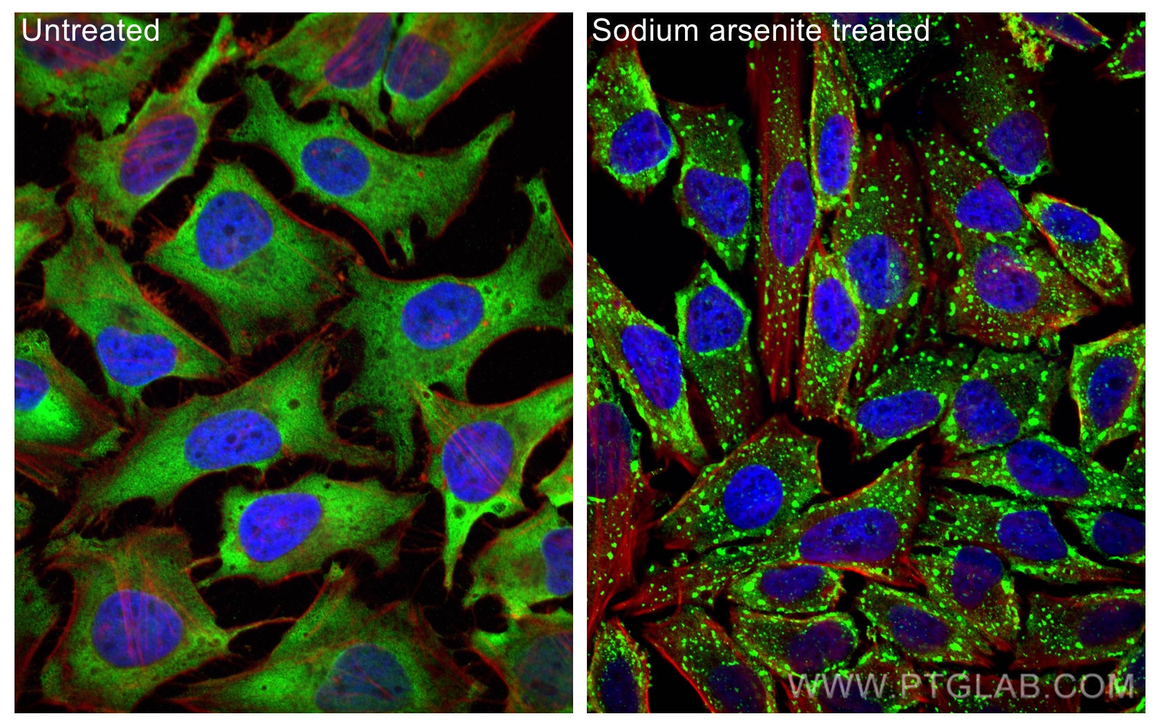
| Positive IF/ICC detected in | sodium arsenite treated HeLa cells |
Recommended dilution
| Application | Dilution |
|---|---|
| Western Blot (WB) | WB : 1:5000-1:50000 |
| Immunohistochemistry (IHC) | IHC : 1:50-1:500 |
| Immunofluorescence (IF)/ICC | IF/ICC : 1:500-1:2000 |
| It is recommended that this reagent should be titrated in each testing system to obtain optimal results. | |
| Sample-dependent, Check data in validation data gallery. | |
Published Applications
| KD/KO | See 10 publications below |
| WB | See 42 publications below |
| IHC | See 1 publications below |
| IF | See 71 publications below |
| IP | See 3 publications below |
| CoIP | See 7 publications below |
| RIP | See 1 publications below |
Product Information
66486-1-Ig targets G3BP1 in WB, IHC, IF/ICC, IP, CoIP, RIP, ELISA applications and shows reactivity with human, mouse, rat, pig samples.
| Tested Reactivity | human, mouse, rat, pig |
| Cited Reactivity | human, mouse, rat, pig, monkey, sheep |
| Host / Isotype | Mouse / IgG1 |
| Class | Monoclonal |
| Type | Antibody |
| Immunogen |
CatNo: Ag3728 Product name: Recombinant human G3BP1 protein Source: e coli.-derived, PGEX-4T Tag: GST Domain: 167-466 aa of BC006997 Sequence: PDDSGTFYDQAVVSNDMEEHLEEPVAEPEPDPEPEPEQEPVSEIQEEKPEPVLEETAPEDAQKSSSPAPADIAQTVQEDLRTFSWASVTSKNLPPSGAVPVTGIPPHVVKVPASQPRPESKPESQIPPQRPQRDQRVREQRINIPPQRGPRPIREAGEQGDIEPRRMVRHPDSHQLFIGNLPHEVDKSELKDFFQSYGNVVELRINSGGKLPNFGFVVFDDSEPVQKVLSNRPIMFRGEVRLNVEEKKTRAAREGDRRDNRLRGPGGPRGGLGGGMRGPPRGGMVQKPGFGVGRGLAPRQ Predict reactive species |
| Full Name | GTPase activating protein (SH3 domain) binding protein 1 |
| Calculated Molecular Weight | 466 aa, 52 kDa |
| Observed Molecular Weight | 68 kDa |
| GenBank Accession Number | BC006997 |
| Gene Symbol | G3BP1 |
| Gene ID (NCBI) | 10146 |
| RRID | AB_2819031 |
| Conjugate | Unconjugated |
| Form | Liquid |
| Purification Method | Protein G purification |
| UNIPROT ID | Q13283 |
| Storage Buffer | PBS with 0.02% sodium azide and 50% glycerol, pH 7.3. |
| Storage Conditions | Store at -20°C. Stable for one year after shipment. Aliquoting is unnecessary for -20oC storage. 20ul sizes contain 0.1% BSA. |
Background Information
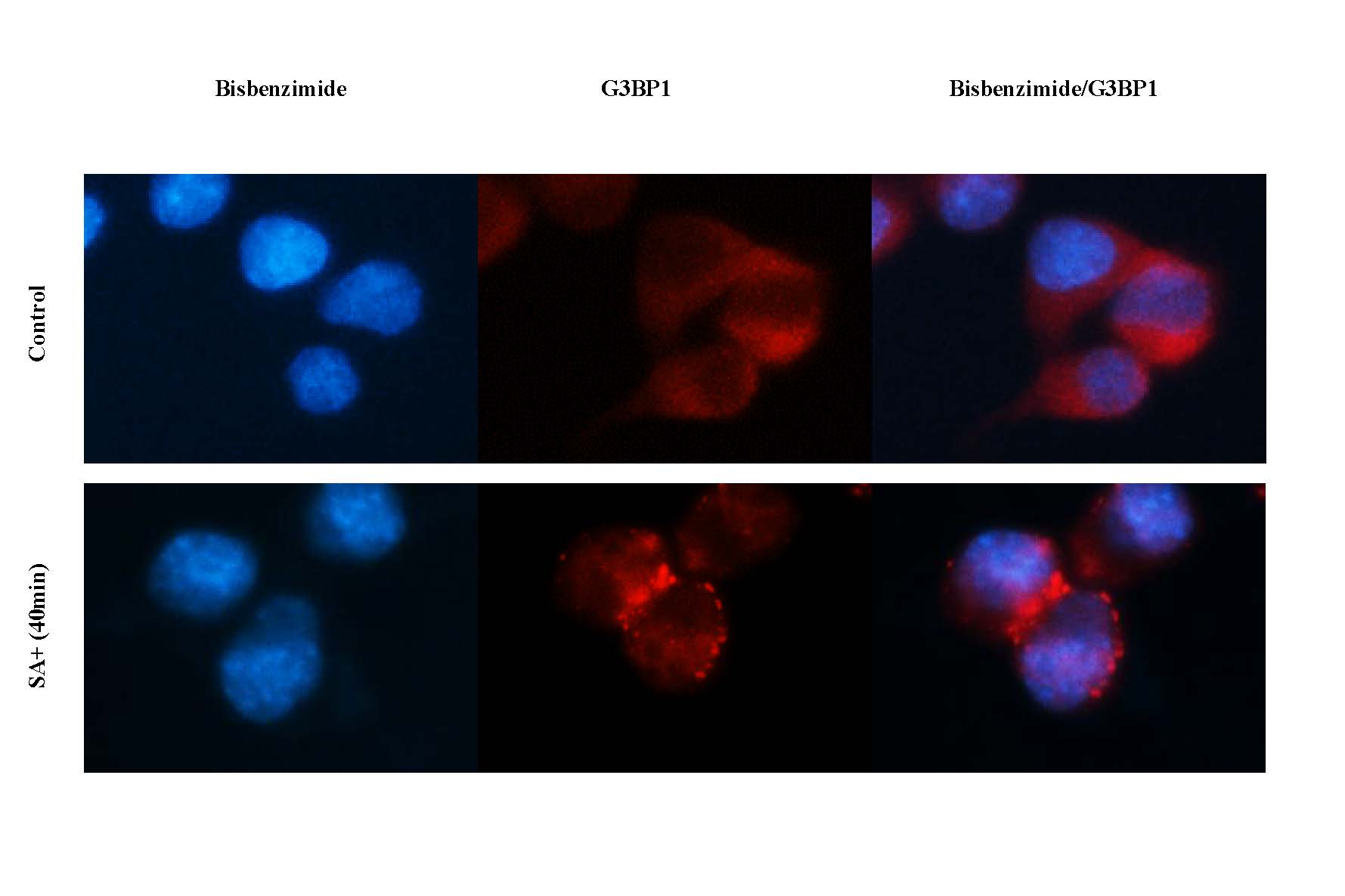
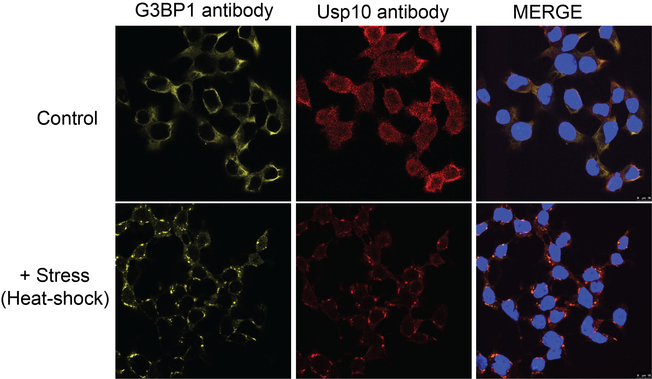
GAP SH3 Binding Protein 1 (G3BP1), also named as G3BP, is an effector of stress granule (SG) assembly. SG biology plays an important role in the pathophysiology of TDP-43 in ALS and FTLD-U. G3BP1 can be used as a marker of SG. It has been shown to function downstream of Ras and play a role in RNA metabolism, signal transduction, and proliferation. G3BP1 is a ubiquitously expressed protein that localizes to the cytoplasm in proliferating cells and to the nucleus in non-proliferating cells. G3BP1 has recently been implicated in cancer biology.
Protocols
| Product Specific Protocols | |
|---|---|
| IF protocol for G3BP1 antibody 66486-1-Ig | Download protocol |
| IHC protocol for G3BP1 antibody 66486-1-Ig | Download protocol |
| WB protocol for G3BP1 antibody 66486-1-Ig | Download protocol |
| Standard Protocols | |
|---|---|
| Click here to view our Standard Protocols |
Publications
| Species | Application | Title |
|---|---|---|
Nature DDX3X acts as a live-or-die checkpoint in stressed cells by regulating NLRP3 inflammasome. | ||
Sci Transl Med Precise genomic editing of pathogenic mutations in RBM20 rescues dilated cardiomyopathy | ||
Nat Struct Mol Biol TDP-43 aggregation induced by oxidative stress causes global mitochondrial imbalance in ALS. | ||
Nat Commun Engineering bi-directional chemically-modulated synthetic condensates for cellular control | ||
Brain Behav Immun Transcriptomic and proteomic profiling of bi-partite and tri-partite murine iPSC-derived neurospheroids under steady-state and inflammatory condition |
Reviews
The reviews below have been submitted by verified Proteintech customers who received an incentive for providing their feedback.
FH Xiaochen (Verified Customer) (11-11-2024) | wonderful image
 |
FH Haibo (Verified Customer) (10-26-2020) | This is a excellent antibody to visualize stress granules tested in HEK293 and human fibroblasts.
|
FH Manohar (Verified Customer) (09-23-2020) | 1% milk is used
|
FH David (Verified Customer) (01-13-2020) | Good signal in control cells and immunopositive puncta seen in response to stress (e.g. sodium arsenite).
|
FH Azita (Verified Customer) (10-04-2019) | Assembly of stress granula upon treatment with sodium arsenite for 40 min. (It works great)
 |
FH Kyosuke (Verified Customer) (06-12-2019) | It works well on HEK293T for Western blot.
|
FH Natalia (Verified Customer) (06-06-2019) | PFA fixated cells
 |