Tested Applications
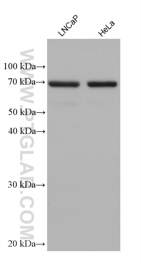
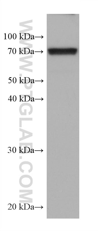
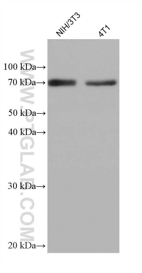
| Positive WB detected in | LNCaP cells, HSC-T6 cells, NIH/3T3 cells, HeLa cells, 4T1 cells |
Recommended dilution
| Application | Dilution |
|---|---|
| Western Blot (WB) | WB : 1:5000-1:50000 |
| It is recommended that this reagent should be titrated in each testing system to obtain optimal results. | |
| Sample-dependent, Check data in validation data gallery. | |
Published Applications
| KD/KO | See 1 publications below |
| WB | See 1 publications below |
| IHC | See 1 publications below |
Product Information
68174-1-Ig targets GPD2 in WB, IHC, ELISA applications and shows reactivity with human, mouse, rat samples.
| Tested Reactivity | human, mouse, rat |
| Cited Reactivity | mouse |
| Host / Isotype | Mouse / IgG1 |
| Class | Monoclonal |
| Type | Antibody |
| Immunogen |
CatNo: Ag11212 Product name: Recombinant human GPD2 protein Source: e coli.-derived, PET28a Tag: 6*His Domain: 5-378 aa of BC019874 Sequence: KAVKGTLVGGGALATVLGLSQFAHYRRKQMNLAYVKAADCISEPVNREPPSREAQLLTLQNTSEFDILVIGGGATGSGCALDAVTRGLKTALVERDDFSSGTSSRSTKLIHGGVRYLQKAIMKLDIEQYRMVKEALHERANLLEIAPHLSAPLPIMLPVYKWWQLPYYWVGIKLYDLVAGSNCLKSSYVLSKSRALEHFPMLQKDKLVGAIVYYDGQHNDARMNLAIALTAARYGAATANYMEVVSLLKKTDPQTGKVHVSGARCKDVLTGQEFDVRAKCVINATGPFTDSVRKMDDKDAAAICQPSAGVHIVMPGYYSPESMGLLDPATSDGRVIFFLPWQKMTIAGTTDTPTDVTHHPIPSEEDINFILNEV Predict reactive species |
| Full Name | glycerol-3-phosphate dehydrogenase 2 (mitochondrial) |
| Calculated Molecular Weight | 41 kDa, 81 kDa |
| GenBank Accession Number | BC019874 |
| Gene Symbol | GPD2 |
| Gene ID (NCBI) | 2820 |
| RRID | AB_3085046 |
| Conjugate | Unconjugated |
| Form | Liquid |
| Purification Method | Protein G purification |
| UNIPROT ID | P43304 |
| Storage Buffer | PBS with 0.02% sodium azide and 50% glycerol, pH 7.3. |
| Storage Conditions | Store at -20°C. Stable for one year after shipment. Aliquoting is unnecessary for -20oC storage. 20ul sizes contain 0.1% BSA. |
Background Information
GPD2(Glycerol-3-phosphate dehydrogenase, mitochondrial) belongs to the FAD-dependent glycerol-3-phosphate dehydrogenase family.It is phosphorylated only in capacitated hamster spermatozoa and is noncanonically localized in the acrosome and principal piece in human, mouse, rat, and hamster spermatozoa, though in somatic cells it is localized in the mitochondria. This noncanonical localization may imply a role of GPD2 in acrosome reaction and hyperactivation(PMID:19333995 ).
Protocols
| Product Specific Protocols | |
|---|---|
| WB protocol for GPD2 antibody 68174-1-Ig | Download protocol |
| Standard Protocols | |
|---|---|
| Click here to view our Standard Protocols |