Tested Applications
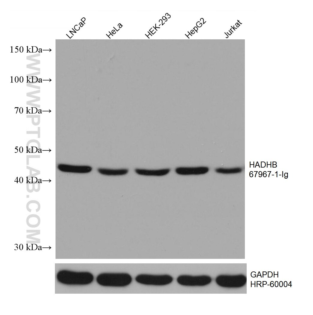
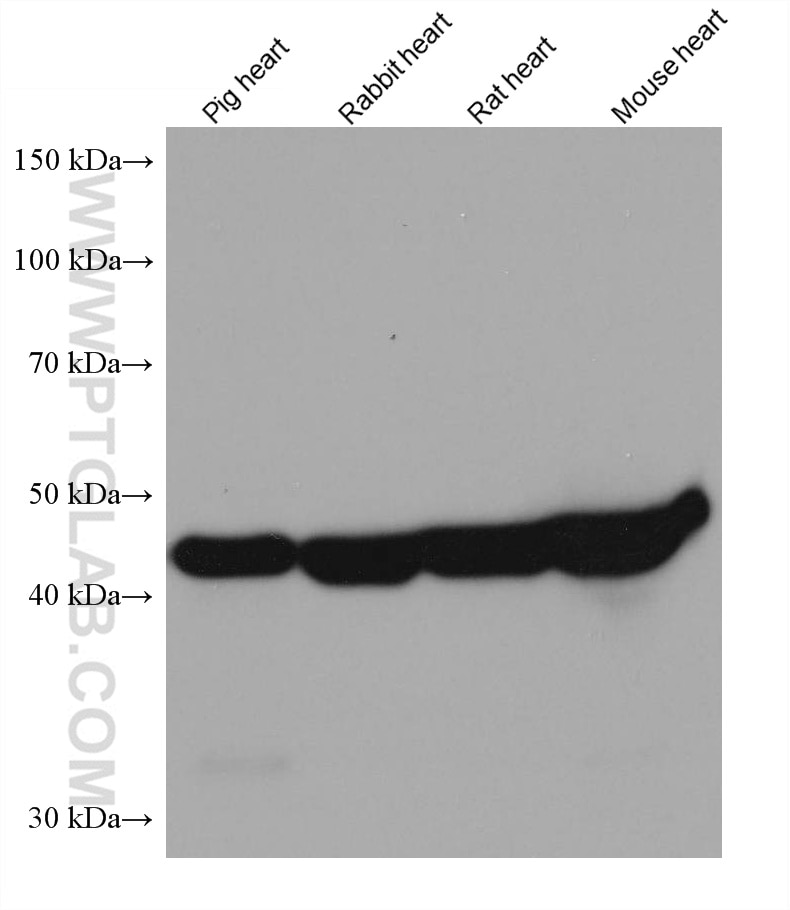
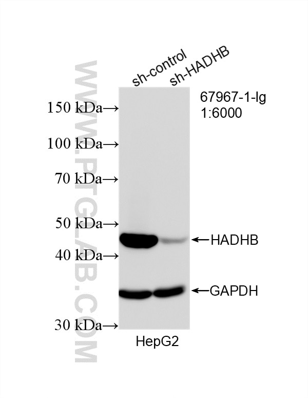
| Positive WB detected in | LNCaP cells, HepG2 cells, pig heart tissue, Hela cells, HEK-293 cells, Jurkat cells, Rabbit heart tissues, Rat heart tissues, Mouse heart tissues |
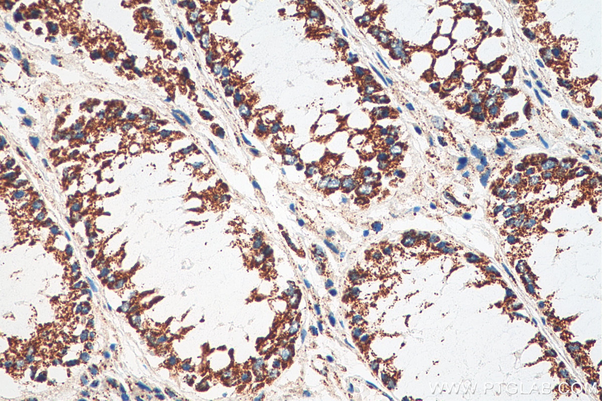
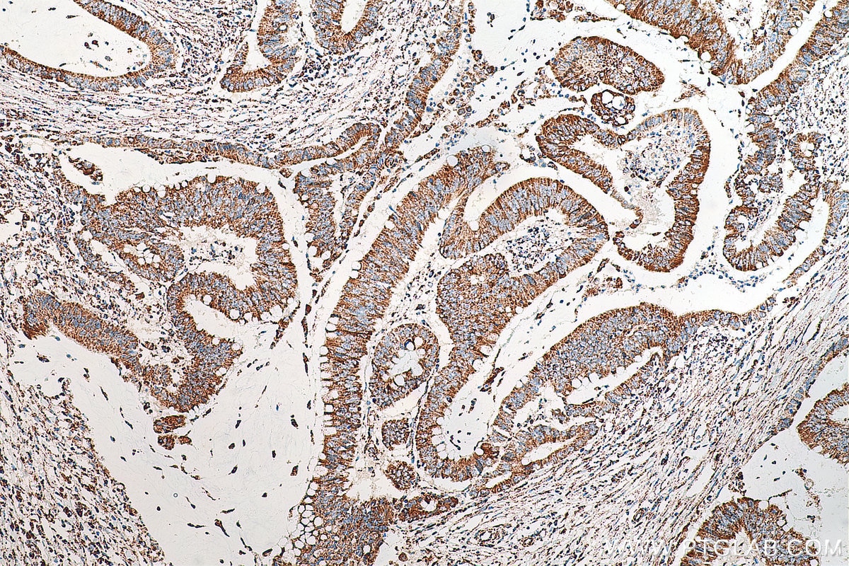
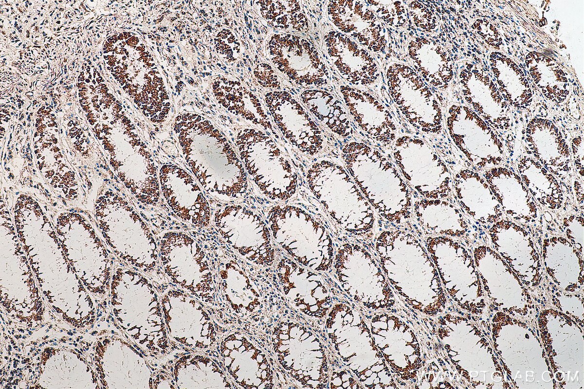
| Positive IHC detected in | human colon cancer tissue Note: suggested antigen retrieval with TE buffer pH 9.0; (*) Alternatively, antigen retrieval may be performed with citrate buffer pH 6.0 |
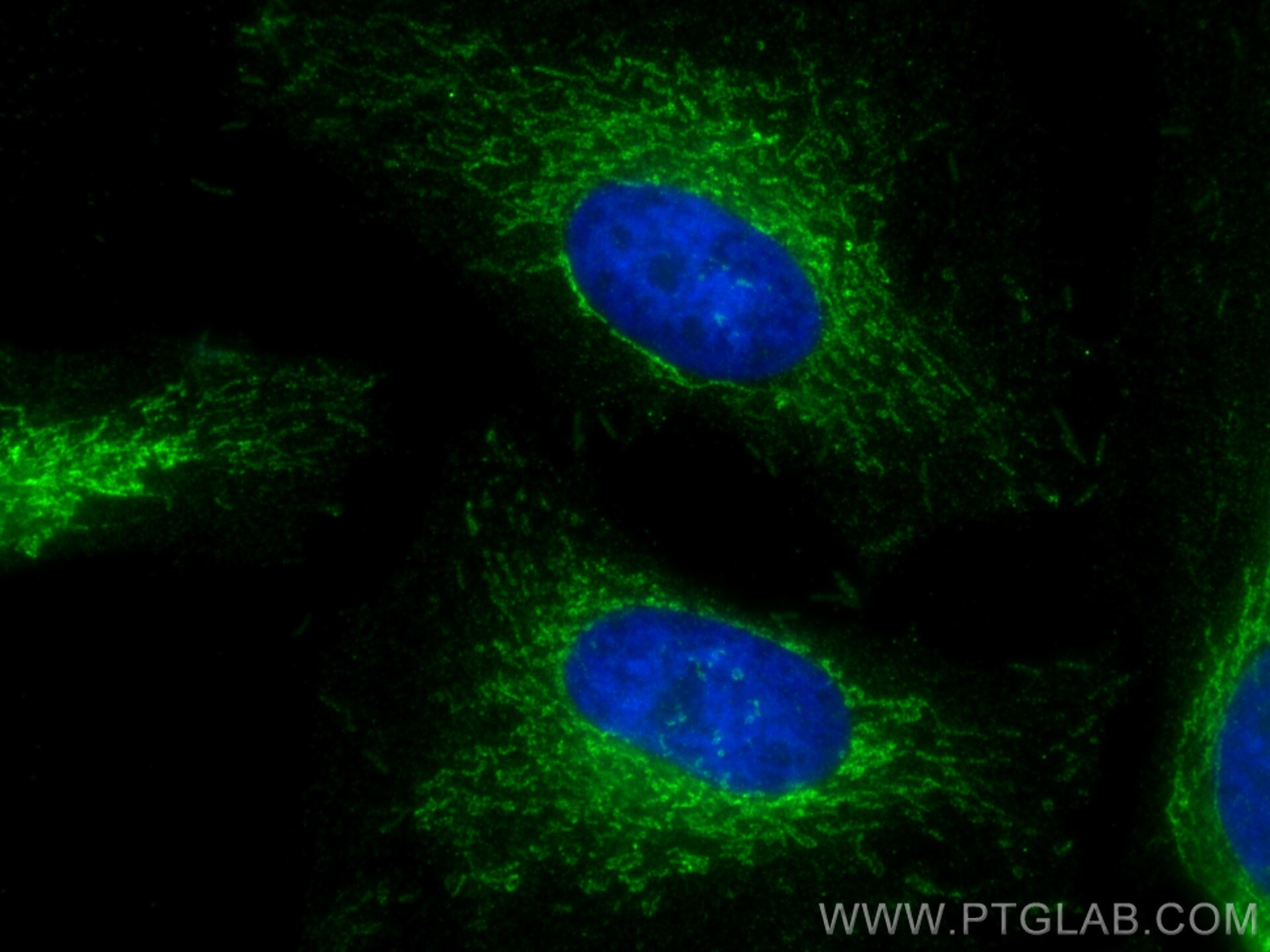
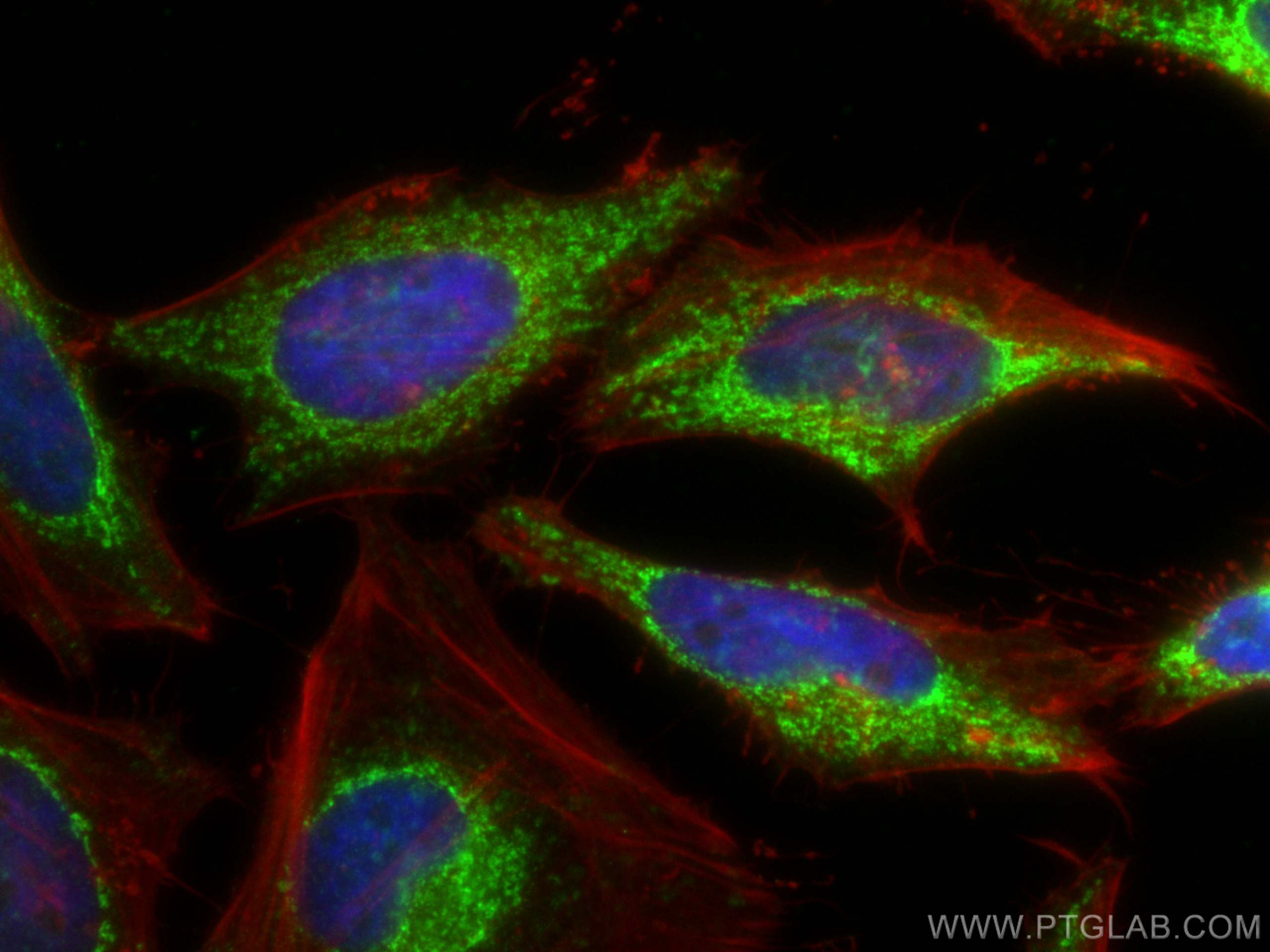
| Positive IF/ICC detected in | HeLa cells |
Recommended dilution
| Application | Dilution |
|---|---|
| Western Blot (WB) | WB : 1:5000-1:50000 |
| Immunohistochemistry (IHC) | IHC : 1:1000-1:4000 |
| Immunofluorescence (IF)/ICC | IF/ICC : 1:200-1:800 |
| It is recommended that this reagent should be titrated in each testing system to obtain optimal results. | |
| Sample-dependent, Check data in validation data gallery. | |
Published Applications
| WB | See 1 publications below |
Product Information
67967-1-Ig targets HADHB in WB, IHC, IF/ICC, ELISA applications and shows reactivity with human, mouse, rat, pig samples.
| Tested Reactivity | human, mouse, rat, pig |
| Cited Reactivity | mouse |
| Host / Isotype | Mouse / IgG2b |
| Class | Monoclonal |
| Type | Antibody |
| Immunogen |
CatNo: Ag30315 Product name: Recombinant human HADHB protein Source: e coli.-derived, PET28a Tag: 6*His Domain: 34-262 aa of BC017564 Sequence: RAAPAVQTKTKKTLAKPNIRNVVVVDGVRTPFLLSGTSYKDLMPHDLARAALTGLLHRTSVPKEVVDYIIFGTVIQEVKTSNVAREAALGAGFSDKTPAHTVTMACISANQAMTTGVGLIASGQCDVIVAGGVELMSDVPIRHSRKMRKLMLDLNKAKSMGQRLSLISKFRFNFLAPELPAVSEFSTSETMGHSADRLAAAFAVSRLEQDEYALRSHSLAKKAQDEGLL Predict reactive species |
| Full Name | hydroxyacyl-Coenzyme A dehydrogenase/3-ketoacyl-Coenzyme A thiolase/enoyl-Coenzyme A hydratase (trifunctional protein), beta subunit |
| Calculated Molecular Weight | 51 kDa |
| Observed Molecular Weight | 47 kDa |
| GenBank Accession Number | BC017564 |
| Gene Symbol | HADHB |
| Gene ID (NCBI) | 3032 |
| RRID | AB_2918718 |
| Conjugate | Unconjugated |
| Form | Liquid |
| Purification Method | Protein A purification |
| UNIPROT ID | P55084 |
| Storage Buffer | PBS with 0.02% sodium azide and 50% glycerol, pH 7.3. |
| Storage Conditions | Store at -20°C. Stable for one year after shipment. Aliquoting is unnecessary for -20oC storage. 20ul sizes contain 0.1% BSA. |
Background Information
HADHB, also named as TP- beta, Acetyl-CoA acyltransferase and Beta-ketothiolase, is a mitochondrial trifunctional enzyme subunit beta. Mitochondrial trifunctional enzyme catalyzes the last three of the four reactions of the mitochondrial beta-oxidation pathway. The mitochondrial beta-oxidation pathway is the major energy-producing process in tissues and is performed through four consecutive reactions breaking down fatty acids into acetyl-CoA. Among the enzymes involved in this pathway, the trifunctional enzyme exhibits specificity for long-chain fatty acids. Mitochondrial trifunctional enzyme is a heterotetrameric complex composed of two proteins, the trifunctional enzyme subunit alpha/HADHA carries the 2,3-enoyl-CoA hydratase and the 3-hydroxyacyl-CoA dehydrogenase activities, while the trifunctional enzyme subunit beta/HADHB described here bears the 3-ketoacyl-CoA thiolase activity. HADHB has 2 isoforms produced by alternative splicing with the MW of 49 kDa and 51 kDa.
Protocols
| Product Specific Protocols | |
|---|---|
| IF protocol for HADHB antibody 67967-1-Ig | Download protocol |
| IHC protocol for HADHB antibody 67967-1-Ig | Download protocol |
| WB protocol for HADHB antibody 67967-1-Ig | Download protocol |
| Standard Protocols | |
|---|---|
| Click here to view our Standard Protocols |