Filter:


Tested Applications
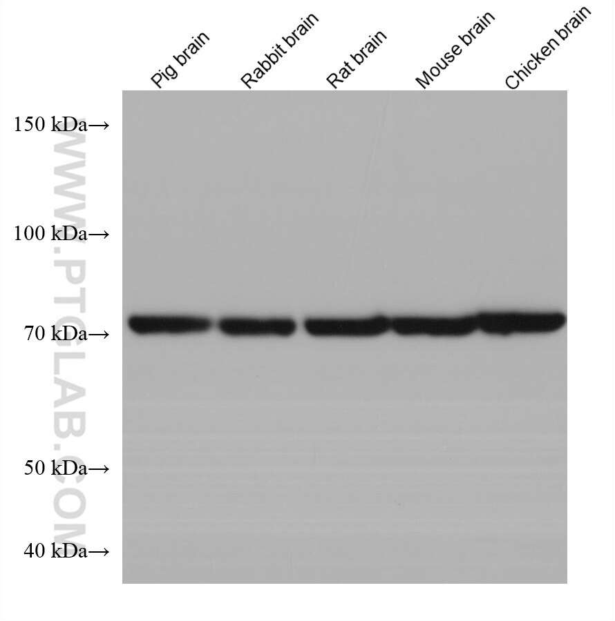
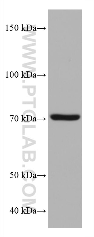
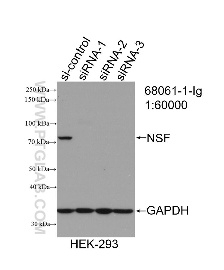
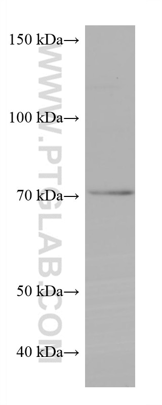
| Positive WB detected in | pig brain tissue, SH-SY5Y cells, PC-12 cells, HEK-293 cells, Rabbit brain tissue, Rat brain tissue, Mouse brain tissue, Chicken brain tissue |
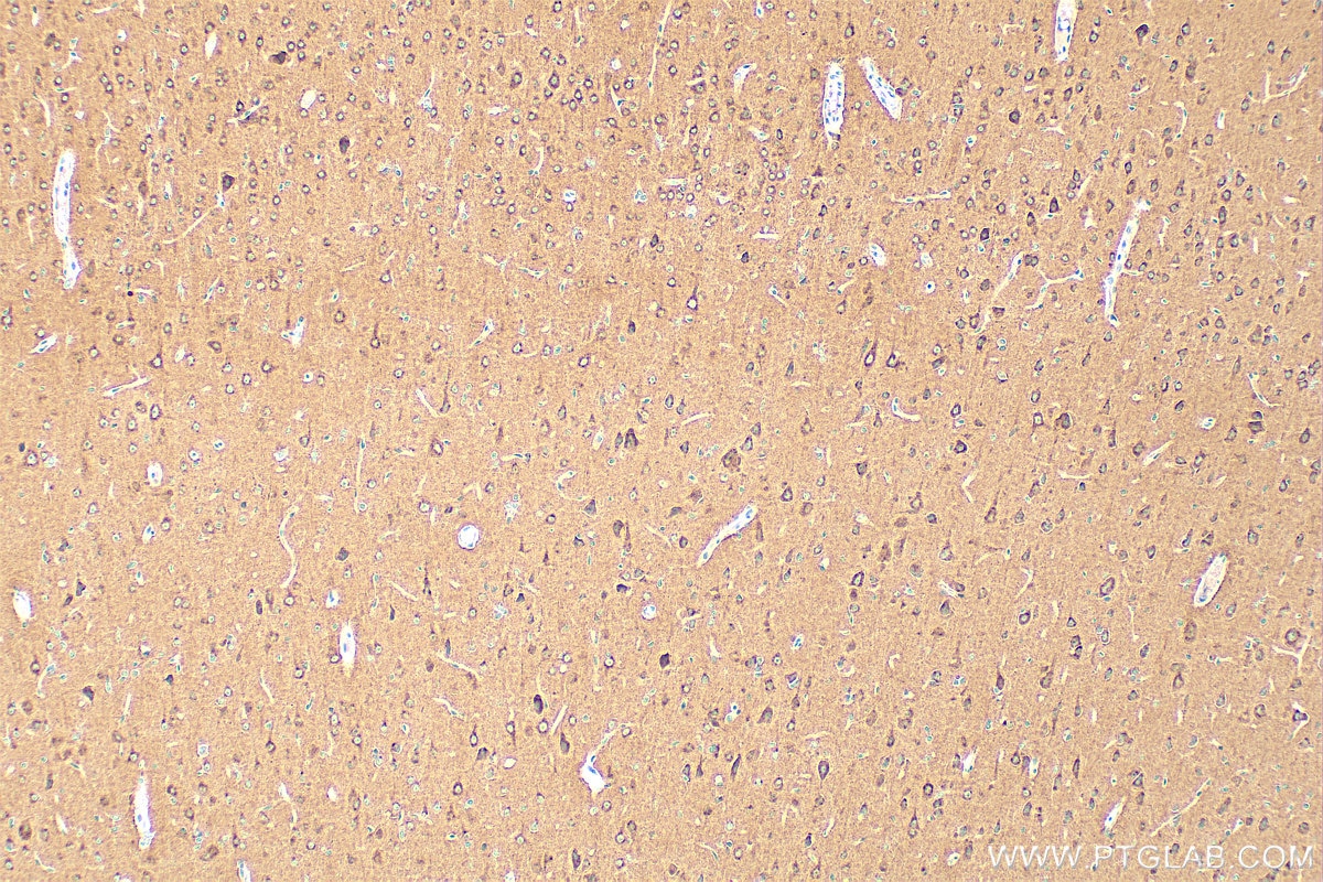
| Positive IHC detected in | rat brain tissue Note: suggested antigen retrieval with TE buffer pH 9.0; (*) Alternatively, antigen retrieval may be performed with citrate buffer pH 6.0 |
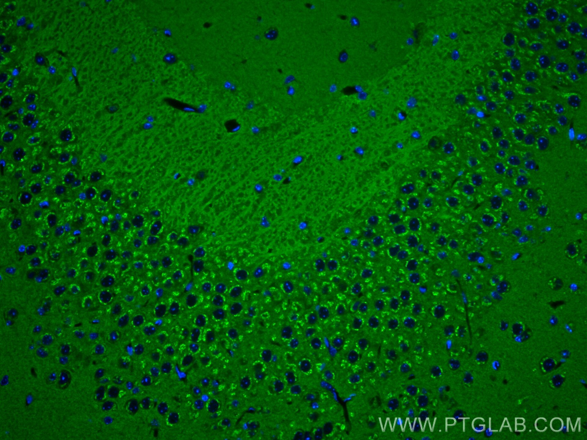
| Positive IF-P detected in | mouse brain tissue |
Recommended dilution
| Application | Dilution |
|---|---|
| Western Blot (WB) | WB : 1:5000-1:50000 |
| Immunohistochemistry (IHC) | IHC : 1:300-1:1200 |
| Immunofluorescence (IF)-P | IF-P : 1:300-1:1200 |
| It is recommended that this reagent should be titrated in each testing system to obtain optimal results. | |
| Sample-dependent, Check data in validation data gallery. | |
Product Information
68061-1-Ig targets NSF in WB, IHC, IF-P, ELISA applications and shows reactivity with human, mouse, rat, pig, rabbit, chicken samples.
| Tested Reactivity | human, mouse, rat, pig, rabbit, chicken |
| Host / Isotype | Mouse / IgG2a |
| Class | Monoclonal |
| Type | Antibody |
| Immunogen |
CatNo: Ag15422 Product name: Recombinant human NSF protein Source: e coli.-derived, T-HIS Tag: 6*His Domain: 394-744 aa of BC030613 Sequence: EIGLPDEKGRLQILHIHTARMRGHQLLSADVDIKELAVETKNFSGAELEGLVRAAQSTAMNRHIKASTKVEVDMEKAESLQVTRGDFLASLENDIKPAFGTNQEDYASYIMNGIIKWGDPVTRVLDDGELLVQQTKNSDRTPLVSVLLEGPPHSGKTALAAKIAEESNFPFIKICSPDKMIGFSETAKCQAMKKIFDDAYKSQLSCVVVDDIERLLDYVPIGPRFSNLVLQALLVLLKKAPPQGRKLLIIGTTSRKDVLQEMEMLNAFSTTIHVPNIATGEQLLEALELLGNFKDKERTTIAQQVKGKKVWIGIKKLLMLIEMSLQMDPEYRVRKFLALLREEGASPLDFD Predict reactive species |
| Full Name | N-ethylmaleimide-sensitive factor |
| Calculated Molecular Weight | 744 aa, 83 kDa |
| Observed Molecular Weight | 70-83 kDa |
| GenBank Accession Number | BC030613 |
| Gene Symbol | NSF |
| Gene ID (NCBI) | 4905 |
| RRID | AB_2918802 |
| Conjugate | Unconjugated |
| Form | Liquid |
| Purification Method | Protein A purification |
| UNIPROT ID | P46459 |
| Storage Buffer | PBS with 0.02% sodium azide and 50% glycerol, pH 7.3. |
| Storage Conditions | Store at -20°C. Stable for one year after shipment. Aliquoting is unnecessary for -20oC storage. 20ul sizes contain 0.1% BSA. |
Protocols
| Product Specific Protocols | |
|---|---|
| IF protocol for NSF antibody 68061-1-Ig | Download protocol |
| IHC protocol for NSF antibody 68061-1-Ig | Download protocol |
| WB protocol for NSF antibody 68061-1-Ig | Download protocol |
| Standard Protocols | |
|---|---|
| Click here to view our Standard Protocols |