Filter:


Tested Applications
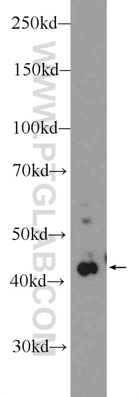
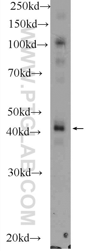
| Positive WB detected in | L02 cells, mouse liver tissue |
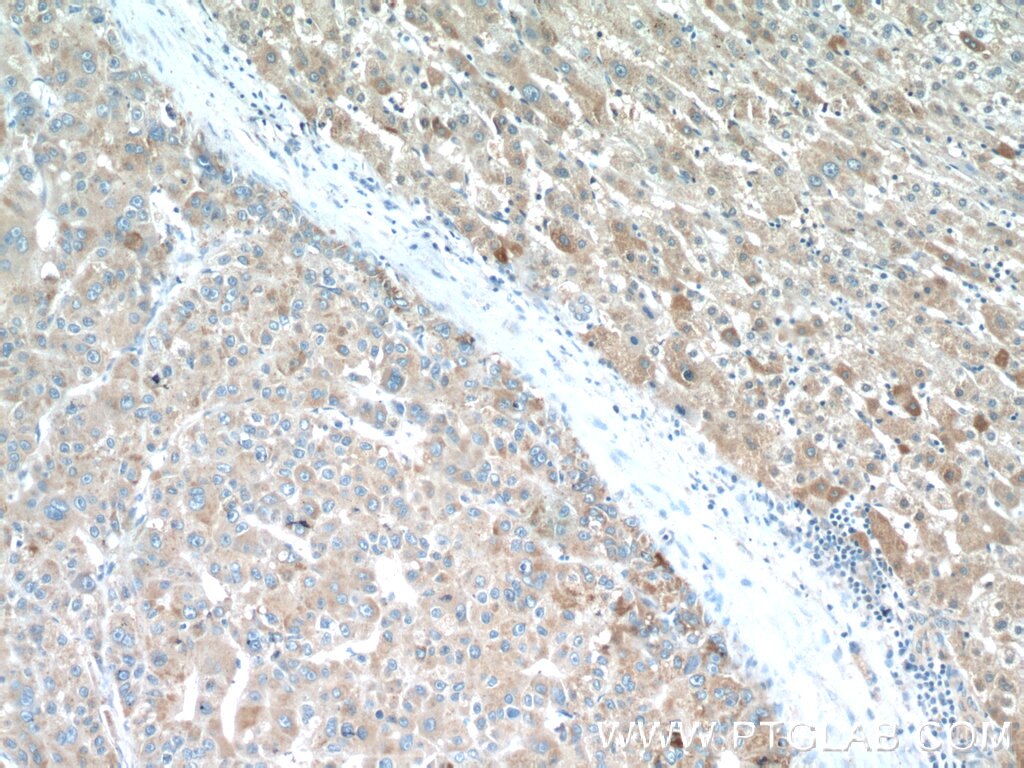
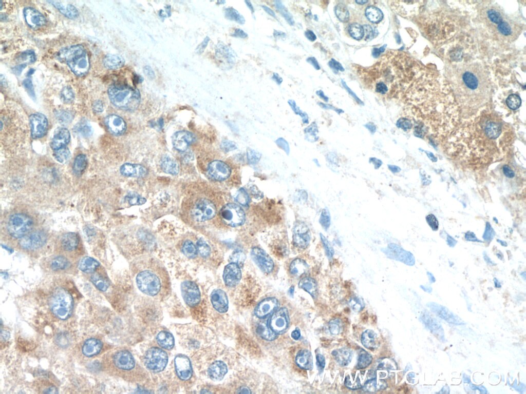
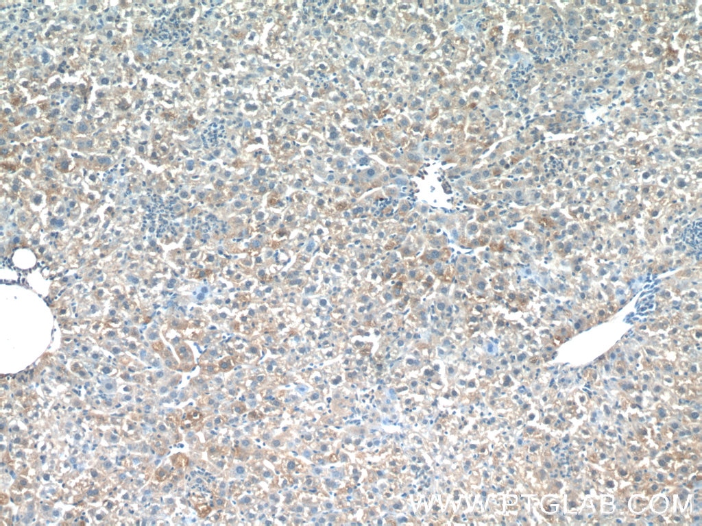
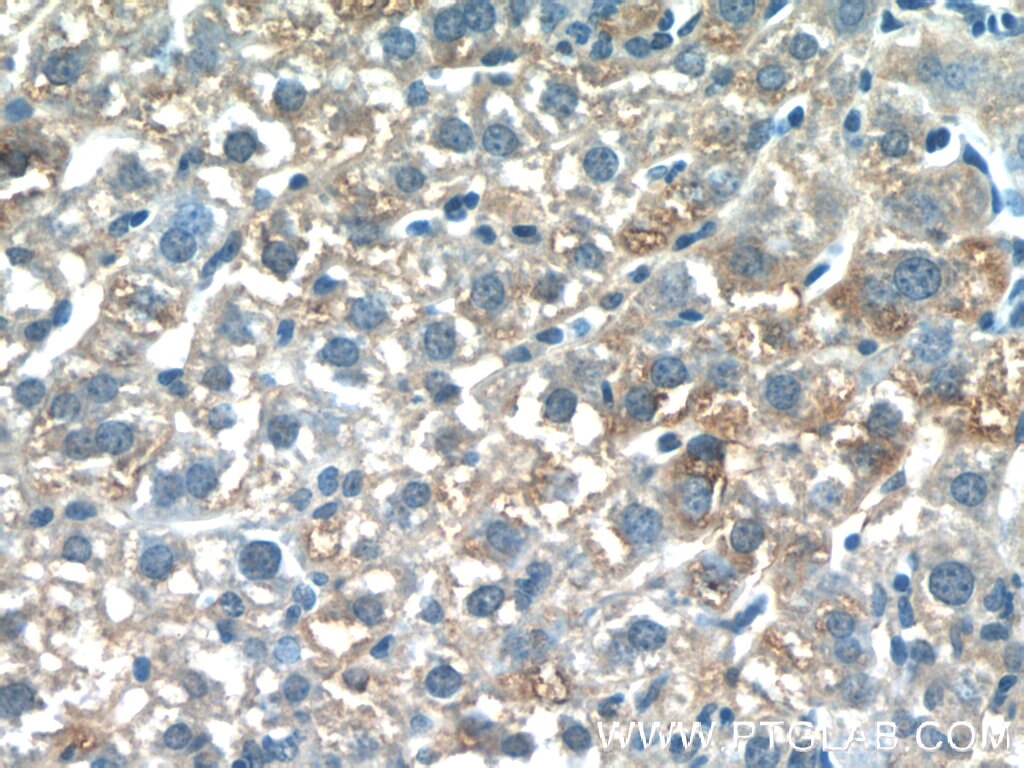
| Positive IHC detected in | human liver cancer tissue, mouse liver tissue Note: suggested antigen retrieval with TE buffer pH 9.0; (*) Alternatively, antigen retrieval may be performed with citrate buffer pH 6.0 |
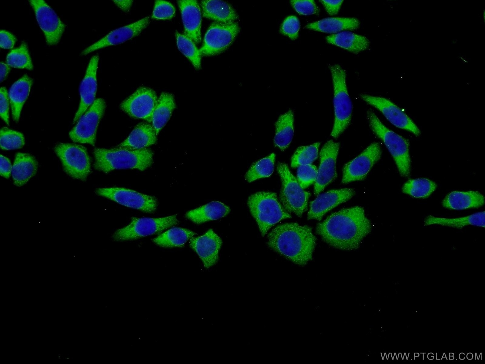
| Positive IF/ICC detected in | L02 cells |
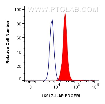
| Positive FC (Intra) detected in | HepG2 cells |
Recommended dilution
| Application | Dilution |
|---|---|
| Western Blot (WB) | WB : 1:500-1:1000 |
| Immunohistochemistry (IHC) | IHC : 1:20-1:200 |
| Immunofluorescence (IF)/ICC | IF/ICC : 1:20-1:200 |
| Flow Cytometry (FC) (INTRA) | FC (INTRA) : 0.25 ug per 10^6 cells in a 100 µl suspension |
| It is recommended that this reagent should be titrated in each testing system to obtain optimal results. | |
| Sample-dependent, Check data in validation data gallery. | |
Published Applications
| KD/KO | See 1 publications below |
| WB | See 2 publications below |
| IHC | See 1 publications below |
Product Information
16217-1-AP targets PDGFRL in WB, IHC, IF/ICC, FC (Intra), ELISA applications and shows reactivity with human, mouse, rat samples.
| Tested Reactivity | human, mouse, rat |
| Cited Reactivity | human |
| Host / Isotype | Rabbit / IgG |
| Class | Polyclonal |
| Type | Antibody |
| Immunogen |
CatNo: Ag9218 Product name: Recombinant human PDGFRL protein Source: e coli.-derived, PGEX-4T Tag: GST Domain: 1-375 aa of BC010927 Sequence: MKVWLLLGLLLVHEALEDVTGQHLPKNKRPKEPGENRIKPTNKKVKPKIPKMKDRDSANSAPKTQSIMMQVLDKGRFQKPAATLSLLAGQTVELRCKGSRIGWSYPAYLDTFKDSRLSVKQNERYGQLTLVNSTSADTGEFSCWVQLCSGYICRKDEAKTGSTYIFFTEKGELFVPSPSYFDVVYLNPDRQAVVPCRVTVLSAKVTLHREFPAKEIPANGTDIVYDMKRGFVYLQPHSEHQGVVYCRAEAGGRSQISVKYQLLYVAVPSGPPSTTILASSNKVKSGDDISVLCTVLGEPDVEVEFTWIFPGQKDERPVTIQDTWRLIHRGLGHTTRISQSVITVEDFETIDAGYYICTAQNLQGQTTVATTVEFS Predict reactive species |
| Full Name | platelet-derived growth factor receptor-like |
| Calculated Molecular Weight | 375 aa, 42 kDa |
| Observed Molecular Weight | 42 kDa |
| GenBank Accession Number | BC010927 |
| Gene Symbol | PDGFRL |
| Gene ID (NCBI) | 5157 |
| RRID | AB_2878230 |
| Conjugate | Unconjugated |
| Form | Liquid |
| Purification Method | Antigen affinity purification |
| UNIPROT ID | Q15198 |
| Storage Buffer | PBS with 0.02% sodium azide and 50% glycerol, pH 7.3. |
| Storage Conditions | Store at -20°C. Stable for one year after shipment. Aliquoting is unnecessary for -20oC storage. 20ul sizes contain 0.1% BSA. |
Protocols
| Product Specific Protocols | |
|---|---|
| FC protocol for PDGFRL antibody 16217-1-AP | Download protocol |
| IF protocol for PDGFRL antibody 16217-1-AP | Download protocol |
| IHC protocol for PDGFRL antibody 16217-1-AP | Download protocol |
| WB protocol for PDGFRL antibody 16217-1-AP | Download protocol |
| Standard Protocols | |
|---|---|
| Click here to view our Standard Protocols |
Publications
| Species | Application | Title |
|---|---|---|
Mol Carcinog The cumulative antitumor effects of regorafenib and radiotherapy in hepatocellular carcinoma | ||
Nat Commun A single cell atlas of frozen shoulder capsule identifies features associated with inflammatory fibrosis resolution | ||
Aging (Albany NY) Identification of a unique stress response state of T cells-related gene signature in patients with gastric cancer
|