Tested Applications
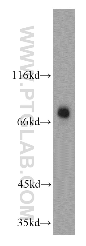
| Positive WB detected in | mouse brain tissue |
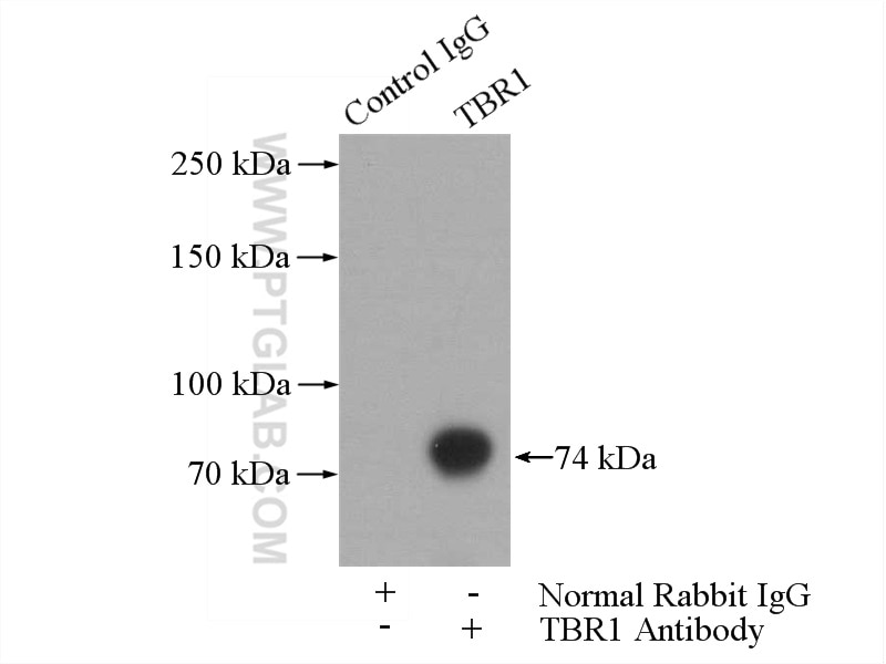
| Positive IP detected in | mouse brain tissue |
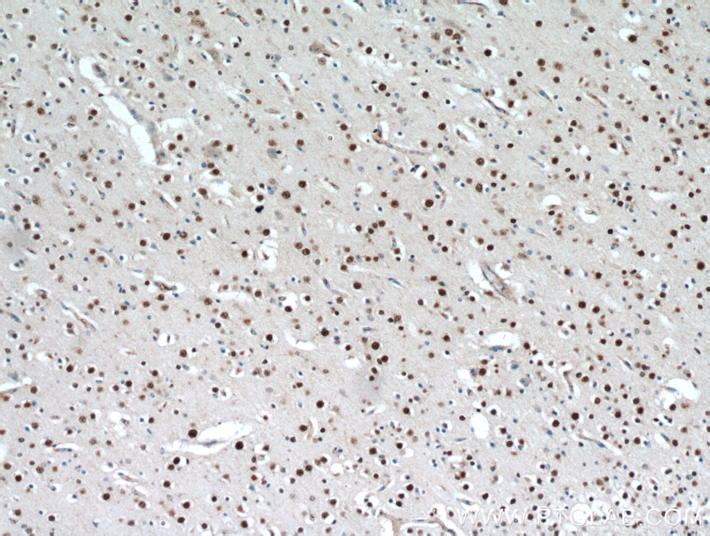
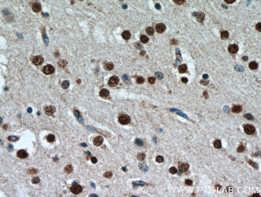
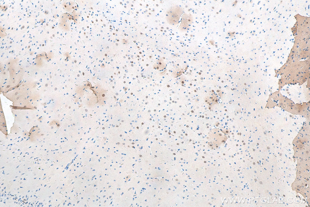
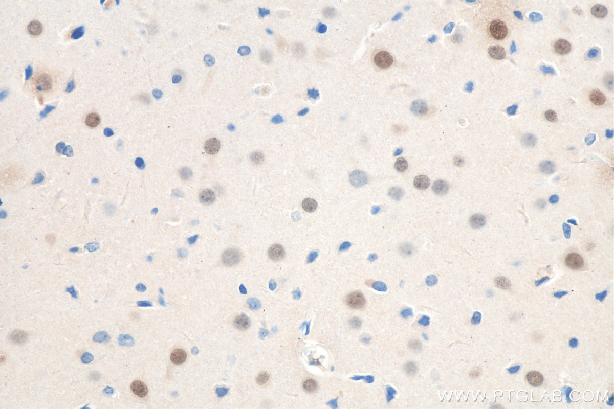
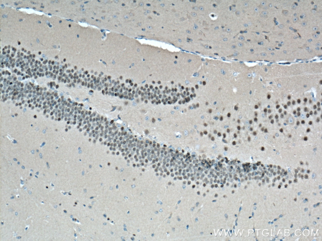
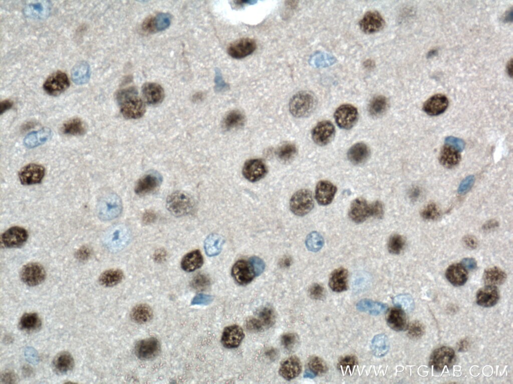
| Positive IHC detected in | human brain tissue, mouse brain tissue, rat brain tissue Note: suggested antigen retrieval with TE buffer pH 9.0; (*) Alternatively, antigen retrieval may be performed with citrate buffer pH 6.0 |
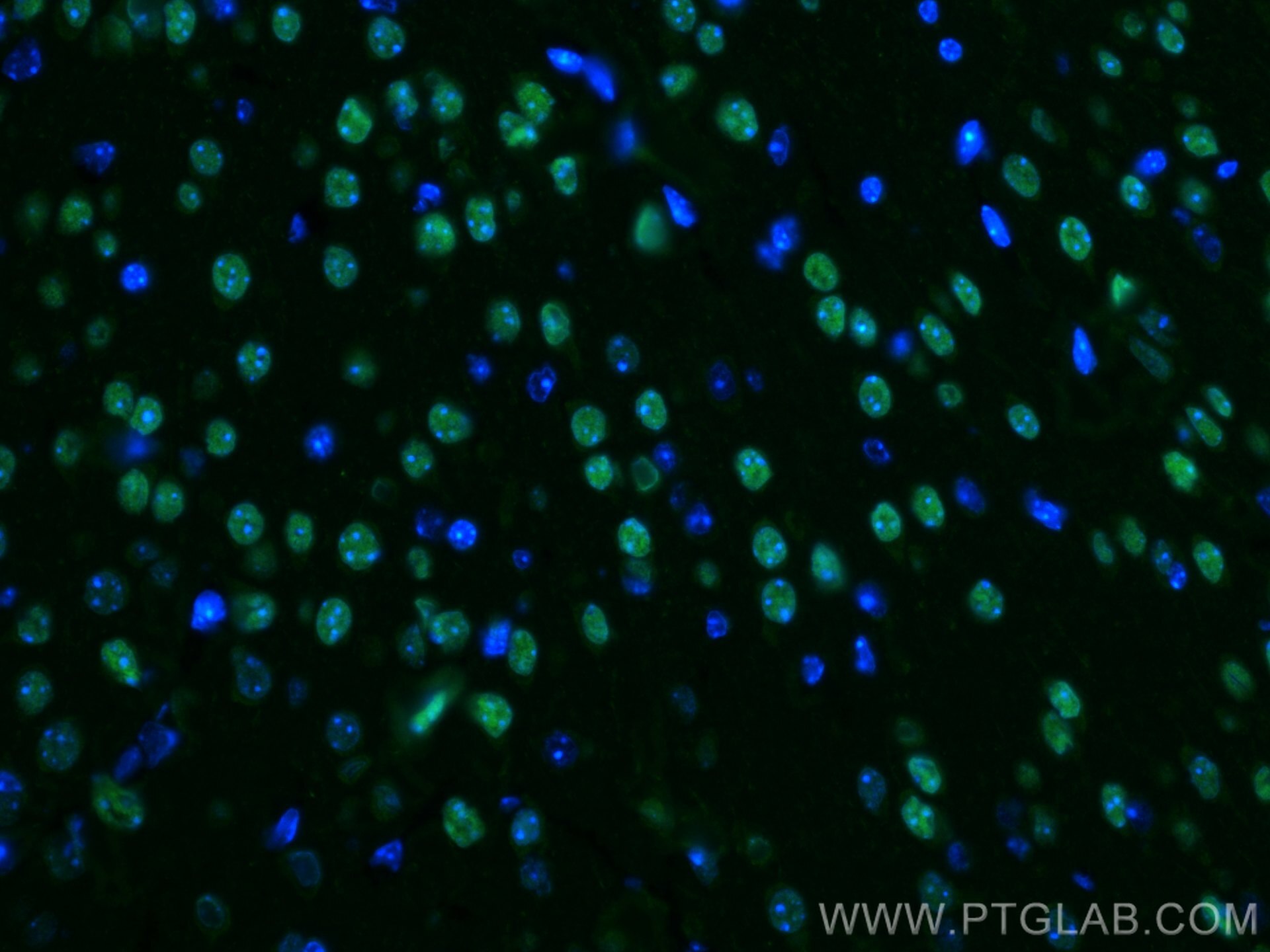
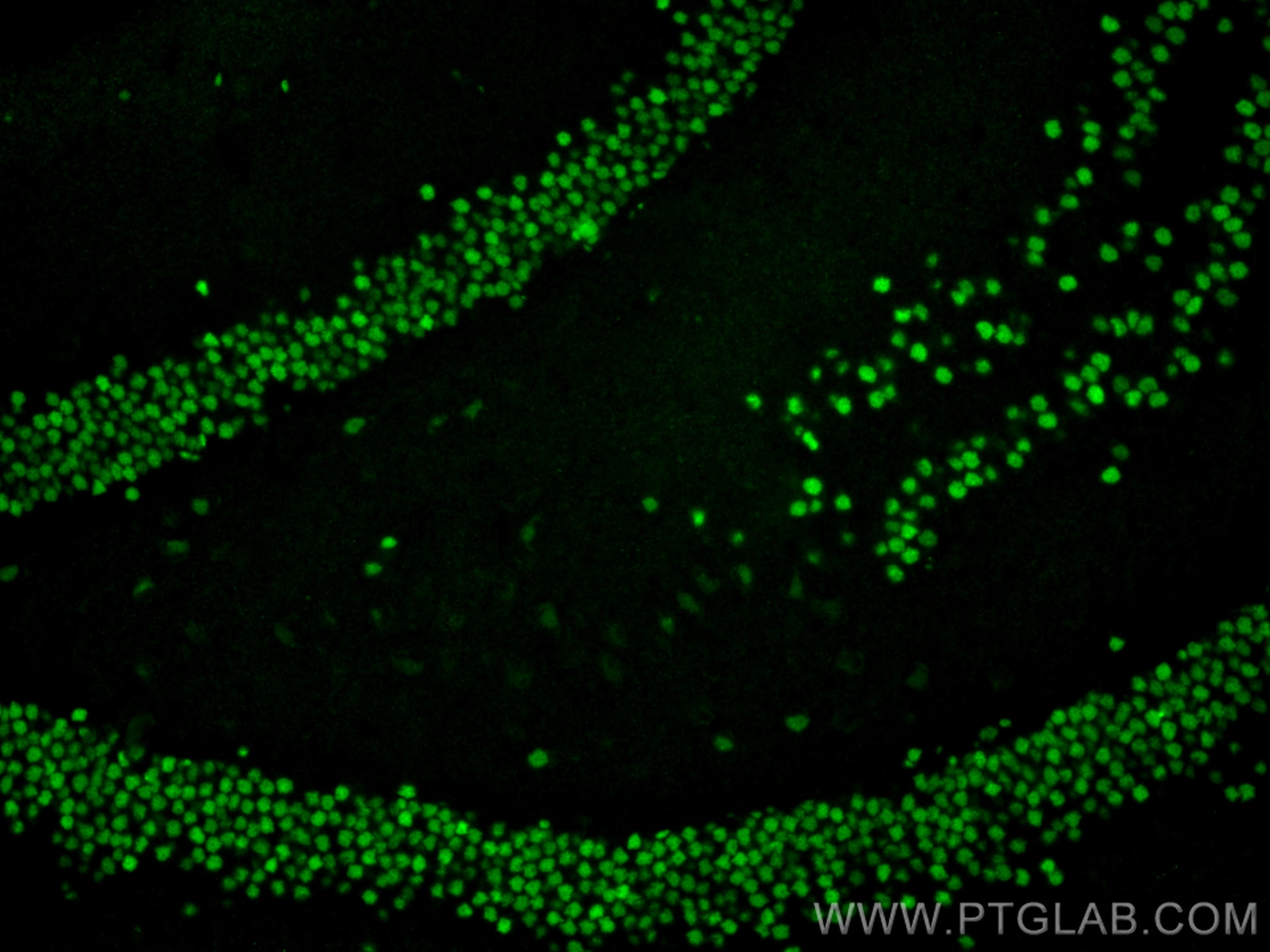
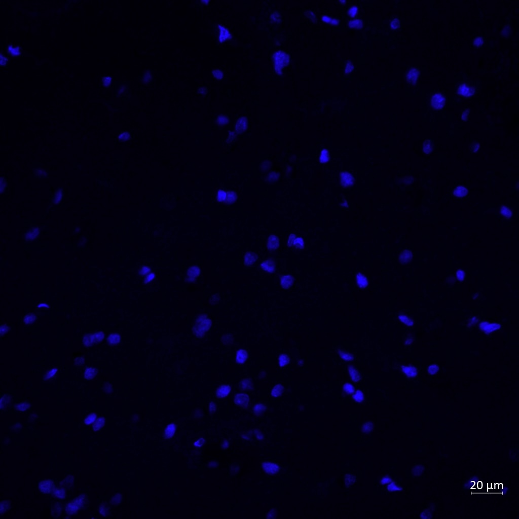
| Positive IF-P detected in | mouse brain tissue |
Recommended dilution
| Application | Dilution |
|---|---|
| Western Blot (WB) | WB : 1:500-1:1000 |
| Immunoprecipitation (IP) | IP : 0.5-4.0 ug for 1.0-3.0 mg of total protein lysate |
| Immunohistochemistry (IHC) | IHC : 1:20-1:200 |
| Immunofluorescence (IF)-P | IF-P : 1:200-1:800 |
| It is recommended that this reagent should be titrated in each testing system to obtain optimal results. | |
| Sample-dependent, Check data in validation data gallery. | |
Published Applications
| WB | See 9 publications below |
| IHC | See 5 publications below |
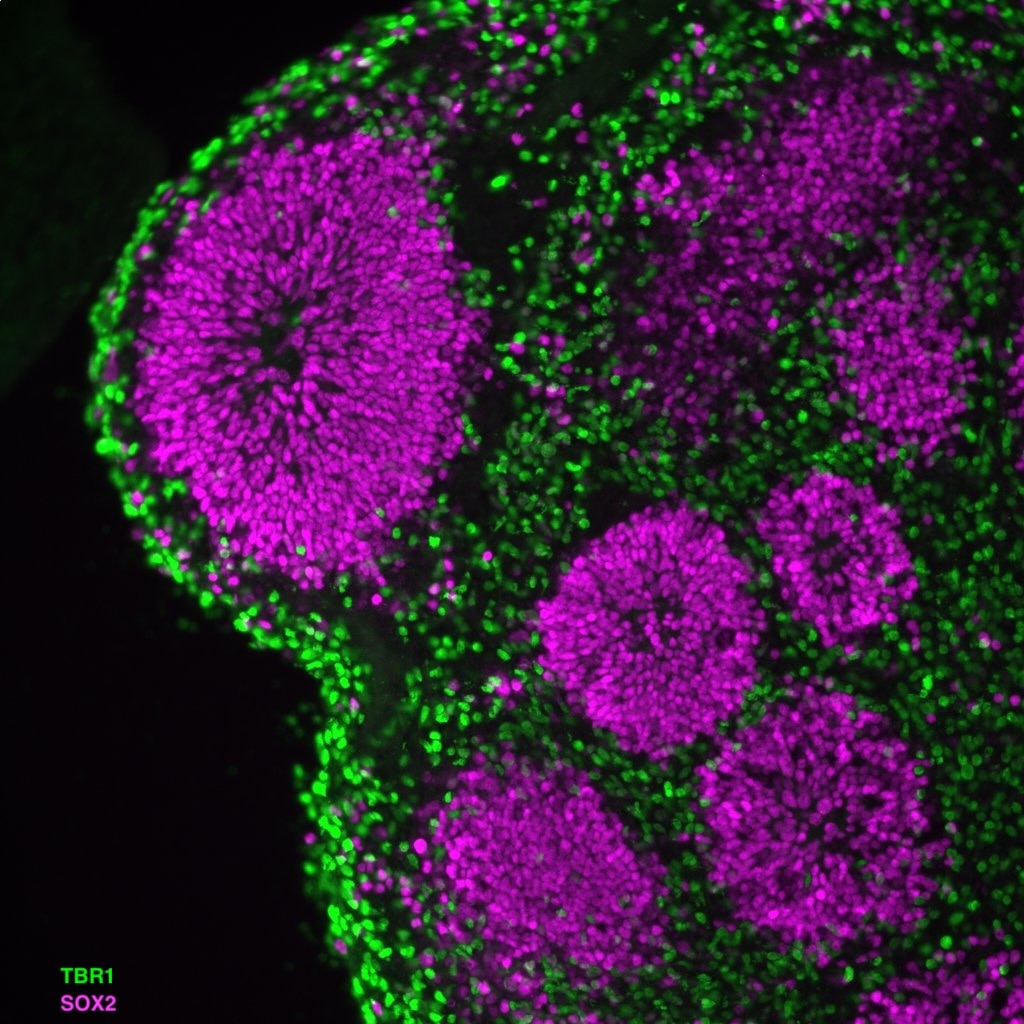
| IF | See 28 publications below |
Product Information
20932-1-AP targets TBR1 in WB, IHC, IF-P, IP, ELISA applications and shows reactivity with human, mouse, rat samples.
| Tested Reactivity | human, mouse, rat |
| Cited Reactivity | human, mouse, rat |
| Host / Isotype | Rabbit / IgG |
| Class | Polyclonal |
| Type | Antibody |
| Immunogen |
CatNo: Ag14935 Product name: Recombinant human TBR1 protein Source: e coli.-derived, PGEX-4T Tag: GST Domain: 1-196 aa of BC104844 Sequence: MQLEHCLSPSIMLSKKFLNVSSSYPHSGGSELVLHDHPIISTTDNLERSSPLKKITRGMTNQSDTDNFPDSKDSPGDVQRSKLSPVLDGVSELRHSFDGSAADRYLLSQSSQPQSAATAPSAMFPYPGQHGPAHPAFSIGSPSRYMAHHPVITNGAYNSLLSNSSPQGYPTAGYPYPQQYGHSYQGAPFYQFSSTQ Predict reactive species |
| Full Name | T-box, brain, 1 |
| Calculated Molecular Weight | 682 aa, 74 kDa |
| Observed Molecular Weight | 74 kDa |
| GenBank Accession Number | BC104844 |
| Gene Symbol | TBR1 |
| Gene ID (NCBI) | 10716 |
| RRID | AB_10695502 |
| Conjugate | Unconjugated |
| Form | Liquid |
| Purification Method | Antigen affinity purification |
| UNIPROT ID | Q16650 |
| Storage Buffer | PBS with 0.02% sodium azide and 50% glycerol, pH 7.3. |
| Storage Conditions | Store at -20°C. Stable for one year after shipment. Aliquoting is unnecessary for -20oC storage. 20ul sizes contain 0.1% BSA. |
Background Information
TBR1, also named as T-box brain protein 1, is a 682 amino acid protein, which contains one T-box DNA-binding domain and localizes in the nucleus. TBR1 is expressed in the brain and as a transcriptional regulator is involved in developmental processes. TBR1 is required for normal brain development.
Protocols
| Product Specific Protocols | |
|---|---|
| IHC protocol for TBR1 antibody 20932-1-AP | Download protocol |
| IP protocol for TBR1 antibody 20932-1-AP | Download protocol |
| WB protocol for TBR1 antibody 20932-1-AP | Download protocol |
| IF protocol for TBR1 antibody 20932-1-AP | Download protocol |
| Standard Protocols | |
|---|---|
| Click here to view our Standard Protocols |
Publications
| Species | Application | Title |
|---|---|---|
Nat Neurosci A tau homeostasis signature is linked with the cellular and regional vulnerability of excitatory neurons to tau pathology. | ||
Nat Commun GRAMD1B is a regulator of lipid homeostasis, autophagic flux and phosphorylated tau | ||
Nat Commun Pathogenic POGZ mutation causes impaired cortical development and reversible autism-like phenotypes. | ||
Nat Commun Disrupted neuronal maturation in Angelman syndrome-derived induced pluripotent stem cells. | ||
Proc Natl Acad Sci U S A Human intermediate progenitor diversity during cortical development. |
Reviews
The reviews below have been submitted by verified Proteintech customers who received an incentive for providing their feedback.
FH Raquel (Verified Customer) (03-04-2026) | Very nice and clean staining of TBR1 positive neurons
 |
FH Reyes (Verified Customer) (03-01-2024) | Used on human brain cortical sections, it marked nicely the nucleus of my neurons.
 |
FH Mandi (Verified Customer) (03-09-2020) | Good staining on iPSC-derived Cortical neurons. Needed a decent amount of optimization to prevent non-specific binding though. Make sure to run primary/secondary deletes.
|