
How To Optimize Your Results With Low MW Proteins
Tips to improve your detection of low molecular weight proteins.
Jump to:
- Western Blot Protocol
- How To Optimize Your Western Blot
- SDS-PAGE Gel Recipes
- How To Optimize Your Results With Low MW Proteins
- Tricine Gel Recipe For Low Molecular Weight Proteins
- Choosing The Right Lysis Buffer
- Choosing The Right Western Blot Detection Method
- Western Blot Troubleshooting: Why Does The Observed Protein Molecular Weight (MW) Differ From The Calculated One?
- Western Blot Troubleshooting: High Background
- Western Blot Troubleshooting: Weak/No Signal & Other
- Western Blot ppt
- Western Blot Video Protocol
At extreme ends of the molecular weight (MW) spectrum, regular SDS-PAGE and Western blotting techniques suffer from limitations: poor separation, signal reduction, or even a total absence of target bands. When blotting for low MW proteins (<20 kDa), protocol modifications can be employed to improve their retention and resolution.
Proteintech control antibodies are $189 each for a 150ul size vial
View all Loading Control AntibodiesUse Tricine
In general, glycine gels are ideal for resolving any proteins that fall within the MW range of 30–250 kDa. However, acrylamide gels based on a Tris-Tricine buffer system will greatly improve your chances of “seeing” your target band(s) if you are working below the 30 kDa range.
(You can find a recipe for a 15% Tricine gel in the 'Tricine Gel Recipe for Low MW Proteins' section.)
|
|
Comparison of Tricine- (A) and glycine-SDS-PAGE (B) separation of myoglobin fragments. Adapted from Schägger and von Jagow 1987. |
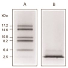
Why It Works
The difference in separation capabilities of glycine and tricine gels is attributed to the differing properties of the glycine and tricine compounds, such as their pK values and ionic mobility. Tricine is more optimal for the separation of low MW proteins because it “stacks” proteins into more uniform bands. A tricine based stacking layer shifts the upper stacking limit down to as low as 30 kDa for the first stack,1 preventing overloading at the interface between the gel layers.
| Ref: 1. H Schägger and G von Jagow. Tricine-sodium dodecyl sulfate-polyacrylamide gel electrophoresis for the separation of proteins in the range from 1 to 100 kDa. Anal Biochem. 1987;166(2):368–79. |
Protein Transfer
As well as ensuring the optimum separation of low MW proteins, you also need to take care at the protein transfer stage. Low MW proteins are susceptible to “over transfer” – loss of sample due to lack of retention by the transfer membrane and/or too-rapid transfer.
Membrane Choice and Pore Size
Most labs have a preferred choice of Western blotting membrane, however PVDF is a better choice in the case of smaller, low MW proteins, due to its greater capacity to bind proteins. There are various membrane pore sizes on offer: 0.45 μm, 0.2 μm, or 0.1 μm versions. Opt for a smaller pore sized version to obtain better transfers of your low MW target proteins – a 0.2 μm membrane is more than adequate for use with any proteins weighing in less than 20 kDa. The Proteintech lab uses Millipore’s Immobilon PSQ PVDF membrane for the transfer of low MW proteins.
Transfer Conditions
In addition to membrane choice, factors such as transfer system, length, temperature, and buffer composition come into play when dealing with low MW proteins. In the case of small proteins, less is often more when it comes to transfer length and voltage. How much you should adjust these by will be down to the brand and model of the system being used.
Other Tips
Be aware some proteins will escape the dye front of the sample loading buffer. You can choose to soak your gel in an SDS-free buffer (or simply H2O) for 5 minutes prior to setting up your transfer. This helps to remove SDS, which coats small proteins and protein fragments in negative charges, increasing their rate of passage through the Western blot membrane.
Able™