"ATP1A1-Specific Antibodies" Comparison
View side-by-side comparison of ATP1A1-Specific antibodies from other vendors to find the one that best suits your research needs.
Tested Applications
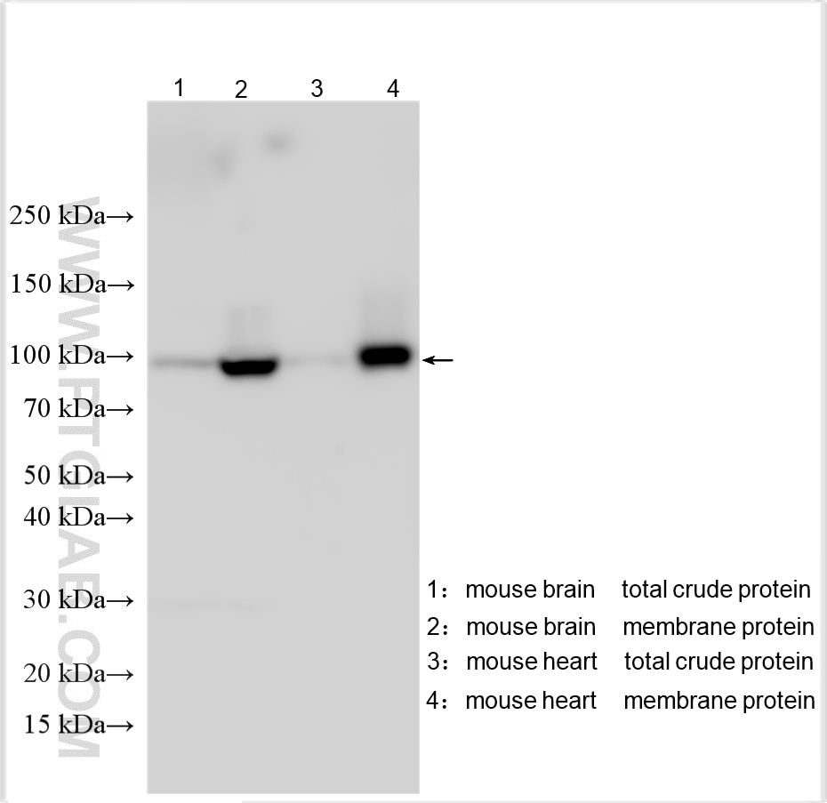
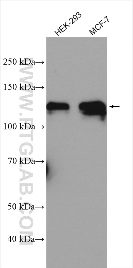
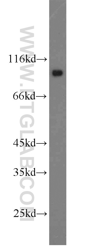
| Positive WB detected in | 37°C incubated mouse brain tissue, HEK-293 cells, mouse brain tissue, mouse heart tissue, Neuro-2a cells, MCF-7 cells |
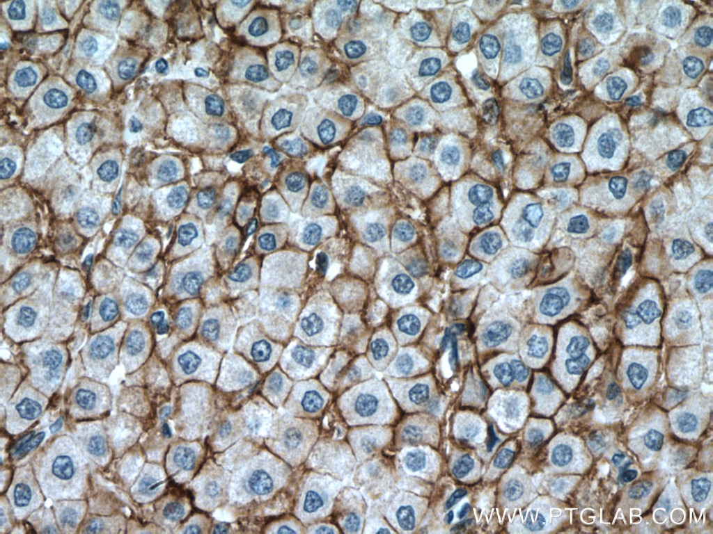
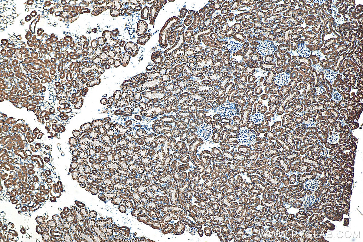
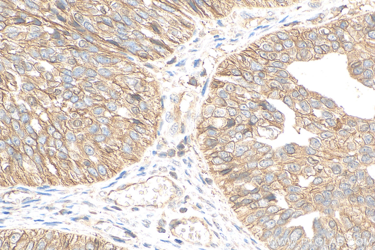
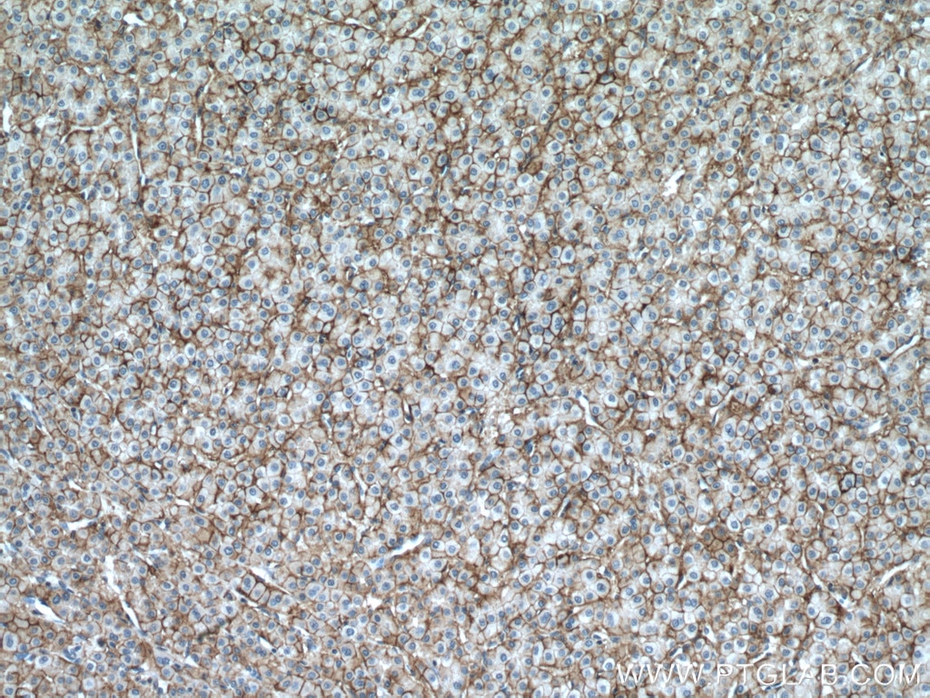
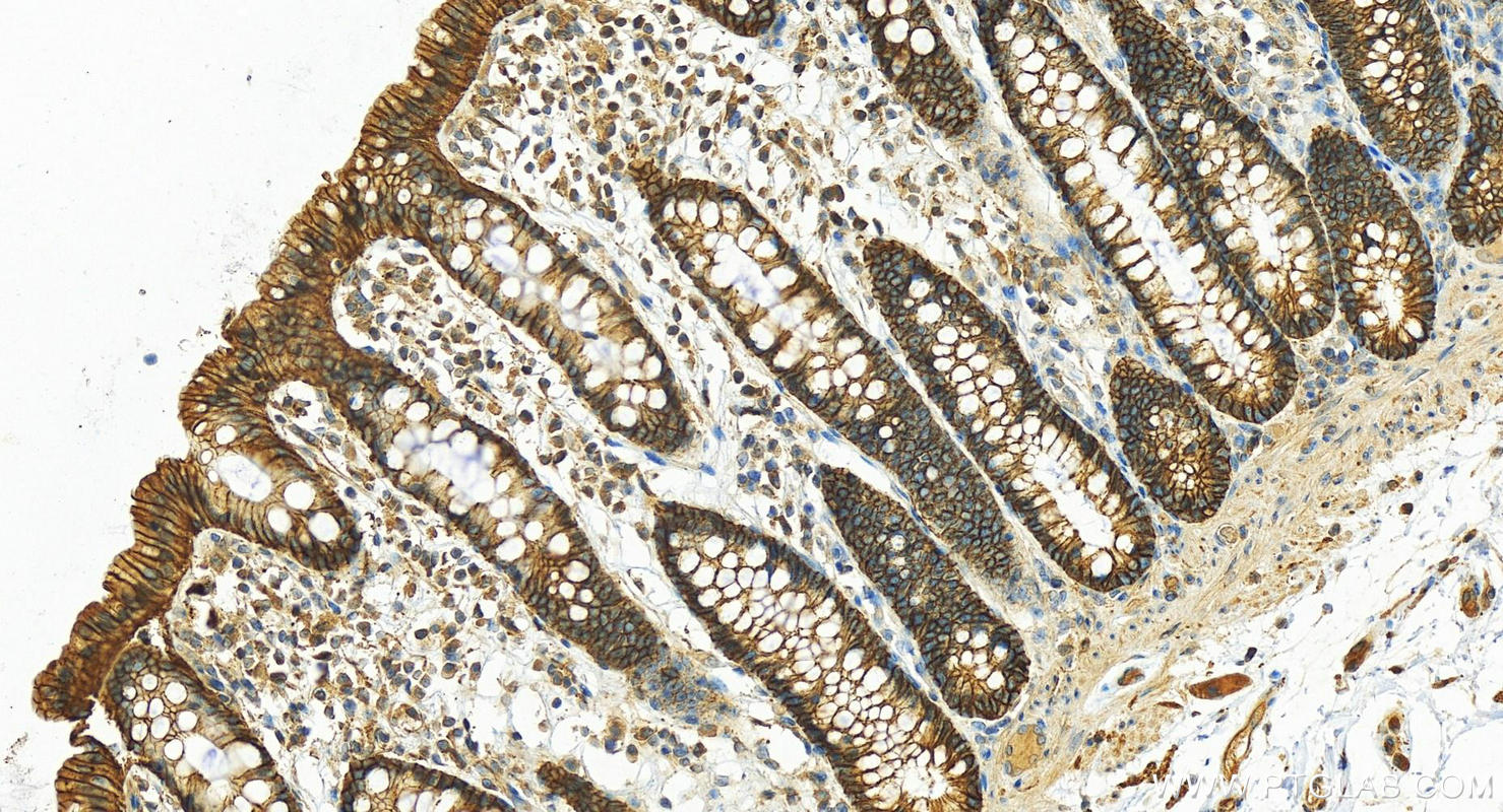
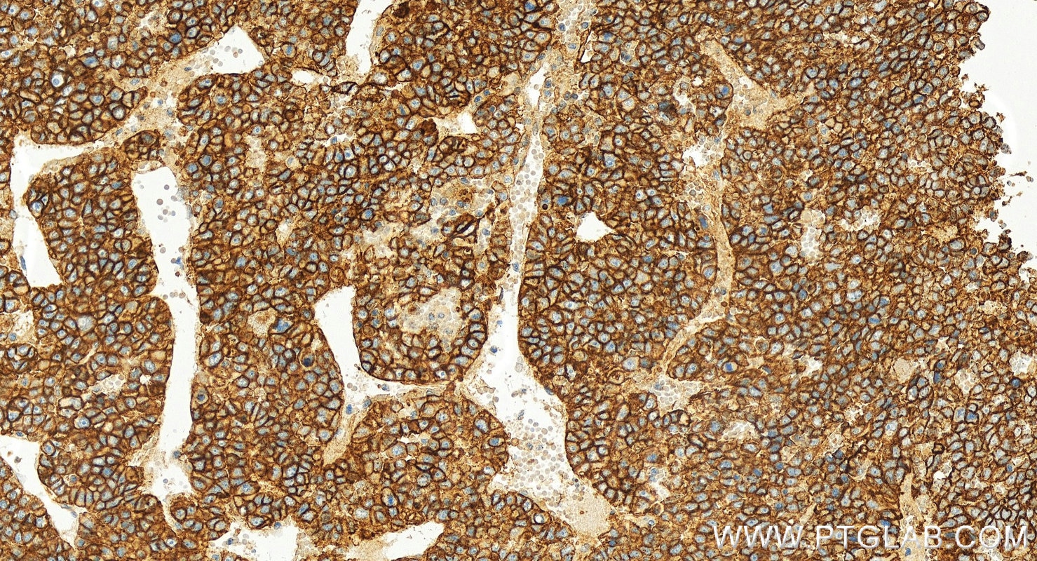
| Positive IHC detected in | human ovary cancer tissue, human colon tissue, human hepatocellular ca, human liver cancer tissue, mouse kidney tissue Note: suggested antigen retrieval with TE buffer pH 9.0; (*) Alternatively, antigen retrieval may be performed with citrate buffer pH 6.0 |
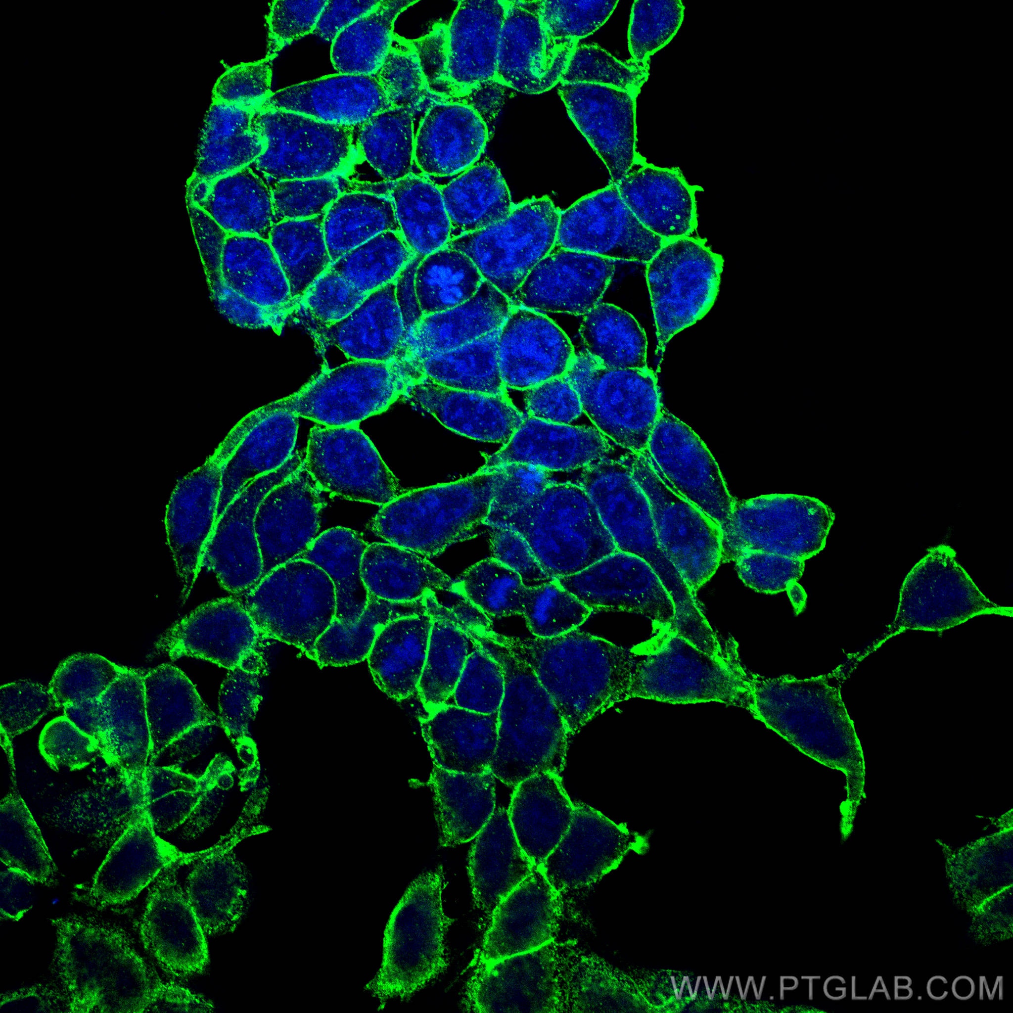
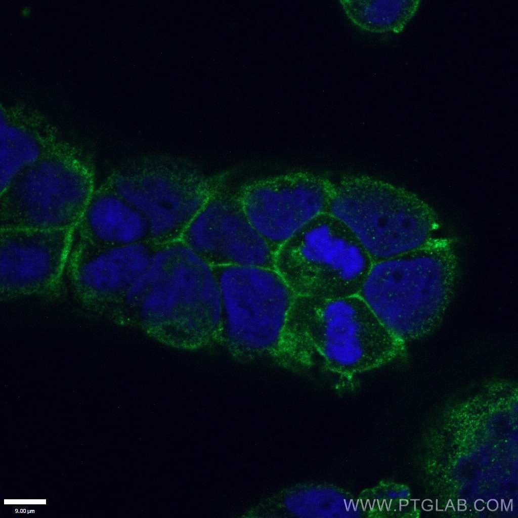
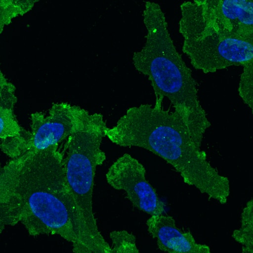
| Positive IF/ICC detected in | HEK-293 cells, Caco-2 cells |
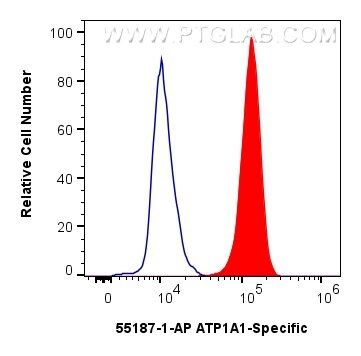
| Positive FC (Intra) detected in | HEK-293 cells |
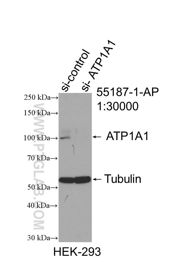
For optimal WB detection with 55187-1-AP, we do not recommend boiling the sample after lysis.
Recommended dilution
| Application | Dilution |
|---|---|
| Western Blot (WB) | WB : 1:5000-1:50000 |
| Immunohistochemistry (IHC) | IHC : 1:500-1:2000 |
| Immunofluorescence (IF)/ICC | IF/ICC : 1:500-1:2000 |
| Flow Cytometry (FC) (INTRA) | FC (INTRA) : 0.40 ug per 10^6 cells in a 100 µl suspension |
| It is recommended that this reagent should be titrated in each testing system to obtain optimal results. | |
| Sample-dependent, Check data in validation data gallery. | |
Published Applications
| WB | See 17 publications below |
| IF | See 9 publications below |
Product Information
55187-1-AP targets ATP1A1-Specific in WB, IHC, IF/ICC, FC (Intra), ELISA applications and shows reactivity with human, mouse samples.
| Tested Reactivity | human, mouse |
| Cited Reactivity | human, mouse, rat |
| Host / Isotype | Rabbit / IgG |
| Class | Polyclonal |
| Type | Antibody |
| Immunogen |
Peptide Predict reactive species |
| Full Name | ATPase, Na+/K+ transporting, alpha 1 polypeptide |
| Calculated Molecular Weight | 113 kDa |
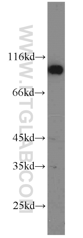
| Observed Molecular Weight | 100-110 kDa |
| GenBank Accession Number | NM_000701 |
| Gene Symbol | ATP1A1 |
| Gene ID (NCBI) | 476 |
| ENSEMBL Gene ID | ENSG00000163399 |
| RRID | AB_10859261 |
| Conjugate | Unconjugated |
| Form | Liquid |
| Purification Method | Antigen affinity purification |
| UNIPROT ID | P05023 |
| Storage Buffer | PBS with 0.02% sodium azide and 50% glycerol, pH 7.3. |
| Storage Conditions | Store at -20°C. Stable for one year after shipment. Aliquoting is unnecessary for -20oC storage. 20ul sizes contain 0.1% BSA. |
Background Information
ATP1A1 is the catalytic component of Na+/K+-ATPase which is a membrane bound enzyme primarily involved in generation of Na+ and K+ gradients across plasma membranes and in determination of cytoplasmic Na+ levels. ATP1A1 is a ubiquitously expressed membrane protein and often used as the marker or internal control for plasma membrane protein. This antibody is specific to ATP1A1.
Protocols
| Product Specific Protocols | |
|---|---|
| FC protocol for ATP1A1-Specific antibody 55187-1-AP | Download protocol |
| IF protocol for ATP1A1-Specific antibody 55187-1-AP | Download protocol |
| IHC protocol for ATP1A1-Specific antibody 55187-1-AP | Download protocol |
| WB protocol for ATP1A1-Specific antibody 55187-1-AP | Download protocol |
| Standard Protocols | |
|---|---|
| Click here to view our Standard Protocols |
Publications
| Species | Application | Title |
|---|---|---|
Autophagy Hepatocyte CD36 modulates UBQLN1-mediated proteasomal degradation of autophagic SNARE proteins contributing to septic liver injury | ||
Nat Commun CLICs-dependent chloride efflux is an essential and proximal upstream event for NLRP3 inflammasome activation. | ||
Adv Healthc Mater Effect of Nanoparticle Rigidity on the Interaction of Stromal Membrane Particles with Leukemia Cells | ||
Hypertension Renal Natriuretic Peptide Receptor-C Deficiency Attenuates NaCl Cotransporter Activity in Angiotensin II-Induced Hypertension. | ||
Cell Chem Biol The HEAT repeat protein MROH7 regulates the inflammatory macrophage response via LBP acetylation | ||
Reviews
The reviews below have been submitted by verified Proteintech customers who received an incentive for providing their feedback.
FH Paulina (Verified Customer) (03-26-2025) | Fixation: 2% PFA for 20min. Permeabilization/Antibodies dilution solution: 0.05% saponin, 5% horse serum in PBS. ATP1A1 antibody was diluted 1:100 in saponin solution and incubated on cells at room temperature for 1 hour, followed by 1 hour incubation with anti-rabbit conjugated to AlexaFluor 488 (1:200).
 |