Tested Applications
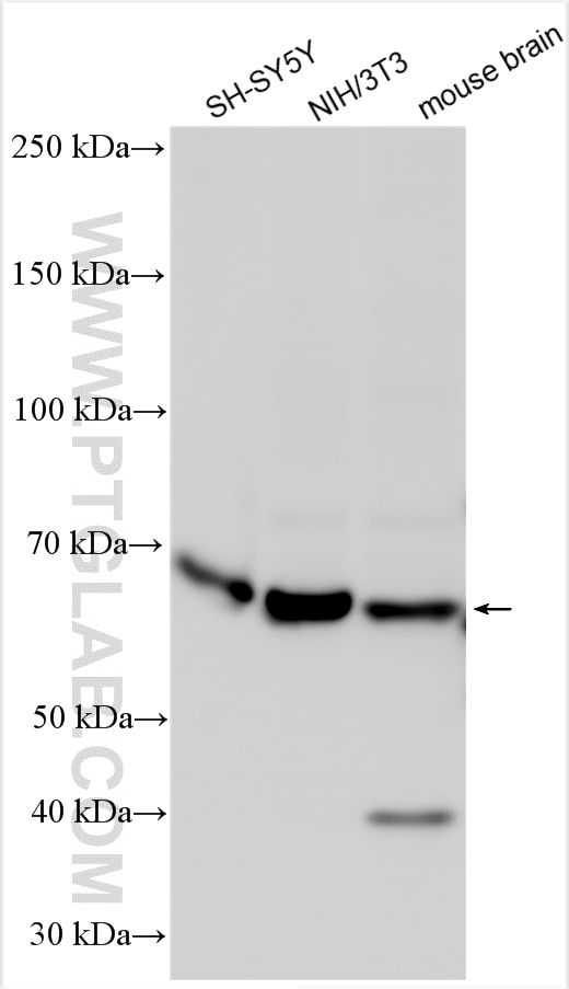
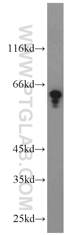
| Positive WB detected in | SH-SY5Y cells, NIH/3T3 cells, mouse brain tissue |
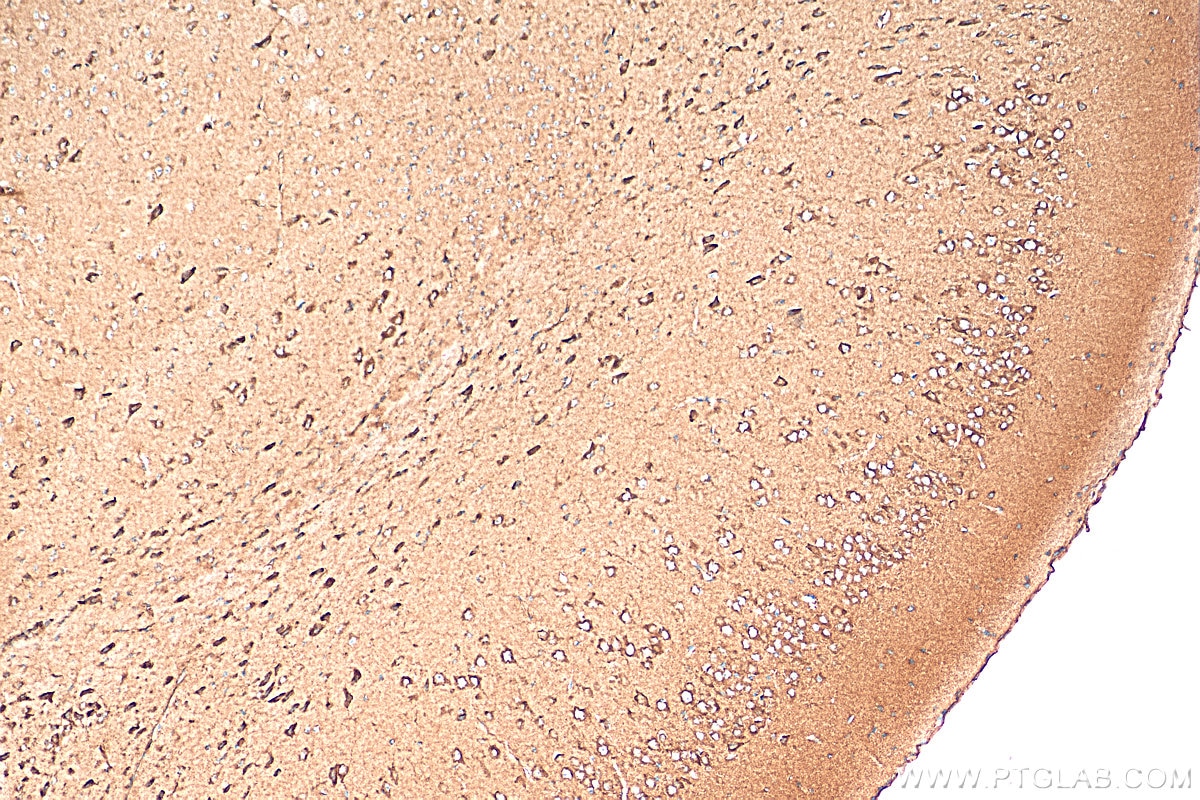
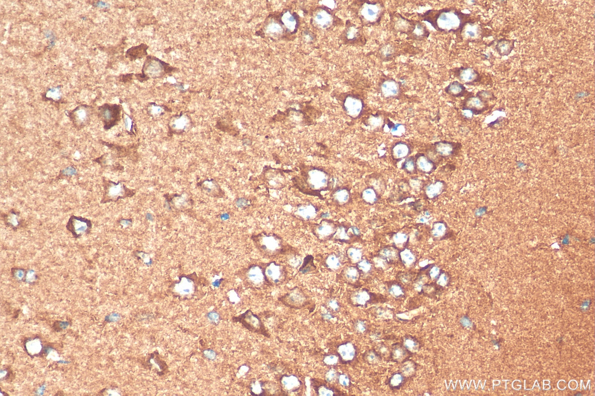
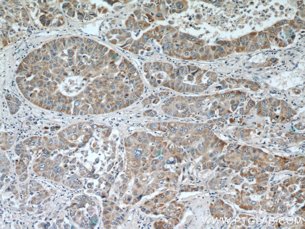
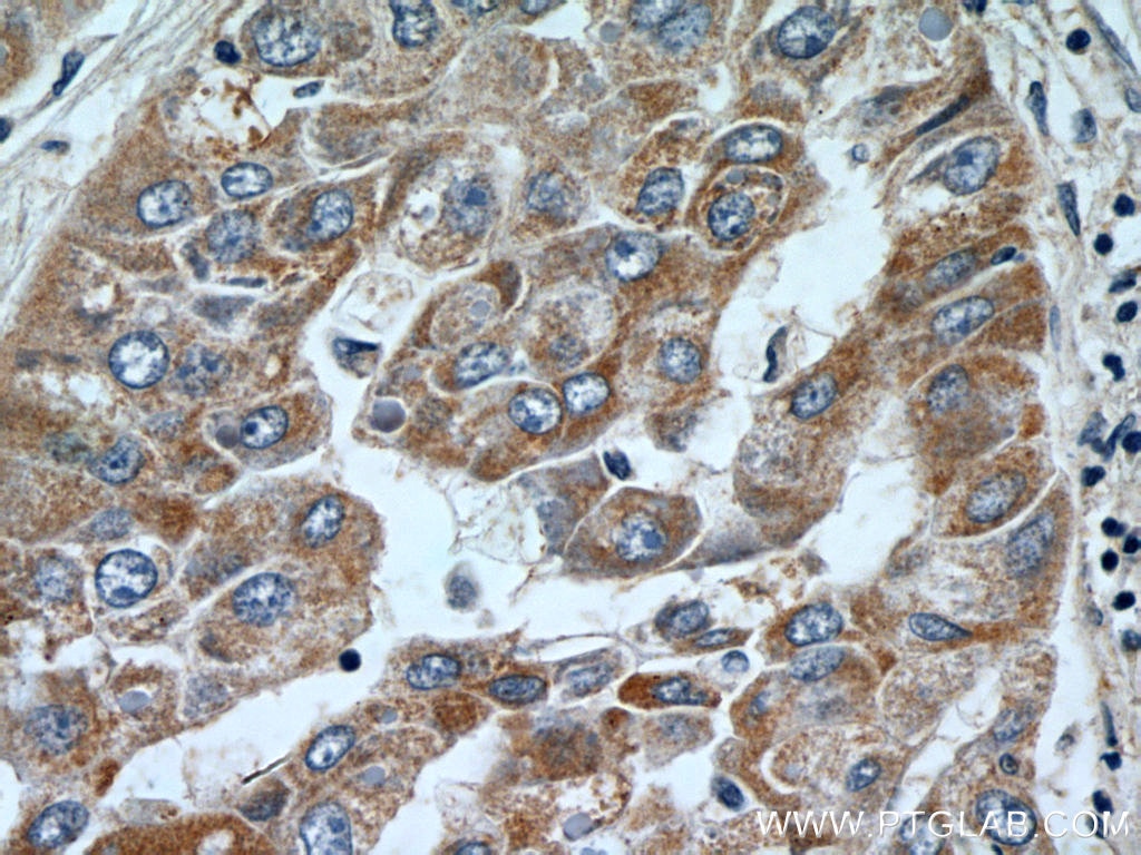
| Positive IHC detected in | mouse brain tissue, human liver cancer tissue Note: suggested antigen retrieval with TE buffer pH 9.0; (*) Alternatively, antigen retrieval may be performed with citrate buffer pH 6.0 |
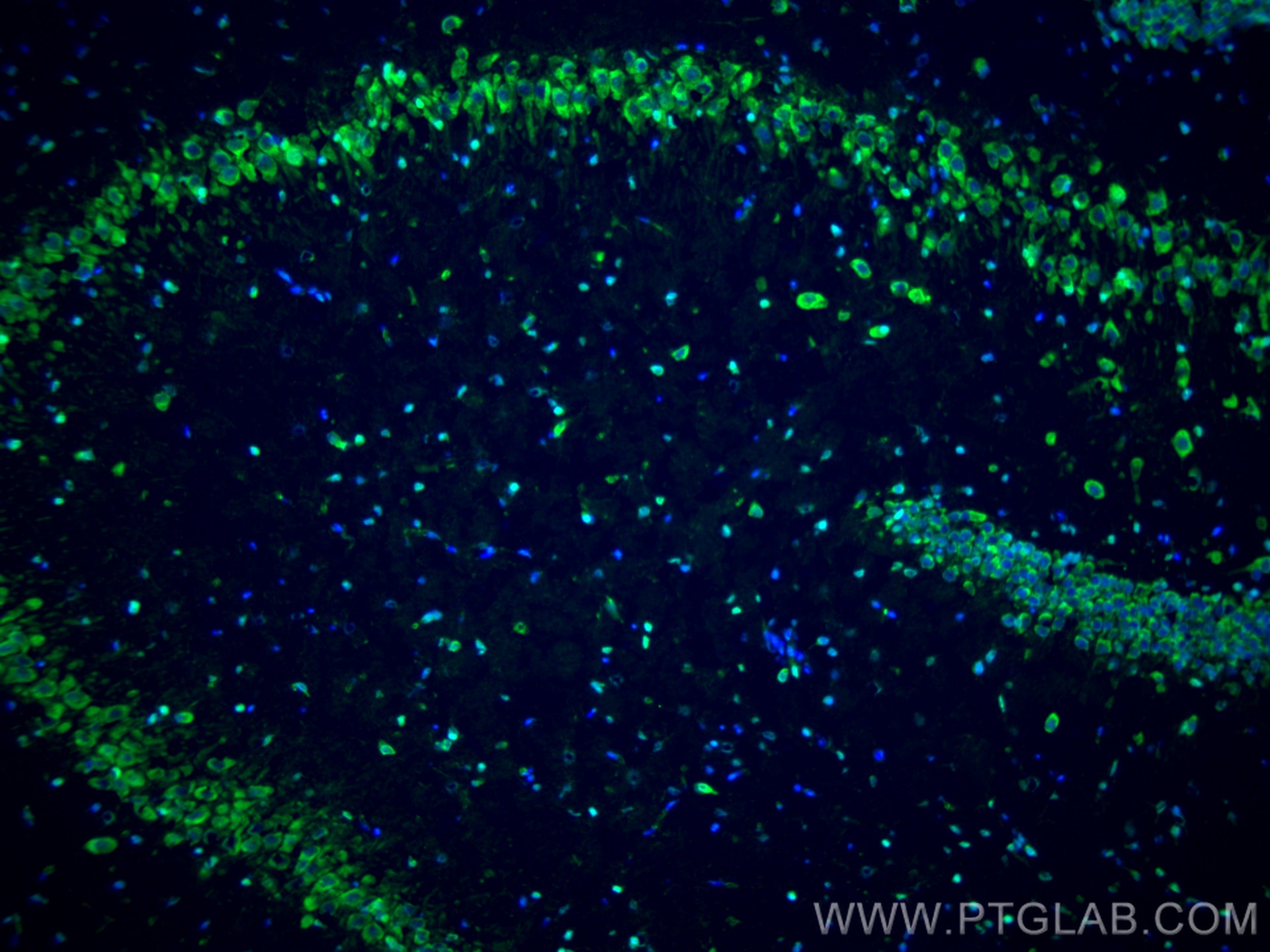
| Positive IF-Fro detected in | rat brain tissue |
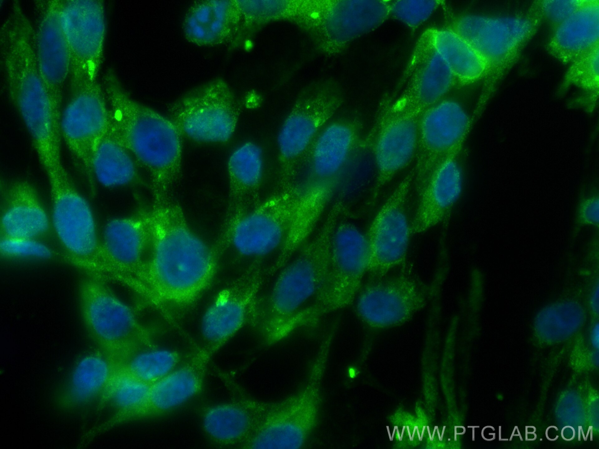
| Positive IF/ICC detected in | SH-SY5Y cells |
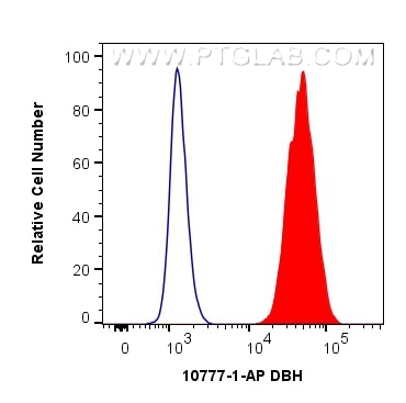
| Positive FC (Intra) detected in | SH-SY5Y cells |
Recommended dilution
| Application | Dilution |
|---|---|
| Western Blot (WB) | WB : 1:1000-1:8000 |
| Immunohistochemistry (IHC) | IHC : 1:50-1:500 |
| Immunofluorescence (IF)-FRO | IF-FRO : 1:50-1:500 |
| Immunofluorescence (IF)/ICC | IF/ICC : 1:200-1:800 |
| Flow Cytometry (FC) (INTRA) | FC (INTRA) : 0.40 ug per 10^6 cells in a 100 µl suspension |
| It is recommended that this reagent should be titrated in each testing system to obtain optimal results. | |
| Sample-dependent, Check data in validation data gallery. | |
Published Applications
| WB | See 4 publications below |
| IHC | See 1 publications below |
| IF | See 3 publications below |
Product Information
10777-1-AP targets Dopamine beta Hydroxylase in WB, IHC, IF/ICC, IF-Fro, FC (Intra), ELISA applications and shows reactivity with human, mouse, rat samples.
| Tested Reactivity | human, mouse, rat |
| Cited Reactivity | human, mouse, zebrafish |
| Host / Isotype | Rabbit / IgG |
| Class | Polyclonal |
| Type | Antibody |
| Immunogen |
CatNo: Ag1196 Product name: Recombinant human DBH protein Source: e coli.-derived, PGEX-4T Tag: GST Domain: 269-603 aa of BC017174 Sequence: CDSKMKPDRLNYCRHVLAAWALGAKAFYYPEEAGLAFGGPGSSRYLRLEVHYHNPLVIEGRNDSSGIRLYYTAKLRRFNAGIMELGLVYTPVMAIPPRETAFILTGYCTDKCTQLALPPSGIHIFASQLHTHLTGRKVVTVLVRDGREWEIVNQDNHYSPHFQEIRMLKKVVSVHPGDVLITSCTYNTEDRELATVGGFGILEEMCVNYVHYYPQTQLELCKSAVDAGFLQKYFHLINRFNNEDVCTCPQASVSQQFTSVPWNSFNRDVLKALYSFAPISMHCNKSSAVRFQGEWNLQPLPKVISTLEEPTPQCPTSQGRSPAGPTVVSIGGGKG Predict reactive species |
| Full Name | DBH |
| Calculated Molecular Weight | 64.8 kDa |
| Observed Molecular Weight | 63 kDa |
| GenBank Accession Number | BC017174 |
| Gene Symbol | DBH |
| Gene ID (NCBI) | 1621 |
| RRID | AB_2277037 |
| Conjugate | Unconjugated |
| Form | Liquid |
| Purification Method | Antigen affinity purification |
| UNIPROT ID | P09172 |
| Storage Buffer | PBS with 0.02% sodium azide and 50% glycerol, pH 7.3. |
| Storage Conditions | Store at -20°C. Stable for one year after shipment. Aliquoting is unnecessary for -20oC storage. 20ul sizes contain 0.1% BSA. |
Background Information
DBH belongs to the copper type II ascorbate-dependent monooxygenase family and catalyzes the oxidative hydroxylation of DBH to norepinephrine. It is almost exclusively located in the adrenal medulla and the synaptic vesicles of postganglionic sympathetic neurons(PMID:11857564).The DBH is glycosylated (63--67 kDa) while still in the membrane(PMID:2325165) and it can form a dimer(PMID:8546710).
Protocols
| Product Specific Protocols | |
|---|---|
| FC protocol for Dopamine beta Hydroxylase antibody 10777-1-AP | Download protocol |
| IF protocol for Dopamine beta Hydroxylase antibody 10777-1-AP | Download protocol |
| IHC protocol for Dopamine beta Hydroxylase antibody 10777-1-AP | Download protocol |
| WB protocol for Dopamine beta Hydroxylase antibody 10777-1-AP | Download protocol |
| Standard Protocols | |
|---|---|
| Click here to view our Standard Protocols |
Publications
| Species | Application | Title |
|---|---|---|
Int J Mol Sci Intermittent Hypoxia Increased the Expression of DBH and PNMT in Neuroblastoma Cells via MicroRNA-375-Mediated Mechanism. | ||
Peptides Markers of the sympathetic, parasympathetic and sensory nervous system are altered in the human diabetic choroid. | ||
Neurobiol Dis Atp7a deficiency induces axonal and myelin developmental defects in zebrafish via ferroptosis. | ||
PLoS One Alteration of BDNF and noradrenergic markers in locus coeruleus in a mouse model of cancer-induced bone pain. | ||
Nat Chem Biol Designing chemigenetic DNA nanotrap for norepinephrine dynamic imaging in organelles. |
Reviews
The reviews below have been submitted by verified Proteintech customers who received an incentive for providing their feedback.
FH Mounika (Verified Customer) (12-25-2025) |
|