

Tested Applications
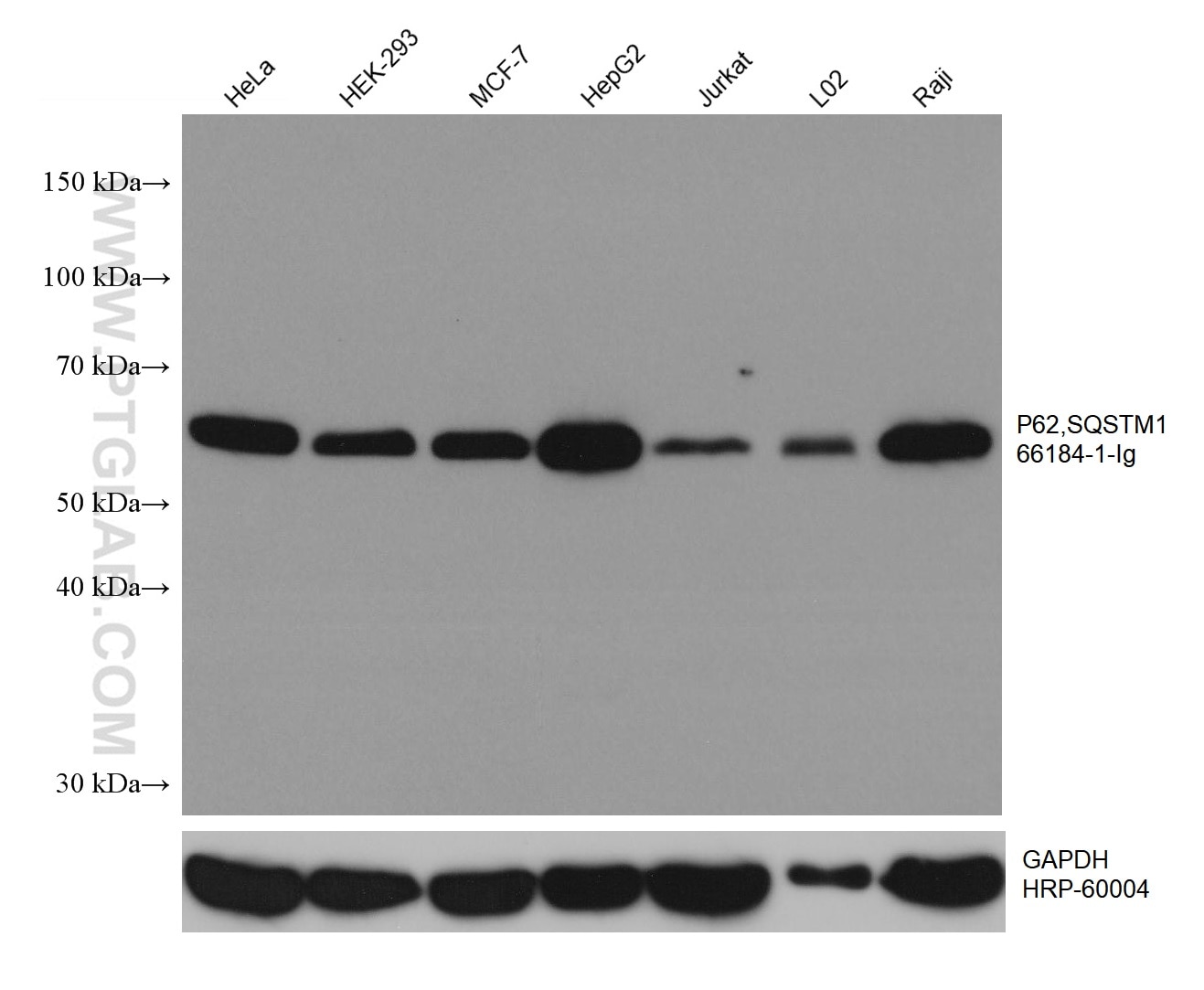
| Positive WB detected in | HeLa cells, U2OS cells, K-562 cells, HEK-293 cells, MCF-7 cells, HepG2 cells, Jurkat cells, L02 cells, Raji cells |
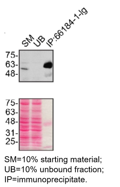
| Positive IP detected in | U2OS cells |
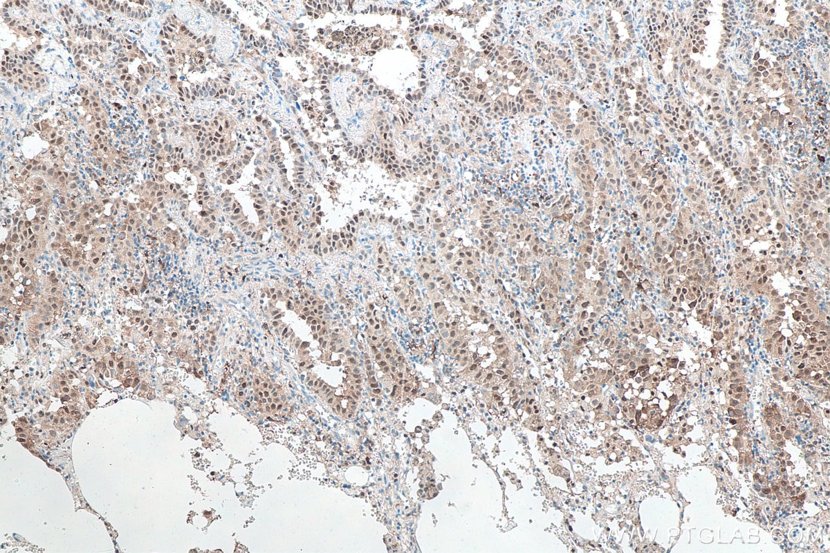
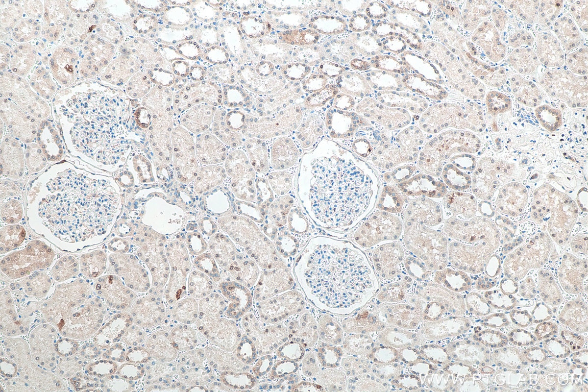
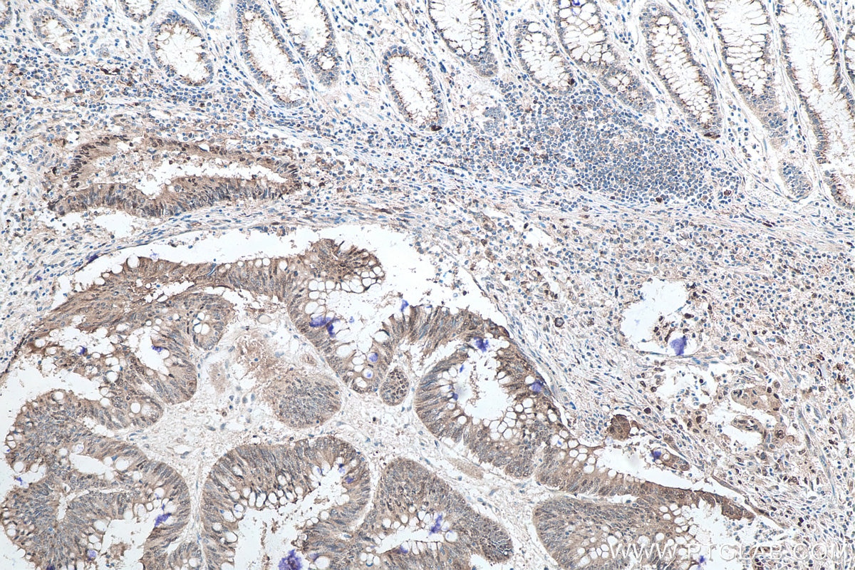
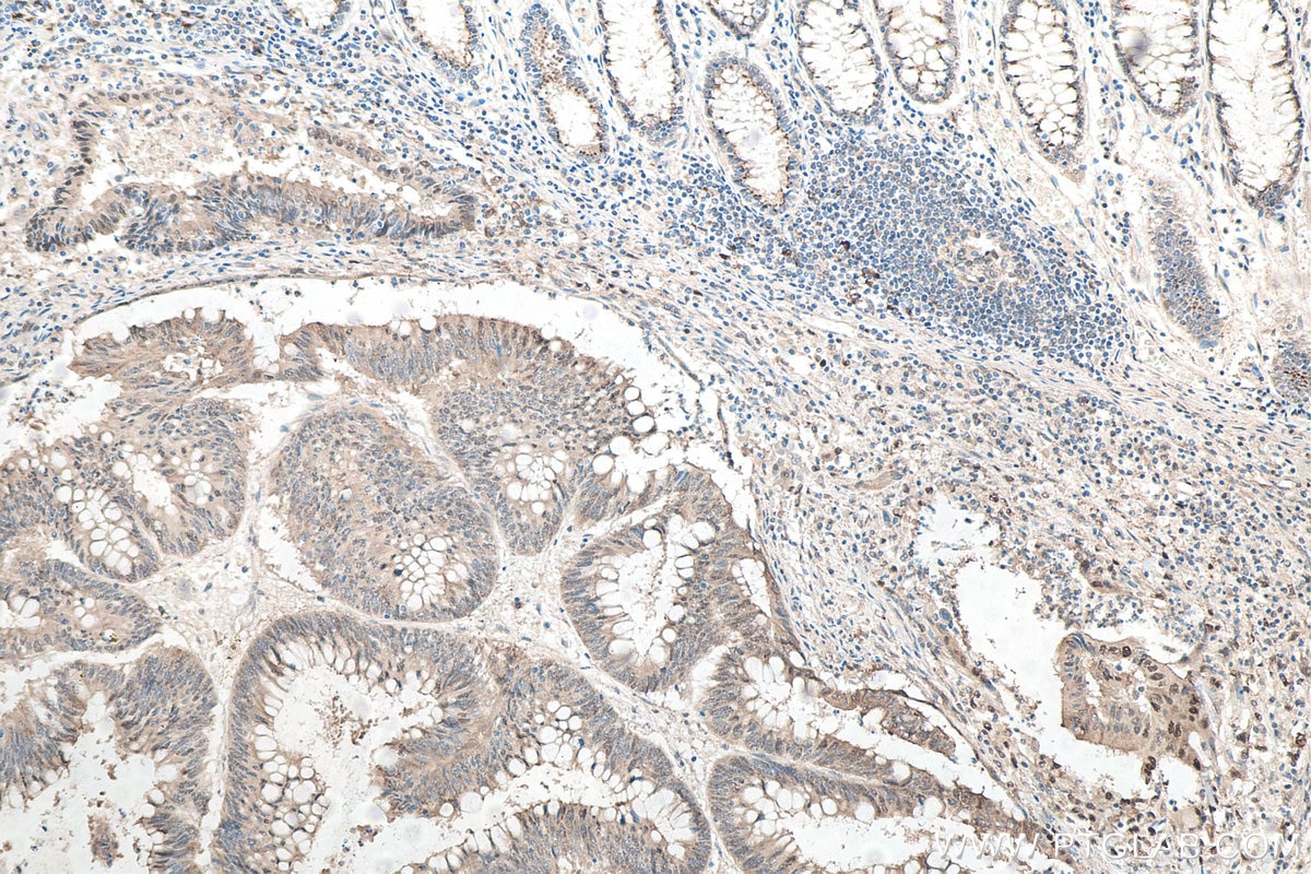
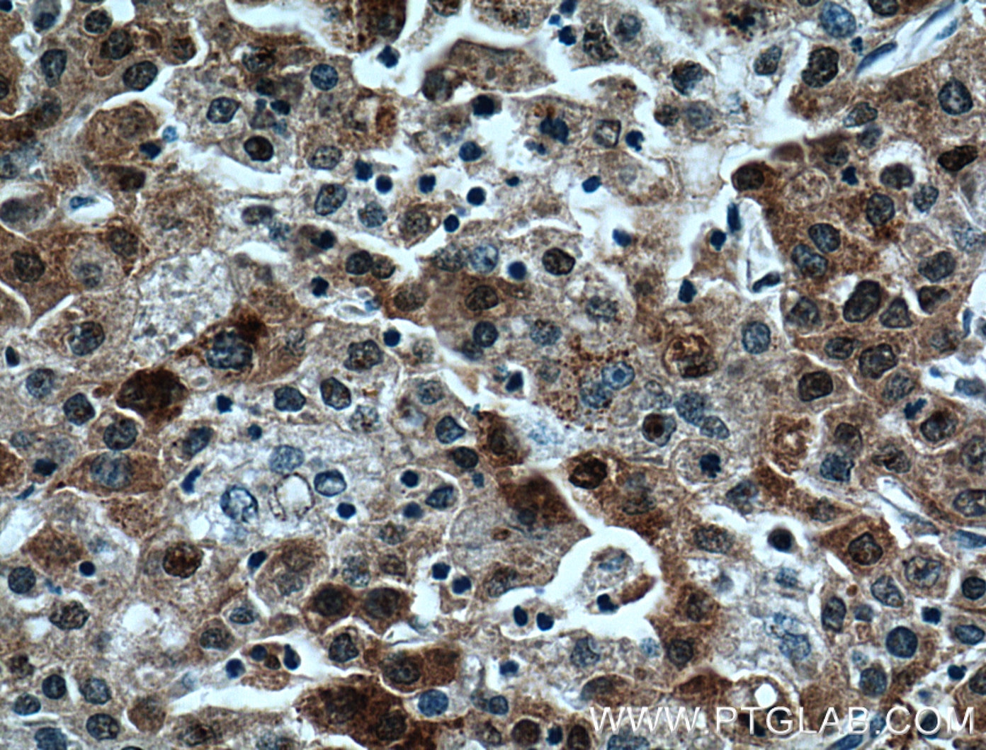
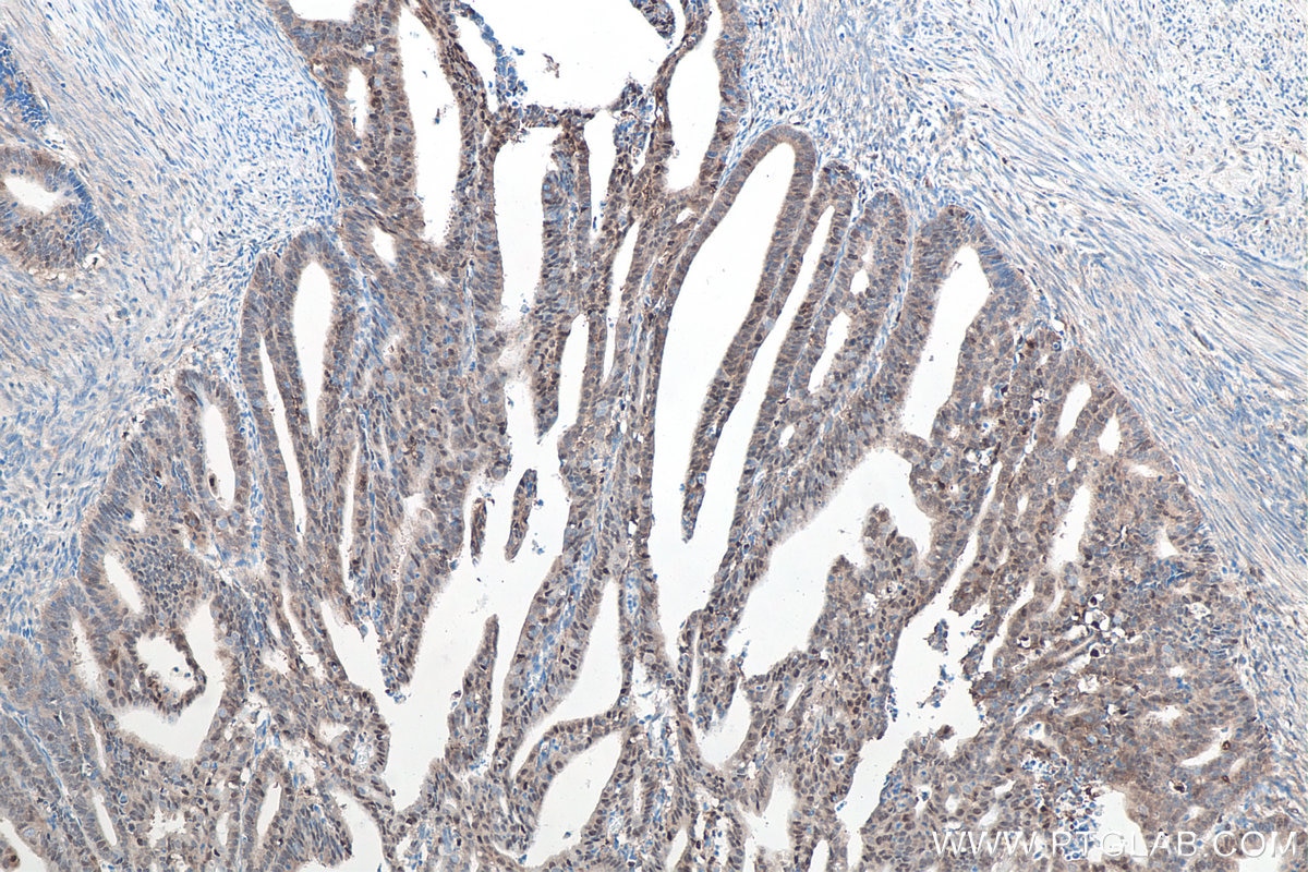
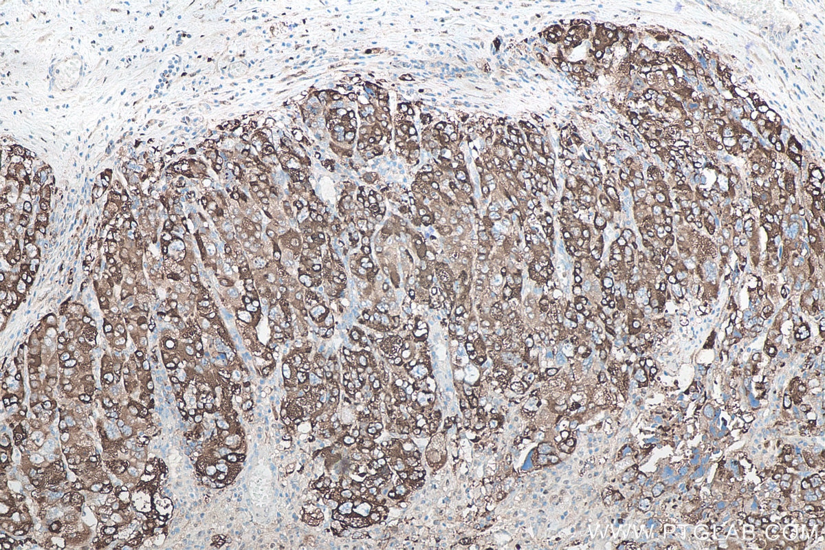
| Positive IHC detected in | human lung cancer tissue, human kidney tissue, human liver cancer tissue, human colon cancer tissue, human endometrial cancer tissue Note: suggested antigen retrieval with TE buffer pH 9.0; (*) Alternatively, antigen retrieval may be performed with citrate buffer pH 6.0 |
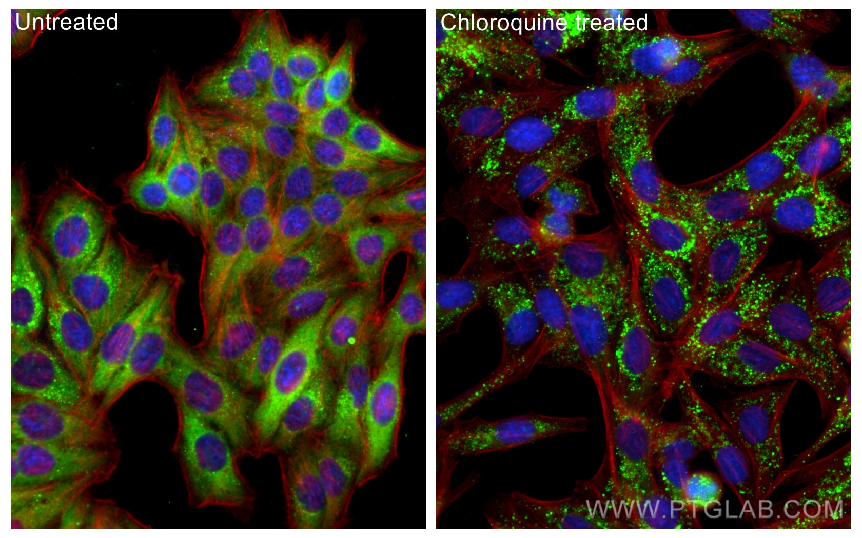
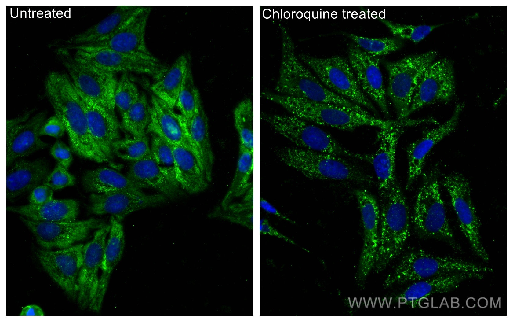
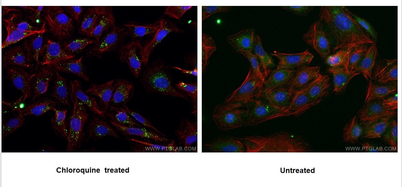
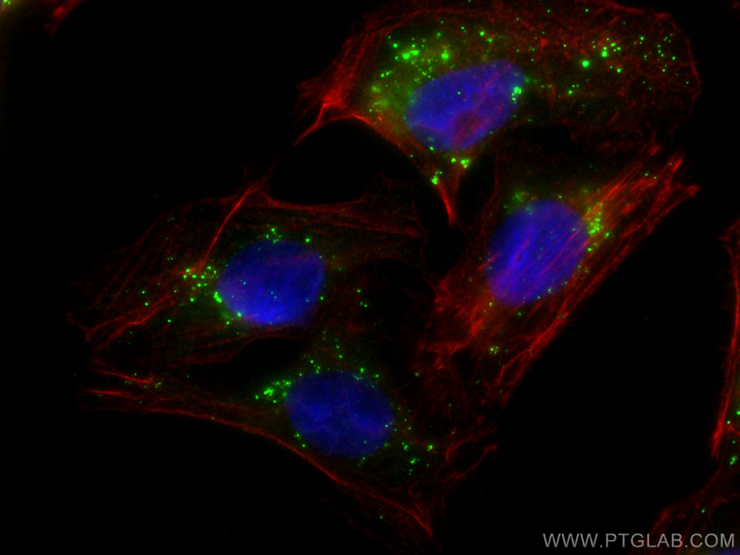
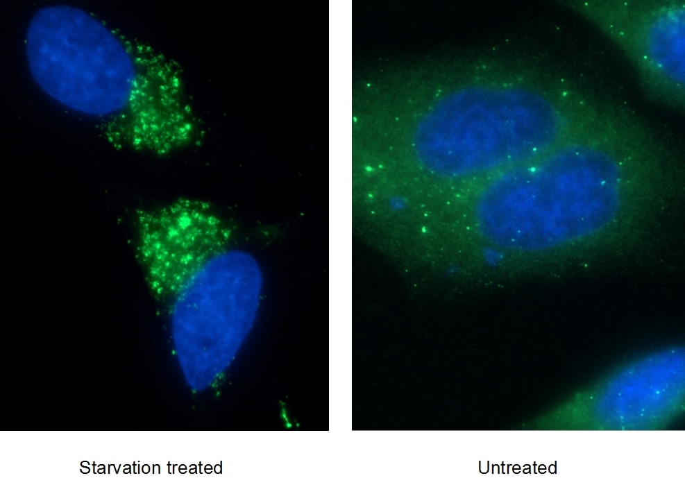
| Positive IF/ICC detected in | Chloroquine treated HepG2 cells, Starvation treated HepG2 cells, U2OS cells, Chloroquine treated U2OS cells |
Recommended dilution
| Application | Dilution |
|---|---|
| Western Blot (WB) | WB : 1:5000-1:50000 |
| Immunoprecipitation (IP) | IP : 0.5-4.0 ug for 1.0-3.0 mg of total protein lysate |
| Immunohistochemistry (IHC) | IHC : 1:2000-1:8000 |
| Immunofluorescence (IF)/ICC | IF/ICC : 1:400-1:1600 |
| It is recommended that this reagent should be titrated in each testing system to obtain optimal results. | |
| Sample-dependent, Check data in validation data gallery. | |
Product Information
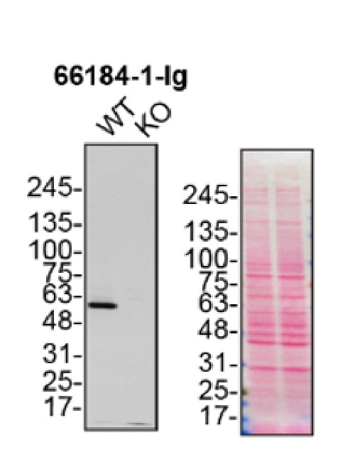
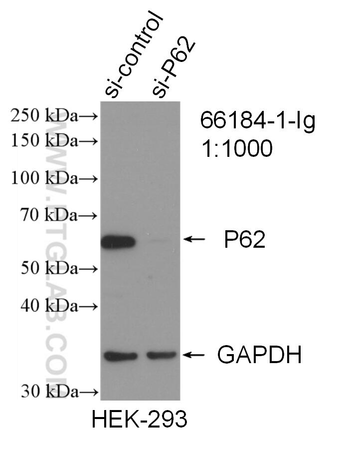
66184-1-Ig targets P62/SQSTM1 in WB, IHC, IF/ICC, IP, CoIP, ELISA applications and shows reactivity with human samples.
| Tested Reactivity | human |
| Cited Reactivity | human, pig, canine, monkey, chicken, bovine, hamster |
| Host / Isotype | Mouse / IgG2b |
| Class | Monoclonal |
| Type | Antibody |
| Immunogen |
CatNo: Ag13131 Product name: Recombinant human P62;SQSTM1 protein Source: e coli.-derived, PGEX-4T Tag: GST Domain: 1-440 aa of BC017222 Sequence: MASLTVKAYLLGKEDAAREIRRFSFCCSPEPEAEAEAAAGPGPCERLLSRVAALFPALRPGGFQAHYRDEDGDLVAFSSDEELTMAMSYVKDDIFRIYIKEKKECRRDHRPPCAQEAPRNMVHPNVICDGCNGPVVGTRYKCSVCPDYDLCSVCEGKGLHRGHTKLAFPSPFGHLSEGFSHSRWLRKVKHGHFGWPGWEMGPPGNWSPRPPRAGEARPGPTAESASGPSEDPSVNFLKNVGESVAAALSPLGIEVDIDVEHGGKRSRLTPVSPESSSTEEKSSSQPSSCCSDPSKPGGNVEGATQSLAEQMRKIALESEGRPEEQMESDNCSGGDDDWTHLSSKEVDPSTGELQSLQMPESEGPSSLDPSQEGPTGLKEAALYPHLPPEADPRLIESLSQMLSMGFSDEGGWLTRLLQTKNYDIGAALDTIQYSKHPPPL Predict reactive species |
| Full Name | sequestosome 1 |
| Calculated Molecular Weight | 48 kDa |
| Observed Molecular Weight | 62 kDa |
| GenBank Accession Number | BC017222 |
| Gene Symbol | P62/SQSTM1 |
| Gene ID (NCBI) | 8878 |
| RRID | AB_2881579 |
| Conjugate | Unconjugated |
| Form | Liquid |
| Purification Method | Protein A purification |
| UNIPROT ID | Q13501 |
| Storage Buffer | PBS with 0.02% sodium azide and 50% glycerol, pH 7.3. |
| Storage Conditions | Store at -20°C. Stable for one year after shipment. Aliquoting is unnecessary for -20oC storage. 20ul sizes contain 0.1% BSA. |
Background Information
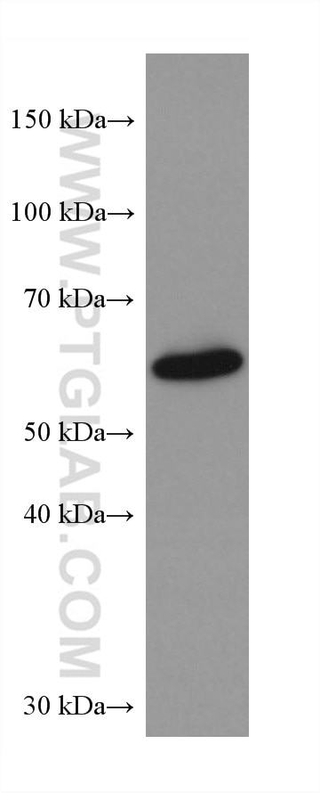
Sequestosome 1 (SQSTM1/p62) is a multifunctional adaptor protein implicated in selective autophagy, cell signaling pathways, and tumorigenesis. p62 has been implicated in shuttling ubiquitinated and sometimes aggregated proteins for autophagic degradation. As a autophagy-specific substrate, p62 is degraded during the autophagic process, which makes intracellular level of p62 as a marker for autophagy flux. p62 is at the cross-roads of several signaling pathways including Ras/ Raf/ MAPK and NFκB and plays important role in cancer. p62 is a component of inclusion bodies/ protein aggregates found in human diseases, including Huntington's disease, Alzheimer's disease, Parkinson's disease in the brain, and nephropathic cystinosis in kidney (PMID: 22074114, 22860231, 22714671). The molecular weight of p62 is predicted as 48/ 38 kDa, while western blot analyses using this antibody demonstrate the major band around 60-62 kDa in various tissues.
Protocols
| Product Specific Protocols | |
|---|---|
| IF protocol for P62/SQSTM1 antibody 66184-1-Ig | Download protocol |
| IHC protocol for P62/SQSTM1 antibody 66184-1-Ig | Download protocol |
| WB protocol for P62/SQSTM1 antibody 66184-1-Ig | Download protocol |
| Standard Protocols | |
|---|---|
| Click here to view our Standard Protocols |
Publications
What published studies show
The anti-p62/SQSTM1 antibody (Cat# 66184-1-Ig) has accumulated 279 citations across journals including Nature Communications, Nature Cell Biology, Autophagy, Cell Reports, Signal Transduction and Targeted Therapy, Advanced Science, Science Advances, Cell Death & Disease, Oncogene, and Free Radical Biology and Medicine. Research fields span autophagy regulation, cancer biology, neurodegeneration, diabetes, cardiovascular disease, inflammation, mitochondrial dynamics, ferroptosis, and drug resistance mechanisms.
| Species | Application | Title |
|---|---|---|
Signal Transduct Target Ther Serine/threonine/tyrosine kinase 1 drives pancreatic carcinogenesis via GSK3β sequestration-mediated Wnt/β-catenin pathway hyperactivation. | ||
Signal Transduct Target Ther Microcolin H, a novel autophagy inducer, exerts potent antitumour activity by targeting PITPα/β | ||
Cell Metab Gut bacteria-driven homovanillic acid alleviates depression by modulating synaptic integrity | ||
Gastroenterology Proteomic characterization identifies clinically relevant subgroups of gastrointestinal stromal tumors | ||
Cell Mol Immunol Dynamic regulation of TBK1 lactylation shapes antiviral immune responses. | ||
Bioact Mater Copper doped bioactive glass promotes matrix vesicles-mediated biomineralization via osteoblast mitophagy and mitochondrial dynamics during bone regeneration |
Reviews
What customers say
Reviews are mixed. One user (5/5) reported excellent results for immunofluorescence on SH-SY5Y cells at 1:100, noting low background in controls and strong p62 puncta upon bafilomycin A treatment. Another user (2/5) found the antibody underperformed on iPSC-derived neurons, with no signal in IF at 1:200 and only a faint band in Western blot at 1:500. Overall, performance appears to vary by cell type and application, with promising results in immunocytochemistry but inconsistent performance in other contexts.
The reviews below have been submitted by verified Proteintech customers who received an incentive for providing their feedback.
Eleonora (Verified Customer) (04-12-2024) | The antibody unfortunately did not work in IF, no signal. In western blot the band is present but very faint.
|
David (Verified Customer) (01-13-2020) | Used for immunocytochemistry. Low signal in control cells, but very strong response when treated with bafilomycin A. Very obvious p62 puncta in cells.
|


















